Major contributions of haematology to genetics - British Society for ...
Major contributions of haematology to genetics - British Society for ...
Major contributions of haematology to genetics - British Society for ...

You also want an ePaper? Increase the reach of your titles
YUMPU automatically turns print PDFs into web optimized ePapers that Google loves.
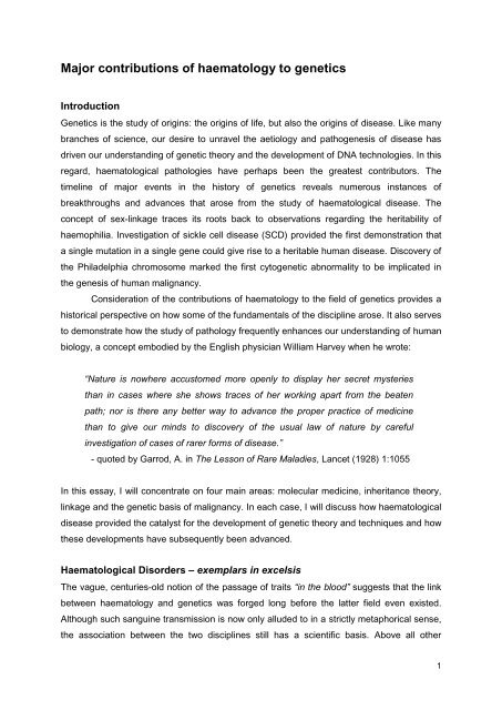
<strong>Major</strong> <strong>contributions</strong> <strong>of</strong> <strong>haema<strong>to</strong>logy</strong> <strong>to</strong> <strong>genetics</strong><br />
Introduction<br />
Genetics is the study <strong>of</strong> origins: the origins <strong>of</strong> life, but also the origins <strong>of</strong> disease. Like many<br />
branches <strong>of</strong> science, our desire <strong>to</strong> unravel the aetiology and pathogenesis <strong>of</strong> disease has<br />
driven our understanding <strong>of</strong> genetic theory and the development <strong>of</strong> DNA technologies. In this<br />
regard, haema<strong>to</strong>logical pathologies have perhaps been the greatest contribu<strong>to</strong>rs. The<br />
timeline <strong>of</strong> major events in the his<strong>to</strong>ry <strong>of</strong> <strong>genetics</strong> reveals numerous instances <strong>of</strong><br />
breakthroughs and advances that arose from the study <strong>of</strong> haema<strong>to</strong>logical disease. The<br />
concept <strong>of</strong> sex-linkage traces its roots back <strong>to</strong> observations regarding the heritability <strong>of</strong><br />
haemophilia. Investigation <strong>of</strong> sickle cell disease (SCD) provided the first demonstration that<br />
a single mutation in a single gene could give rise <strong>to</strong> a heritable human disease. Discovery <strong>of</strong><br />
the Philadelphia chromosome marked the first cy<strong>to</strong>genetic abnormality <strong>to</strong> be implicated in<br />
the genesis <strong>of</strong> human malignancy.<br />
Consideration <strong>of</strong> the <strong>contributions</strong> <strong>of</strong> <strong>haema<strong>to</strong>logy</strong> <strong>to</strong> the field <strong>of</strong> <strong>genetics</strong> provides a<br />
his<strong>to</strong>rical perspective on how some <strong>of</strong> the fundamentals <strong>of</strong> the discipline arose. It also serves<br />
<strong>to</strong> demonstrate how the study <strong>of</strong> pathology frequently enhances our understanding <strong>of</strong> human<br />
biology, a concept embodied by the English physician William Harvey when he wrote:<br />
“Nature is nowhere accus<strong>to</strong>med more openly <strong>to</strong> display her secret mysteries<br />
than in cases where she shows traces <strong>of</strong> her working apart from the beaten<br />
path; nor is there any better way <strong>to</strong> advance the proper practice <strong>of</strong> medicine<br />
than <strong>to</strong> give our minds <strong>to</strong> discovery <strong>of</strong> the usual law <strong>of</strong> nature by careful<br />
investigation <strong>of</strong> cases <strong>of</strong> rarer <strong>for</strong>ms <strong>of</strong> disease.”<br />
- quoted by Garrod, A. in The Lesson <strong>of</strong> Rare Maladies, Lancet (1928) 1:1055<br />
In this essay, I will concentrate on four main areas: molecular medicine, inheritance theory,<br />
linkage and the genetic basis <strong>of</strong> malignancy. In each case, I will discuss how haema<strong>to</strong>logical<br />
disease provided the catalyst <strong>for</strong> the development <strong>of</strong> genetic theory and techniques and how<br />
these developments have subsequently been advanced.<br />
Haema<strong>to</strong>logical Disorders – exemplars in excelsis<br />
The vague, centuries-old notion <strong>of</strong> the passage <strong>of</strong> traits “in the blood” suggests that the link<br />
between <strong>haema<strong>to</strong>logy</strong> and <strong>genetics</strong> was <strong>for</strong>ged long be<strong>for</strong>e the latter field even existed.<br />
Although such sanguine transmission is now only alluded <strong>to</strong> in a strictly metaphorical sense,<br />
the association between the two disciplines still has a scientific basis. Above all other<br />
1
tissues, blood has two key properties which render it the ideal research subject. Firstly, it is<br />
readily accessible via venepuncture and secondly, it is constantly replenished by the bone<br />
marrow. Thus, it is unsurprising that new techniques and ideas have <strong>of</strong>ten been applied <strong>to</strong>,<br />
or arisen from, investigation <strong>of</strong> blood physiology and pathology be<strong>for</strong>e other tissues. As new<br />
knowledge is generally founded on old, some haema<strong>to</strong>logical disorders have found<br />
themselves de fac<strong>to</strong> paradigms <strong>for</strong> our understanding <strong>of</strong> disease as it has evolved from<br />
simple phenotypic observations <strong>to</strong> include his<strong>to</strong>logical, cellular, proteomic and genetic<br />
aspects. SCD is one such disorder and elucidating its origin heralded the birth <strong>of</strong> molecular<br />
and genetic medicine.<br />
Blood was one <strong>of</strong> the first tissues <strong>to</strong> be put under <strong>to</strong> the microscope by its inven<strong>to</strong>r,<br />
seventeenth century Dutch scientist An<strong>to</strong>nie van Leeuwenhoek. In 1840, Hünefeld used a<br />
(much improved) version <strong>of</strong> van Leeuwenhoek’s instrument <strong>to</strong> observe the first crystals <strong>of</strong> a<br />
protein, haemoglobin (Hb) 1 . In 1910, the American cardiologist James Herrick described<br />
“peculiar elongated and sickle-shaped red blood corpuscles in a case <strong>of</strong> severe anaemia”<br />
(figure 1) 2 . Irving Sherman later observed birefringence <strong>of</strong> such sickled red cells under<br />
polarised light demonstrating that the cause <strong>of</strong> such peculiar corpuscles was the<br />
crystallisation <strong>of</strong> haemoglobin 3 . In his seminal 1949 paper, “Sickle Cell Anaemia, a Molecular<br />
Disease” Linus Pauling demonstrated a difference in electrophoretic mobility between<br />
haemoglobin from SCD patients (HbS) and from healthy controls (HbA) 4 . He suggested that<br />
this was due <strong>to</strong> a difference in the number <strong>of</strong> ionisable amino acids in the globin portion – the<br />
concept <strong>of</strong> molecular medicine was born. Eight years later, Vernon Ingram finished what<br />
Pauling had started and identified the single amino acid substitution in β-globin responsible<br />
<strong>for</strong> the sickling <strong>of</strong> red blood cells – glutamate <strong>to</strong> valine at position 6 (figure 2) 5, 6 . Ingram’s<br />
work demonstrated that a difference <strong>of</strong> just one amino acid (and he postulated, one DNA<br />
base – a missense mutation) was enough <strong>to</strong> cause a disease, earning him the moniker, the<br />
“Father <strong>of</strong> Molecular Medicine”.<br />
2
Direction <strong>of</strong> Chroma<strong>to</strong>graphy →<br />
Figure 1: Original drawings from James<br />
Herrick’s 1910 paper describing “peculiar<br />
elongated and sickle-shaped red blood<br />
corpuscles in a case <strong>of</strong> severe anaemia” 2 .<br />
Sickle Cell Haemoglobin (HbS)<br />
Wild-type Haemoglobin (HbA)<br />
Figure 2: These haemoglobin “fingerprints” demonstrate the presence <strong>of</strong> an amino<br />
acid difference between sickle cell and wild-type haemoglobin 5 . Vernon Ingram used<br />
trypsin <strong>to</strong> digest both types <strong>of</strong> haemoglobin in<strong>to</strong> approximately 30 peptide fragments<br />
(each containing ~10 amino acids). Using a combination <strong>of</strong> electrophoresis and<br />
chroma<strong>to</strong>graphy the fragments could be separated <strong>to</strong> produce the molecular<br />
fingerprints shown above. The shaded (sickle cell) and stippled (wild type) spots<br />
belong <strong>to</strong> the peptide fragment showing the difference between the two<br />
haemoglobins. This fragment was then isolated and its amino acids sequenced <strong>to</strong><br />
reveal the specific, single amino acid substitution 6 .<br />
Since Ingram’s discovery, thousands <strong>of</strong> single-gene disorders have been identified<br />
and in many, such as cystic fibrosis, the relevant gene and molecular basis are known. In<br />
addition, Ingram’s work contributed <strong>to</strong> unravelling the genetic code. At the time, it was known<br />
that a triplet <strong>of</strong> DNA bases coded <strong>for</strong> a single amino acid, but early models employed an<br />
overlapping code, with one base contributing <strong>to</strong> more than one codon. Ingram had proved<br />
3
that such a code was impossible as a substitution <strong>of</strong> one or more bases would have affected<br />
several adjacent amino acids, not just one as he had found.<br />
It was fitting that the study <strong>of</strong> a haema<strong>to</strong>logical disorder should usher in the medical<br />
genetic era because we now appreciate that haema<strong>to</strong>logical diseases comprise the second<br />
largest group <strong>of</strong> inherited single-gene disorders (after neurological and neuromuscular<br />
disorders) and that haemoglobinopathies are the commonest Mendelian diseases<br />
worldwide. The very prevalence <strong>of</strong> haemoglobinopathies is in itself intriguing and uncovering<br />
this mystery made a significant contribution <strong>to</strong> <strong>genetics</strong> – specifically, <strong>to</strong> inheritance theory.<br />
Inheritance Theory – Passage <strong>of</strong> Traits in the Blood<br />
Like many diseases, the aetiology <strong>of</strong> SCD was originally woven in<strong>to</strong> a fabric <strong>of</strong> mythology<br />
and local medical understanding. Until recently the Igbo people <strong>of</strong> Nigeria believed that a<br />
second child born with SCD was a reincarnation – or ogbanje – <strong>of</strong> a previous sibling who<br />
had died from the disease 7 . A genetic basis was suspected following the investigation <strong>of</strong> a<br />
family demonstrating the sickling phenomenon and with a his<strong>to</strong>ry <strong>of</strong> multiple deaths from<br />
severe anaemia 8 . Although asymp<strong>to</strong>matic sickle cell trait had been identified, early theories<br />
proposed that SCD was caused by a single dominant gene, expressed strongly in some<br />
(SCD) and weakly in others (trait). Strong evidence that the inheritance was au<strong>to</strong>somal<br />
recessive emerged in 1949 when prospective analysis <strong>of</strong> the parental blood <strong>of</strong> known SCD<br />
patients demonstrated the sickling phenomenon in all cases 9 . This was also indicated by<br />
Pauling’s a<strong>for</strong>ementioned work, in which patients with the trait phenotype had an equal<br />
proportion <strong>of</strong> both HbS and HbA 4 . Even then there was confusion as the prevalence <strong>of</strong> SCD<br />
amongst African populations was lower than expected from the trait prevalence. The<br />
subsequent realisation that children were dying undiagnosed be<strong>for</strong>e they could be included<br />
in prevalence surveys was a sobering solution <strong>to</strong> the discrepancy. Although Mendel’s laws<br />
were well known, their practical application <strong>to</strong> human disease, away from the controlled<br />
Drosophilia-based labora<strong>to</strong>ry environment, proved difficult. SCD was a learning curve and<br />
working through these difficulties provided the academic keys <strong>to</strong> unlock the genetic<br />
mysteries <strong>of</strong> other disorders.<br />
Having established the inheritance pattern <strong>of</strong> SCD, the question remained as <strong>to</strong> why<br />
the HbS gene was so frequent in certain, largely African, populations when the lethality <strong>of</strong><br />
the homozygous state severely curtailed its vertical transmission? The incidence <strong>of</strong> novel<br />
HbA mutation was not high and could not explain the intriguing geographic distribution <strong>of</strong> the<br />
HbS gene described by Anthony Allison. He found high frequencies <strong>of</strong> sickle cell trait carriers<br />
(20-30%) along the cost <strong>of</strong> Kenya and near Lake Vic<strong>to</strong>ria but low frequencies in the<br />
intervening highlands (
parasite, providing a selection pressure <strong>to</strong> maintain its frequency in these populations. This<br />
hypothesis was backed up by population studies and an ethically dubious investigation <strong>of</strong><br />
parasitaemia levels following malaria inoculation <strong>of</strong> individuals with and without sickle cell<br />
trait 10 . Allison concluded that:<br />
“Genetically speaking, this is a balanced polymorphism where the heterozygote<br />
has an advantage over either homozygote.”<br />
Allison’s ground-breaking work made a major contribution <strong>to</strong> the theoretical basis <strong>of</strong><br />
population <strong>genetics</strong> and how we understand gene-environment interactions.<br />
Linkage and the Royal Disease<br />
Just as SCD is perhaps the archetypal au<strong>to</strong>somal recessive disorder and provided the<br />
disease model from which the concept <strong>of</strong> heterozygous advantage arose, so the study <strong>of</strong><br />
haemophilia was invaluable <strong>for</strong> the development <strong>of</strong> theories <strong>of</strong> linkage. Christian Friedrich<br />
Nasse first commentated on sex-linkage in his 1820 treatise on hereditary bleeding<br />
disorders 11 . Nasse observed that the hereditary disposition <strong>to</strong> fatal bleeding (the term<br />
haemophilia did not arise until 1828) only occurred in males and that these males did not<br />
transmit the disease themselves, rather it was transmitted by their unaffected female<br />
relations. An experimentally validated theory <strong>of</strong> sex-linkage arose from Thomas Hunt<br />
Morgan’s observations in 1910 that the white eye phenotype <strong>of</strong> Drosophilia followed patterns<br />
<strong>of</strong> sex chromosome inheritance 12 . A year later the genes causing both colour blindness and<br />
haemophilia were mapped <strong>to</strong> the X chromosome, resulting in the development <strong>of</strong> one <strong>of</strong> the<br />
major techniques in human genome analysis – genetic linkage (the tendency <strong>for</strong> certain<br />
alleles <strong>to</strong> be inherited <strong>to</strong>gether). In 1937, Bell and Haldane reported the first genetic linkage<br />
in humans, between haemophilia and colour blindness 13 . This linkage was <strong>of</strong> practical value<br />
in itself as it allowed the prediction <strong>of</strong> carrier status in females <strong>of</strong> families displaying both<br />
traits. In addition, Bell and Haldane’s research methodology paved the way <strong>for</strong> the<br />
identification <strong>of</strong> other genetically linked traits which facilitated early attempts at antenatal<br />
diagnosis and is still employed in the localisation <strong>of</strong> genes <strong>for</strong> specific genetic disorders.<br />
In the mid-19 th century, haemophilia was described as “the most hereditary <strong>of</strong> all<br />
diseases” and its prevalence amongst the European monarchy served <strong>to</strong> frame haemophilia<br />
as the exemplary hereditary disorder amongst both experts and laypeople 11 . Given such<br />
status, it was natural that questions <strong>of</strong> reproductive control concerning haemophilia should<br />
arise following the popularisation <strong>of</strong> eugenics in the early 1900s. The principal debate<br />
surrounded whether the reproduction <strong>of</strong> females from haemophilia families should be<br />
prohibited due <strong>to</strong> their carrier status. Although opposed <strong>to</strong> sterilisation, Haldane was a<br />
5
proponent <strong>of</strong> such negative eugenics 14 and it is ironic that it should be a study by Haldane<br />
himself that undermined the notion that haemophilia could be eradicated by preventing<br />
carriers from reproducing. In an insightful paper, Haldane calculated the mutation rate <strong>for</strong> the<br />
haemophilia gene, providing the first such (accurate) estimate <strong>for</strong> a human gene 15 . The<br />
haemophilia gene was found <strong>to</strong> have a high rate <strong>of</strong> mutation and Haldane concluded that<br />
approximately one third <strong>of</strong> haemophilia cases arise de novo, from spontaneous mutation.<br />
Haldane then remarked that haemophilia in Queen Vic<strong>to</strong>ria’s children most probably arose<br />
from a gene mutation,<br />
“in the nucleus <strong>of</strong> a cell in one <strong>of</strong> the testicles <strong>of</strong> Edward, Duke <strong>of</strong> Kent,<br />
Vic<strong>to</strong>ria’s Father, in the year 1818.” 11<br />
While the exactitude <strong>of</strong> his statement remains unknown, we do know that with haemophilia<br />
as his muse, Haldane’s insights in<strong>to</strong> X-linked inheritance, genetic linkage and mutation rates<br />
made lasting <strong>contributions</strong> <strong>to</strong> genetic theory and were a rational <strong>for</strong>ce against fundamentalist<br />
eugenic principals.<br />
Molecular Carcinogenesis – the Cy<strong>to</strong>genetic Revolution<br />
Although <strong>genetics</strong> as a discipline originally arose from the study <strong>of</strong> heredity it was not long<br />
be<strong>for</strong>e we began <strong>to</strong> appreciate the effects <strong>of</strong> acquired genetic mutation. One <strong>of</strong> the first<br />
proposals <strong>of</strong> a genetic basis <strong>for</strong> cancer came from biologist Theodore Boveri. In 1914, he<br />
hypothesised that human malignancies originated from mi<strong>to</strong>tic abnormalities that resulted in<br />
aneuploidy or other, more subtle genetic abnormalities that did not involve the entire<br />
chromosome 16 . Definitive pro<strong>of</strong> <strong>of</strong> Boveri’s theories necessitated the development <strong>of</strong> more<br />
sophisticated molecular genetic techniques and had <strong>to</strong> wait 45 years <strong>for</strong> Nowell and<br />
Hunger<strong>for</strong>d’s <strong>of</strong>t-cited Science abstract describing the presence <strong>of</strong> “A minute chromosome in<br />
human chronic granulocytic leukaemia” 17 . This distinctive chromosome was only observed in<br />
neoplastic leukocytes and was designated the Philadelphia chromosome, after the city in<br />
which it was discovered (figure 3a). This landmark “paper” is recognised as the first<br />
example <strong>of</strong> a consistent chromosome abnormality in a human neoplasm. Further support <strong>for</strong><br />
the cy<strong>to</strong>genetic basis <strong>of</strong> cancer came from studies demonstrating increased spontaneous<br />
breakage <strong>of</strong> chromosomes prepared from normal circulating lymphocytes from patients with<br />
a hereditary leukaemic disposition, such as Fanconi anaemia. Despite this evidence, other<br />
consistent cy<strong>to</strong>genetic abnormalities associated with tumours failed <strong>to</strong> materialise and the<br />
general consensus was that these anomalies were the result, rather than the cause, <strong>of</strong><br />
malignancies. The development <strong>of</strong> chromosome banding in 1970 allowed the accurate<br />
identification <strong>of</strong> individual chromosomes and the recognition <strong>of</strong> small translocations and<br />
6
other rearrangements. Application <strong>of</strong> banding techniques <strong>to</strong> human malignancies led <strong>to</strong> a<br />
rapidly expanding list <strong>of</strong> specific tumour-associated cy<strong>to</strong>genetic alterations, with the<br />
Philadelphia chromosome sitting proudly at the <strong>to</strong>p. Rowley et al. demonstrated that the<br />
“minute chromosome” is actually a truncated chromosome 22, resulting from a reciprocal<br />
translocation between chromosomes 22 and 9 (figure 3b) 18 . Further work in the mid-80s and<br />
early 90s identified the resultant fusion gene as BCR-ABL. The product <strong>of</strong> this genetic hybrid<br />
was an aberrant protein tyrosine kinase that gave leukaemic myeloid cells a survival<br />
advantage over their non-leukaemic counterparts.<br />
A<br />
B<br />
Figure 3: A, shows a karyotype from one <strong>of</strong> the<br />
early studies on chromosome abnormalities in<br />
CML 19 . The minute, Philadelphia (Ph)<br />
chromosome is labelled. Note that the Ph<br />
chromosome was first assigned <strong>to</strong> chromosome<br />
21 and later changed, by convention, <strong>to</strong> 22. B,<br />
depicts the reciprocal translocation between<br />
chromosomes 9 and 22. The similarity between<br />
the additional material at the end <strong>of</strong> the long<br />
arm <strong>of</strong> chromosome 9 (arrow) and that missing<br />
from the long arm <strong>of</strong> chromosome 22 suggested<br />
the translocation.<br />
Chronic myeloid leukaemia (CML) is another example <strong>of</strong> a haema<strong>to</strong>logical disorder<br />
whose pathophysiology serves as the archetype <strong>for</strong> other diseases. The discovery and study<br />
<strong>of</strong> the Philadelphia chromosome and its tumourigenic product proved the genetic origin <strong>of</strong><br />
cancer and laid the foundation <strong>for</strong> our current understanding <strong>of</strong> molecular carcinogenesis.<br />
Nowell’s work also left a legacy that occupies researchers <strong>to</strong> this day - the concept <strong>of</strong> cancer<br />
stem cells, the discovery <strong>of</strong> countless cy<strong>to</strong>genetic abnormalities and the appreciation <strong>of</strong> the<br />
crucial role <strong>of</strong> kinases in malignancy. Interestingly, his work also gave rise <strong>to</strong> one example <strong>of</strong><br />
a growing phenomenon – the <strong>contributions</strong> <strong>of</strong> <strong>genetics</strong> <strong>to</strong> <strong>haema<strong>to</strong>logy</strong>.<br />
7
Concluding Remarks on a Symbiotic Relationship<br />
Blood is <strong>of</strong>ten seen as the essence <strong>of</strong> an individual which, along with its accessibility,<br />
rendered it the subject <strong>of</strong> much early scientific enquiry. Consequently haema<strong>to</strong>logical<br />
disorders have found themselves pathophysiological exemplars in numerous fields, but<br />
particularly <strong>genetics</strong>. Sanguine disease has been a foil <strong>for</strong> <strong>genetics</strong>; contributing <strong>to</strong> many <strong>of</strong><br />
its sub-disciplines, including molecular medicine, inheritance theory, eugenics and<br />
carcinogenesis. However, <strong>genetics</strong> has now started <strong>to</strong> repay its debt through the<br />
development <strong>of</strong> novel diagnostic tests and targeted therapies. Techniques such as<br />
polymerase chain reaction have allowed the prenatal diagnosis <strong>of</strong> a variety <strong>of</strong> disorders,<br />
including haemoglobinopathies, and have significantly reduced the number <strong>of</strong> children born<br />
with thalassaemia in certain parts <strong>of</strong> the world. Recombinant human fac<strong>to</strong>r VIII has obviated<br />
the need <strong>for</strong> haemophiliacs <strong>to</strong> use allogeneic blood products, dramatically improving<br />
haemophilia therapy. Following the identification <strong>of</strong> the BCR-ABL protein, rational drug<br />
design led <strong>to</strong> the development <strong>of</strong> a specific tyrosine kinase inhibi<strong>to</strong>r – imatinib – which<br />
revolutionised the treatment <strong>of</strong> CML and increased 5-year survival from 70 <strong>to</strong> 89% 20 . We can<br />
even dare <strong>to</strong> hope that gene therapy may <strong>of</strong>fer a cure <strong>for</strong> monogenic disorders. The<br />
reciprocal <strong>contributions</strong> between <strong>haema<strong>to</strong>logy</strong> and <strong>genetics</strong> serve as a prime example <strong>of</strong><br />
what can be achieved by cooperation between disciplines and the translation <strong>of</strong> basic<br />
science <strong>to</strong> clinical medicine. It seems likely that such symbiosis will continue <strong>to</strong> benefit both<br />
parties well in<strong>to</strong> the distant future.<br />
Word Count: 2,494<br />
8
References<br />
1. Hünefeld FL. 1840. Die Chemismus in der Thienschen Organization, Leipzig,. 160<br />
[Taken from Reichert and Brown, 1909].<br />
2. Herrick JB. 1910. Peculiar elongated and sickle-shaped red blood corpuscles in a<br />
case <strong>of</strong> severe anaemia. Archives <strong>of</strong> Internal Medicine. 6:517-21.<br />
3. Sherman IJ. 1940. Bulletin <strong>of</strong> Johns Hopkins Hospital. 67:309.<br />
4. Pauling L, Itano HA, et al. 1949. Sickle cell anemia, a molecular disease. Science.<br />
110:543-8.<br />
5. Ingram VM. 1956. A specific chemical difference between the globins <strong>of</strong> normal<br />
human and sickle-cell anaemia haemoglobin. Nature. 178:792-4.<br />
6. Ingram VM. 1957. Gene mutations in human haemoglobin: the chemical difference<br />
between normal and sickle cell haemoglobin. Nature. 180:326-8.<br />
7. Nzewi E. 2001. Malevolent ogbanje: recurrent reincarnation or sickle cell disease?<br />
Social Science and Medicine. 52:1403-16.<br />
8. Emmel VE. 1917. A study <strong>of</strong> the erythrocytes in a case <strong>of</strong> severe anaemia with<br />
elongated and sickle-shaped red blood corpuscles. Archives <strong>of</strong> Internal Medicine. 20:586-98.<br />
9. Neel JV. 1949. The Inheritance <strong>of</strong> Sickle Cell Anemia. Science. 110:64-6.<br />
10. Allison AC. 2004. Two lessons from the interface <strong>of</strong> <strong>genetics</strong> and medicine. Genetics.<br />
166:1591-9.<br />
11. Pember<strong>to</strong>n S. 2011. The Bleeding Disease: Hemophilia and the Unintended<br />
Consequences <strong>of</strong> Medical Progress. 1 ed: The Johns Hopkins University Press.<br />
12. Morgan TH. 1910. Sex-limited inheritance in Drosophilia. Science. 32:120-2.<br />
13. Bell J. 1937. The Linkage between the Genes <strong>for</strong> Colour-Blindness and Haemophilia<br />
in Man. Proceedings <strong>of</strong> the Royal <strong>Society</strong> <strong>of</strong> London Series B, Biological sciences. 123:119-<br />
50.<br />
14. Sarkar S. 1992. Science, philosophy, and politics in the work <strong>of</strong> JBS Haldane, 1922–<br />
1937. Biology & Philosophy. 7:385-409.<br />
15. Haldane JBS. 1946. The mutation rate <strong>of</strong> the gene <strong>for</strong> haemophilia, and its<br />
segregation ratios in males and females. Annals <strong>of</strong> Human Genetics. 13:262-71.<br />
16. Bovari T. 1914. Zur Frage der Entstehung maligner Tumoren. Gustav Fischer Jena,<br />
Germany. 64 pp.<br />
17. Nowell PC, Hunger<strong>for</strong>d D. 1960. A minute chromosome in human chronic<br />
granulocytic leukemia. Science. 132:1497.<br />
18. Rowley JD. 1973. Letter: A new consistent chromosomal abnormality in chronic<br />
myelogenous leukaemia identified by quinacrine fluorescence and Giemsa staining. Nature.<br />
243:290-3.<br />
9
19. Tough IM, Court Brown WM, Baikie AG, et al. 1961. Cy<strong>to</strong>genetic studies in chronic<br />
myeloid leukaemia and acute leukaemia associated with monogolism. Lancet. 1:411-7.<br />
20. Sherbenou DW and Drucker BJ. 2007. Applying the Discovery <strong>of</strong> the Philadelphia<br />
Chromosome. Journal <strong>of</strong> Clinical Investigation. 116: 2067-2074.<br />
10
<strong>Major</strong><br />
Contributions<br />
<strong>of</strong><br />
Haema<strong>to</strong>logy<br />
<strong>to</strong> Genetics<br />
Arun Arujun Bhaskaran<br />
Word count: 2,494 excluding<br />
references and figures<br />
Ask anyone <strong>to</strong> name a common genetic disorder and their answer would probably be<br />
sickle cell anaemia. Indeed, sickle cell anaemia was the first diagnosed disease <strong>to</strong> be<br />
genetically characterised when Guthrie and Huck <strong>of</strong> John Hopkins University Medical<br />
School, analysed pedigree charts from two families in 1923. However, sickle cell<br />
anaemia is only one <strong>of</strong> many blood disorders with a genetic basis. Haema<strong>to</strong>logy has<br />
made pr<strong>of</strong>ound <strong>contributions</strong> <strong>to</strong> <strong>genetics</strong>, furthering our understanding <strong>of</strong> the field. This<br />
essay aims <strong>to</strong> explore how <strong>haema<strong>to</strong>logy</strong> has helped in our quest <strong>to</strong> make sense <strong>of</strong> the<br />
‘stuff <strong>of</strong> life’ using various examples from haemophilia <strong>to</strong> thalassemia.
HAEMATOLOGY AND AUTOSOMAL DISORDERS<br />
Figure 1 – Gregor Mendel; an<br />
Austrian monk considered the<br />
‘father <strong>of</strong> <strong>genetics</strong>’<br />
Genetics is arguably the ‘baby’ <strong>of</strong> modern day science, still in<br />
its developing stages. However, we have made overwhelming<br />
medical advances with what little we know. It began with<br />
Gregor Mendel who proposed all organisms possess 2<br />
hereditary ‘fac<strong>to</strong>rs’ <strong>for</strong> any given characteristic; one <strong>of</strong> which is<br />
‘dominant’ and overrides the effect <strong>of</strong> a ‘recessive’ fac<strong>to</strong>r on<br />
the organism’s physical traits. These fac<strong>to</strong>rs come from the<br />
organism’s parents and are passed down through generations<br />
in a randomised fashion 1 . The discovery <strong>of</strong> sickle cell<br />
anaemia and various other blood disorders provided further<br />
evidence <strong>for</strong> this and helped shape our understanding <strong>of</strong><br />
various inheritance patterns.<br />
Erythrocytes contain haemoglobin, which transports oxygen. Normal adult haemoglobin<br />
consists <strong>of</strong> 2 α and 2 β globin chains. In sickle cell anaemia, a glutamate residue is<br />
substituted <strong>for</strong> a valine on one <strong>of</strong> the β-globin chains <strong>for</strong>ming haemoglobin S. This is prone<br />
<strong>to</strong> polymerisation. Polymerised haemoglobin dis<strong>to</strong>rts erythrocytes, making them rigid and<br />
sickled (crescent shaped). These rigid and sickled cells can occlude vessels and block blood<br />
flow causing problems like gall s<strong>to</strong>nes, hypertension, retinopathy and renal failure. The allele<br />
responsible is located on chromosome 11. Because it is recessive, only homozygotes<br />
(individuals carrying 2 copies <strong>of</strong> the mutated allele) are affected 2 .<br />
Figure 2 – Electron micrographs comparing normal and sickled erythrocytes<br />
P a g e | 1
Figure 3 – Original pedigree chart created by Guthrie and Huck illustrating the recessive<br />
nature <strong>of</strong> sickle cell anaemia<br />
Sickle cell anaemia also exhibits overdominance. Since it was first described, clinicians<br />
noted that the disease ran most frequently in families <strong>of</strong> African descent. Indeed, the<br />
prevalence <strong>of</strong> sickle cell anaemia is greatest in tropical and sub-tropical regions <strong>of</strong> the world<br />
such as Sub-Saharan Africa. 3<br />
Figure 4 – Figure illustrating the prevalence <strong>of</strong> sickle cell anaemia worldwide (darker colour<br />
indicates greater prevalence)<br />
P a g e | 2
It was later revealed that the geographical distribution <strong>of</strong> sickle cell anaemia corresponded<br />
with that <strong>of</strong> malaria. Studies found that the sickle cell trait is actually protective against<br />
malaria though exact mechanisms are yet <strong>to</strong> be determined. It may be that the Plasmodium<br />
falciparum parasites which cause malaria are unable <strong>to</strong> grow and reproduce effectively<br />
inside HbS-containing erythrocytes. However, a recent study by Williams et al. at the Kenya<br />
Medical Research Institute found that while children with the sickle cell trait have enhanced<br />
immunity <strong>to</strong> the parasite, the level <strong>of</strong> which increases with age, suggesting a role <strong>for</strong><br />
acquired immune processes <strong>to</strong>o 4 .<br />
So what’s all this got <strong>to</strong> do with genes? Heterozygotes (carriers <strong>of</strong> sickle cell anaemia)<br />
produce small amounts <strong>of</strong> HbS erythrocytes, though not enough <strong>to</strong> produce symp<strong>to</strong>ms.<br />
However, they are still resistant <strong>to</strong> malaria, meaning they have a survival advantage over<br />
both homozygous dominant individuals (with no malaria resistance) and homozygous<br />
recessive individuals (with sickle cell anaemia). This phenomenon where homozygotes ‘get<br />
the best <strong>of</strong> both worlds’ is known as overdominance and is described using the Gillespie<br />
model, which is used widely in the field <strong>of</strong> population <strong>genetics</strong> 5 .<br />
HAEMATOLOGY AND SEX-LINKED DISORDERS<br />
Haema<strong>to</strong>logy has helped us understand sexlinked<br />
disorders, as well as au<strong>to</strong>somal ones.<br />
Haemophilia A is a sex-linked disorder caused<br />
by a loss <strong>of</strong> function mutation in the gene<br />
encoding fac<strong>to</strong>r VIII near the tip <strong>of</strong> the q arm <strong>of</strong><br />
the X-chromosome. Commonly patients have<br />
deletions in the fac<strong>to</strong>r VIII gene but some severe<br />
cases exhibit a ‘flip-flop’ inversion where the<br />
gene is disrupted by an inversion at the end <strong>of</strong><br />
the X chromosome. These mutations result in<br />
little or no synthesis <strong>of</strong> fac<strong>to</strong>r VIII, which is<br />
needed in the blood coagulation cascade.<br />
Without it, clots cannot <strong>for</strong>m effectively following<br />
blood vessel damage and pr<strong>of</strong>use bleeding<br />
occurs. 6<br />
Figure 5 – Diagram illustrating the so called<br />
‘flip-flop’ inversion in some severe cases <strong>of</strong><br />
haemophilia A<br />
Abu al-Qasim al-Zahrawi, in the 10 th century AD, was the first physician <strong>to</strong> document men<br />
bleeding <strong>to</strong> death following minor injuries 7 . It was not until sometime later when the <strong>genetics</strong><br />
<strong>of</strong> the disorder became clear. It was frequently observed that males were more commonly<br />
P a g e | 3
affected by this bleeding disorder than women, even in members <strong>of</strong> the European royal<br />
family.<br />
Figure 6 – Pedigree chart showing the inheritance <strong>of</strong> haemophilia A in the <strong>British</strong> Royal Family<br />
In X-linked disorders like haemophilia, men are more affected than females. This is due <strong>to</strong> a<br />
process known as X inactivation. Females possess 2 copies <strong>of</strong> the X chromosome; during<br />
development, in utero, females silence one copy in all body cells. This process is completely<br />
random. Usually though, copies with mutations such as fac<strong>to</strong>r VIII deletions or inversions are<br />
‘inactivated’ giving rise <strong>to</strong> no or very mild symp<strong>to</strong>ms. In some exceptional cases this doesn’t<br />
work out well and the ‘good’ X chromosome is turned <strong>of</strong>f giving rise <strong>to</strong> symp<strong>to</strong>ms - skewed<br />
X-inactivation. Females with Turner’s syndrome with only 1 X-chromosome are also at risk.<br />
Figure 7 – Young girl with Turner’s Syndrome; symp<strong>to</strong>ms<br />
include short stature, infertility, skin folds, cardiac<br />
mal<strong>for</strong>mations and characteristic facial features.<br />
Sufferers only have 1 X chromosome. Thus, those which<br />
carry mutations suffer from X-linked disorders like<br />
haemophilia<br />
P a g e | 4
HAEMATOLOGY, MULTIPLE ALLELES AND CODOMINANCE<br />
As mentioned, Mendel believed all phenotypes are controlled by a pair <strong>of</strong> alleles, the<br />
dominant <strong>of</strong> which is actually expressed. However, we now know this is not completely true<br />
thanks <strong>to</strong> the heritability <strong>of</strong> blood type. There are 4 major blood groups: A, B, AB and O,<br />
which are defined by the carbohydrate antigens expressed on the surface <strong>of</strong> an individual’s<br />
erythrocytes and antibodies in their blood. Hirszfeld discovered blood type was determined<br />
by, not 2 but, 3 alleles – I A , I B and I O . This phenomenon is now known as ‘multiple alleles’<br />
and is seen with other phenotypes including coat colour in certain animals 8 . Relatively few<br />
physical traits are in fact ‘di-allelic’. Blood group heritability also highlights another flaw in<br />
Mendel’s work. In some instances, alleles at specific loci are equally dominant and one does<br />
not overpower the other. In such circumstances, which one determines the actual<br />
phenotype? The answer is both - <strong>for</strong> example, in some plant species, pink flowers result<br />
from a cross between red and white parents. I A and I B are co-dominant alleles. Thusly,<br />
heterozygotes have the AB blood group 9 .<br />
Figure 8 – Table illustrating the antigens and antibodies present in the blood <strong>of</strong> individuals<br />
with each <strong>of</strong> the 4 blood groups<br />
HAEMATOLOGY AND MUTATIONS<br />
Thalassemia is a common blood disorder where there is a reduced synthesis <strong>of</strong> the globin<br />
chains needed <strong>to</strong> make haemoglobin. Thalassemia has helped promote our understanding<br />
<strong>of</strong> mutations. Mutations are spontaneous changes in an organism’s DNA, which can affect<br />
transcription and translation. Sickle cell anaemia has only one known cause – a glutamate <strong>to</strong><br />
valine substitution at position 6 <strong>of</strong> the β-globin sequence. Thalassemia, however, can arise<br />
as a result <strong>of</strong> many different types <strong>of</strong> mutation. Examples <strong>of</strong> mutations that produce β-<br />
P a g e | 5
thalassaemia range from deletions and insertions <strong>of</strong> several bases right through <strong>to</strong><br />
polyadenylation-signal mutations that result in production <strong>of</strong> unstable mRNA. 10<br />
Figure 9 – Diagram depicting some <strong>of</strong> the many mutations that can potentially give rise <strong>to</strong> β-<br />
thalassemia<br />
Understanding mutations have improved our knowledge<br />
<strong>of</strong> selection and evolution and helped us make sense <strong>of</strong><br />
protein synthesis. Base triplets in genes code <strong>for</strong> amino<br />
acids, which join <strong>to</strong> <strong>for</strong>m polypeptides. Mutations cause a<br />
change in the base sequence which can produce<br />
different amino acids. Mutations can be harmful in the<br />
case <strong>of</strong> thalassemia but not always. Studies have<br />
revealed marked variations in haemoglobin amino acid<br />
sequences <strong>of</strong> different mammals. S<strong>to</strong>rz et al. identified 4<br />
mutations in the haemoglobin genes <strong>of</strong> deer mice that<br />
dwell in mountainous areas, which increase their affinity<br />
<strong>for</strong> oxygen, and is extremely advantageous at high<br />
altitudes where oxygen is scarce 11 . Another interesting<br />
study found mutations in mammoth haemoglobin that<br />
allowed them <strong>to</strong> extract oxygen from the air even at<br />
below freezing temperatures, enabling them <strong>to</strong> survive<br />
during the Ice Age 12 .<br />
Figure 10 – Artist’s depiction <strong>of</strong> a<br />
woolly mammoth, whose<br />
haemoglobin was adapted <strong>to</strong><br />
extract oxygen from the<br />
atmosphere in extremely cold<br />
conditions<br />
P a g e | 6
HAEMATOLOGY AND EPISTASIS<br />
Mendel also proposed that genes encoding different characteristics were passed down<br />
through generations independently <strong>of</strong> one another (independent assortment). We now know<br />
this is not entirely true, thanks <strong>to</strong> <strong>haema<strong>to</strong>logy</strong>. We now know that genes at different loci can<br />
and do interact with each other. This phenomenon is known as epistasis. One <strong>of</strong> the first<br />
examples <strong>of</strong> epistasis <strong>to</strong> be described was the Bombay phenotype. As mentioned, there are<br />
4 major blood groups. Individuals with the Bombay phenotype may have A and/or B alleles<br />
but appear <strong>to</strong> be blood group O, due <strong>to</strong> the absence <strong>of</strong> a H protein needed <strong>to</strong> <strong>for</strong>m A and B<br />
antigens. 13<br />
HAEMATOLOGY AND GENE TECHNOLOGY<br />
Gene technology is a rapidly evolving field dealing with understanding how genes work and<br />
using this knowledge <strong>to</strong> our advantage. A new exciting branch <strong>of</strong> gene technology is genetic<br />
modification, in particular gene therapy. Gene therapy involves editing faulty base<br />
sequences. Haemophilia A and B have been extensively researched in this field and we are<br />
now closer than ever in finding appropriate treatment. As discussed, in haemophilia A<br />
excessive bleeding occurs following injury due <strong>to</strong> a lack <strong>of</strong> fac<strong>to</strong>r VIII. In haemophilia B<br />
inappropriate haemorrhage also occurs but due <strong>to</strong> the absence <strong>of</strong> fac<strong>to</strong>r IX.<br />
Safe, long-term expression <strong>of</strong> fac<strong>to</strong>rs VIII and IX has already been demonstrated in murine<br />
models using different gene transfer strategies. Initially adenovirus vec<strong>to</strong>rs were used.<br />
Despite being effective, they were associated with hepa<strong>to</strong><strong>to</strong>xicity. Recently attention has<br />
turned <strong>to</strong> lentiviral vec<strong>to</strong>rs. Unlike adenoviruses, these contain RNA. Using the enzyme<br />
reverse-transcriptase, lentiviruses can create a cDNA strand that can be inserted in<strong>to</strong> host<br />
DNA using ligases. When the cell undergoes protein synthesis, it transcribes the viral DNA<br />
<strong>to</strong>o. For retroviruses <strong>to</strong> have effect, the cell must be actively dividing. This is perfect <strong>for</strong><br />
haema<strong>to</strong>logical disorders because haema<strong>to</strong>poietic stem cells divide rapidly. However,<br />
insertional mutagenesis is a problem. Inserting viral DNA in<strong>to</strong> the genome can disrupt other<br />
gene sequences and increase susceptibility <strong>to</strong> various cancers. Clinical trials on healthy<br />
volunteers and patients are yet <strong>to</strong> be done <strong>to</strong>o. 14<br />
HAEMATOLOGY AND THE GENETIC BASIS OF CANCER<br />
Cancer is a family <strong>of</strong> around 200 diseases affecting virtually every organ in the human body.<br />
In cancer, cells divide uncontrollably <strong>for</strong>ming tumours. Some tumours, known as malignant<br />
neoplasms, can invade neighbouring tissues and spread around the body via blood or<br />
lymphatics wreaking havoc. Cancer has been around <strong>for</strong> a long time – it was even observed<br />
P a g e | 7
y the Ancient Greeks but the aetiology was widely unknown. It was not until 1914 and the<br />
ingenuity <strong>of</strong> German biologist Theodor Boveri that the genetic basis <strong>of</strong> cancer was unveiled.<br />
Since Boveri first proposed that ‘malignant tumours might be the result <strong>of</strong> a certain abnormal<br />
condition <strong>of</strong> the chromosomes, which may arise from multipolar (abnormal) mi<strong>to</strong>sis’, a lot has<br />
been discovered. Studies <strong>of</strong> various haema<strong>to</strong>logical malignancies have helped us better our<br />
understanding <strong>of</strong> the aetiology <strong>of</strong> cancer and the role genes play 15 .<br />
Figure 11 – Illustration from Theodor Boveri’s original work on malignant tumours and their<br />
cause; diagram shows his chromosome studies with eggs <strong>of</strong> the roundworm Ascaris<br />
Two major families <strong>of</strong> genes have been implicated in cancer: oncogenes and tumoursuppressor<br />
genes. Pro<strong>to</strong>-oncogenes are needed <strong>for</strong> normal cell function. However, gain <strong>of</strong><br />
function mutations can produce oncogenes that promote excessive cell division and failure<br />
<strong>of</strong> cell apop<strong>to</strong>sis (programmed cell death). Tumour suppressor genes control the rate <strong>of</strong> cell<br />
division. They repair damaged DNA or encourage apop<strong>to</strong>sis <strong>of</strong> affected cells. Loss <strong>of</strong><br />
function mutations can result in inactivation <strong>of</strong> such genes, increasing the risk <strong>of</strong> abnormal<br />
division.<br />
P a g e | 8
Figure 12 – Figure <strong>to</strong> illustrate the<br />
role <strong>of</strong> oncogenes and tumour<br />
suppressor genes in tumour<br />
<strong>for</strong>mation<br />
There are various haema<strong>to</strong>logical malignancies that arise as a result <strong>of</strong> defective oncogenes<br />
including leukaemia. Leukaemia is a groups <strong>of</strong> disorders in which white blood cells divide<br />
uncontrollably and become malignant, invading the bone marrow (myeloid) and lymphatic<br />
system (lymphoid). It can be acute or chronic. Chronic myeloid leukaemia (CML) is<br />
associated with various mutations including mutations <strong>of</strong> the c-kit pro<strong>to</strong>-oncogene. This<br />
normally codes <strong>for</strong> a tyrosine kinase recep<strong>to</strong>r on haema<strong>to</strong>poietic stem cells used in cell<br />
signalling but if defective can increase cell proliferation. The evidence <strong>for</strong> its involvement in<br />
carcinogenesis is undeniable. In fact, tyrosine kinase inhibi<strong>to</strong>rs such as Imatinib are<br />
mainstay treatment <strong>for</strong> CML and have proven efficacious in all patients at just 400 mg per<br />
day 16 .<br />
Another characteristic feature <strong>of</strong> CML is the Philadelphia chromosome. Part <strong>of</strong> the pro<strong>to</strong>oncogene,<br />
ABL1, on chromosome 9 is transferred <strong>to</strong> the BCR gene on chromosome 22<br />
<strong>for</strong>ming the BCR-ABL1 fusion protein. This is believed <strong>to</strong> modulate tyrosine kinase activity<br />
and is responsible <strong>for</strong> promoting rapid cell division. Tumour cells <strong>of</strong> CML patients express<br />
high levels <strong>of</strong> this protein and detection <strong>of</strong> the Philadelphia chromosome on a karyotype aids<br />
diagnosis. Fusion proteins also crop up in other malignancies such as Burkitt’s and follicular<br />
lymphoma. In Burkitt’s, promo<strong>to</strong>r regions <strong>of</strong> genes encoding the heavy chains on antibodies<br />
join with the transcription fac<strong>to</strong>r gene myc <strong>for</strong>ming c-myc. High levels <strong>of</strong> the c-myc protein<br />
can promote excessive B-lymphocyte proliferation. In follicular lymphoma, a t(14;18)<br />
translocation augments bcl-2 activity. This encodes anti-apop<strong>to</strong>tic fac<strong>to</strong>rs that are handy <strong>for</strong><br />
tumour cells trying <strong>to</strong> evade bodily controls 17 .<br />
P a g e | 9
Figure 13 – Formation <strong>of</strong> the Philadelphia chromosome – there is a 9;22 translocation resulting<br />
in the <strong>for</strong>mation <strong>of</strong> a fusion protein<br />
The table below (adapted from Essential Haema<strong>to</strong>logy, 6 th Edn © A.V. H<strong>of</strong>fbrand & P.A.H.<br />
Moss; published 2011 by Blackwell Publishing Ltd.) shows some <strong>of</strong> the oncogenes involved<br />
in various other haema<strong>to</strong>logical cancers:<br />
Disease Mutations Oncogene<br />
Acute myeloid leukaemia Translocations [t(8;21)] ETO/AML1<br />
Myelodysplasia Deletions on 5q and 7q RPS 14<br />
N RAS<br />
B-acute lymphoblastic<br />
leukaemia<br />
Translocations [t(12;21),<br />
t(9;22), t(4,11)]<br />
TEL/AML1<br />
BCR-ABL1<br />
AF4/MLL<br />
Myeloproliferative Point mutations JAK-2<br />
TET-2<br />
Chronic lymphoid<br />
leukaemia<br />
Deletions on 17p and 11q<br />
P53<br />
ATM<br />
Malfunctioning tumour-suppressor genes are also seen in various haema<strong>to</strong>logical<br />
malignancies. p53 is a protein essential <strong>to</strong> the running <strong>of</strong> the cell cycle. It is produced in<br />
response <strong>to</strong> DNA damage during interphase. It ‘pauses’ the cycle until the DNA is repaired<br />
or if it cannot be repaired, it promotes cell suicide. Mutations <strong>of</strong> p53 are seen in conditions<br />
such as Li-Fraumeni where sufferers have an abnormally high risk <strong>of</strong> developing cancers<br />
such as acute leukaemias 18 .<br />
P a g e | 10
Now that we know more about the causes <strong>of</strong> various cancers we can go about finding<br />
suitable treatments. As mentioned earlier, tyrosine kinase inhibi<strong>to</strong>rs are extremely valuable<br />
in the treatment <strong>of</strong> CML. Recently interest has shifted <strong>to</strong> epigenetic alterations in certain<br />
cancers like myelodysplasia (MDS) and acute myeloid leukaemia. Epi<strong>genetics</strong> is concerned<br />
with how genes are transcribed. Various transcription fac<strong>to</strong>rs are needed <strong>to</strong> initiate the<br />
process and produce stable mRNA. However, in conditions like MDS and AML processes<br />
are at work, which hinder this. Common mechanisms include DNA methylation or<br />
deacetylation <strong>of</strong> the his<strong>to</strong>nes that support DNA. These mechanisms interfere with the binding<br />
<strong>of</strong> transcription fac<strong>to</strong>rs and ultimately block gene transcription. In AML and MDS,<br />
transcription <strong>of</strong> tumour suppressor genes is inhibited in this way leading <strong>to</strong> dysregulated cell<br />
growth and tumour <strong>for</strong>mation. Pharmaceutical companies have developed demethylating<br />
agents which reverse these epigenetic changes and have been very effective in AML and<br />
MDS sufferers. 19<br />
HAEMATOLOGY, GENETIC MARKERS AND DIAGNOSIS<br />
By learning so much about <strong>genetics</strong> from <strong>haema<strong>to</strong>logy</strong>, we are now able <strong>to</strong> test individuals<br />
<strong>for</strong> various abnormalities. Fluorescence in situ hybridisation analysis uses fluorescently<br />
labelled gene probes <strong>to</strong> detect mutated base sequences. It is commonly used <strong>to</strong> diagnose<br />
CML. Flow cy<strong>to</strong>metry is used commonly in B-lymphocyte malignancies <strong>to</strong> count and<br />
examine leukemic cells. It involves using different fluorescent labels (flourophores) that<br />
attach <strong>to</strong> antigens on the surface <strong>of</strong> either normal or malignant cells. Other diagnostic <strong>to</strong>ols<br />
include karyotype analysis, immunohis<strong>to</strong>logy and DNA microarrays. New diagnostic methods<br />
are been discovered all the time. MicroRNAs are short, non- coding RNA sequences which<br />
are thought <strong>to</strong> be involved in carcinogenesis. The microRNA mir-17-92 was shown <strong>to</strong><br />
behave as an oncogene in individuals with B-cell lymphoma. Analysis <strong>of</strong> over 300<br />
candidates found that microRNA pr<strong>of</strong>iles could be <strong>of</strong> use in cancer diagnosis. These <strong>to</strong>ols<br />
are not only used <strong>for</strong> cancers. Carriers <strong>of</strong> haemophilia can be detected with gene probes.<br />
Testing can be done in utero <strong>to</strong>o simply using a sample <strong>of</strong> chorionic villus. As our knowledge<br />
<strong>of</strong> <strong>haema<strong>to</strong>logy</strong> and <strong>genetics</strong> grow, we could potentially use these <strong>to</strong>ols <strong>to</strong> screen healthy<br />
individuals and identify those who are predisposed <strong>to</strong> certain diseases. In the UK, there is a<br />
big drive <strong>to</strong> prevent disease rather than treat it and such screening programmes could aid<br />
this re<strong>for</strong>m. 6<br />
CONCLUSION<br />
Haema<strong>to</strong>logy has contributed massively <strong>to</strong> the field <strong>of</strong> <strong>genetics</strong>. Thanks <strong>to</strong> <strong>haema<strong>to</strong>logy</strong>, we<br />
have built upon Mendel’s foundations and have better insight in<strong>to</strong> how certain traits are<br />
P a g e | 11
passed down from generation <strong>to</strong> generation. We recognise when things go wrong and are<br />
taking steps <strong>to</strong> correct mistakes in the inheritance process. Haema<strong>to</strong>logy has helped get<br />
<strong>genetics</strong> from the labora<strong>to</strong>ry <strong>to</strong> the bedside and actually helping patients, including those<br />
with cancer – the world’s biggest premature killer 20 . Nonetheless, <strong>haema<strong>to</strong>logy</strong> will continue<br />
<strong>to</strong> inspire the world <strong>of</strong> <strong>genetics</strong> and we can hope <strong>to</strong> see even more future advances.<br />
REFERENCES & FIGURES<br />
All figures other than those taken from Essential Haema<strong>to</strong>logy, 6 th Edn © A.V. H<strong>of</strong>fbrand &<br />
P.A.H. Moss; published 2011 by Blackwell Publishing Ltd are courtesy <strong>of</strong> Google Images<br />
1. Westerlund, J. F. and Fairbanks, D. J. (2010), Gregor Mendel's classic paper and the<br />
nature <strong>of</strong> science in <strong>genetics</strong> courses. Hereditas, 147: 293–303. doi: 10.1111/j.1601-<br />
5223.2010.02199.x<br />
2. Steinberg, M.H. In the clinic: sickle cell anaemia. Ann Intern Med September 6, 2011<br />
155:ITC3-1<br />
3. WHO. Sickle cell anaemia. 59 th World Health Assembly. Provisional agenda item 11.4.<br />
24 April 2006<br />
4. Relation between falciparum malaria and bacteraemia in Kenyan children: a<br />
population-based, case-control study and a longitudinal study<br />
Dr J Anthony G Scott FRCP,James A Berkley MD,Isaiah Mwangi MMed,Lucy Ochola<br />
PhD,Sophie Uyoga MSc,Alexander Macharia MSc,Carolyne Ndila MSc,Brett S Lowe<br />
MPhil,Salim Mwarumba MSc,Evasius Bauni PhD,Kevin Marsh FRCP,Thomas N<br />
Williams MRCP<br />
The Lancet - 8 Oc<strong>to</strong>ber 2011 ( Vol. 378, Issue 9799, Pages 1316-1323 )<br />
DOI: 10.1016/S0140-6736(11)60888-X<br />
5. Gillespie, John (2004). Population Genetics: A Concise Guide, Second Edition. Johns<br />
Hopkins University Press. ISBN 0-8018-8008-4.<br />
6. Essential Haema<strong>to</strong>logy, 6 th Edn © A.V. H<strong>of</strong>fbrand & P.A.H. Moss; published 2011 by<br />
Blackwell Publishing Ltd<br />
7. Cosman, Madeleine Pelner; Jones, Linda Gale. Handbook <strong>to</strong> life in the medieval world.<br />
Infobase Publishing. p. 528–529. ISBN 0816048878.<br />
8. Crow J (1993). "Felix Bernstein and the first human marker locus". Genetics 133 (1): 4–7<br />
9. http://anthro.palomar.edu/blood/ABO_system.htm - written by Dennis O’Neil; last<br />
updated on Saturday, August 20, 2011<br />
10. Olivieri, N.F. The β-thalassemias. The New Journal <strong>of</strong> Medicine. Volume 341. Number 2.<br />
July 8, 1999.<br />
P a g e | 12
11. Genetic differences in hemoglobin function between highland and lowland deer mice,<br />
Jay F. S<strong>to</strong>rz, Amy M. Runck, Hideaki Moriyama, Roy E. Weber, and Angela Fago<br />
12. Campbell, K.L., J.E.E. Roberts, L.N. Watson, J. Stetefeld, A.M. Sloan, A.V. Signore, J.W.<br />
Howatt, J.R.H. Tame, N. Rohland, T-J. Shen, J.J. Austin, M. H<strong>of</strong>reiter, C. Ho, R.E.<br />
Weber† and A. Cooper†. 2010. Substitutions in woolly mammoth hemoglobin confer<br />
biochemical properties adaptive <strong>for</strong> cold <strong>to</strong>lerance. Nature Genetics, 42(6):536-540.<br />
13. Bhende YM, Deshpande CK, Bhatia HM, Sanger R, Race RR, Morgan WT, Watkins<br />
WM. (May 1952). "A "new" blood group character related <strong>to</strong> the ABO system". Lancet. 1<br />
(6714): 903–4<br />
14. Murphy SL, High KA. Gene therapy <strong>for</strong> haemophilia. Br J Haema<strong>to</strong>l. 2008;140:479–487.<br />
15. Boveri, Theodor (2008). "Concerning The Origin <strong>of</strong> Malignant Tumours". Journal <strong>of</strong> Cell<br />
Science 121 (Supplement 1): 1–84<br />
16. Deininger MW, Druker BJ (September 2003). "Specific targeted therapy <strong>of</strong> chronic<br />
myelogenous leukemia with imatinib". Pharmacol. Rev. 55 (3): 401–23<br />
17. Kurzrock, R.; Kantarjian, H. M.; Druker, B. J.; Talpaz, M. (2003). "Philadelphia<br />
chromosome-positive leukemias: From basic mechanisms <strong>to</strong> molecular therapeutics".<br />
Annals <strong>of</strong> internal medicine 138 (10): 819–830<br />
18. http://www.ncbi.nlm.nih.gov/books/NBK22268/ The p53 tumour suppressor protein.<br />
Created: March 28, 2011; Last Update: August 11, 2011<br />
19. Epigenomics <strong>of</strong> leukemia: from mechanisms <strong>to</strong> therapeutic applications. Cristina Florean,<br />
Michael Schnekenburger, Cindy Grandjenette, Mario Dica<strong>to</strong>, and Marc Diederich.<br />
Epigenomics. Oc<strong>to</strong>ber 2011, Vol. 3, No. 5, Pages 581-609<br />
20. Office <strong>for</strong> National Statistics Mortality Statistics: Deaths registered in 2009, England and<br />
Wales (PDF 798KB) 2010, National Statistics: London<br />
P a g e | 13
Chris<strong>to</strong>pher Tang – BSH Essay Prize 2011<br />
<strong>British</strong> <strong>Society</strong> <strong>for</strong> Haema<strong>to</strong>logy Essay Prize 2011: <strong>Major</strong><br />
Contributions <strong>of</strong> Haema<strong>to</strong>logy <strong>to</strong> Genetics<br />
Chris<strong>to</strong>pher Tang, King’s College London<br />
Word count: 2475<br />
1
Chris<strong>to</strong>pher Tang – BSH Essay Prize 2011<br />
<strong>Major</strong> Contributions <strong>of</strong> Haema<strong>to</strong>logy <strong>to</strong> Genetics<br />
Modern <strong>genetics</strong> arises from the work <strong>of</strong> Mendel in the 19 th century, which detailed<br />
the inheritance patterns <strong>of</strong> specific traits in pea plants. The initial study <strong>of</strong> <strong>genetics</strong>, known as<br />
classical <strong>genetics</strong>, there<strong>for</strong>e focussed on exploring the inheritance <strong>of</strong> physical traits between<br />
generations. With the discovery <strong>of</strong> chromosomes and DNA, research within <strong>genetics</strong> shifted<br />
<strong>to</strong>wards exploring the concepts <strong>of</strong> mutations and chromosomal abnormalities, and in recent<br />
years the field <strong>of</strong> molecular <strong>genetics</strong> has grown increasingly important; this field pertains <strong>to</strong><br />
the study <strong>of</strong> <strong>genetics</strong> at a molecular level, and is focussed on understanding the<br />
mechanisms <strong>of</strong> gene expression.<br />
Haema<strong>to</strong>logy is the study <strong>of</strong> blood and blood-related diseases, and is relevant <strong>to</strong> all<br />
aspects <strong>of</strong> <strong>genetics</strong>, given that many haema<strong>to</strong>logical disorders have a genetic basis. In<br />
particular, it is arguable that <strong>haema<strong>to</strong>logy</strong> has made its greatest <strong>contributions</strong> <strong>to</strong>wards<br />
cy<strong>to</strong><strong>genetics</strong> and the growing field <strong>of</strong> molecular <strong>genetics</strong>; indeed, in striving <strong>to</strong> understand<br />
the underlying pathological basis <strong>of</strong> various haema<strong>to</strong>logical disorders, we have also greatly<br />
aided our understanding <strong>of</strong> <strong>genetics</strong> in general. Both fields are there<strong>for</strong>e closely intertwined,<br />
and are likely <strong>to</strong> remain so in future, with many issues and questions still <strong>to</strong> be resolved. This<br />
essay will there<strong>for</strong>e examine the key <strong>contributions</strong> <strong>of</strong> <strong>haema<strong>to</strong>logy</strong> <strong>to</strong> our knowledge <strong>of</strong><br />
<strong>genetics</strong>, beginning with <strong>contributions</strong> <strong>to</strong> cy<strong>to</strong><strong>genetics</strong>, be<strong>for</strong>e exploring <strong>contributions</strong> <strong>to</strong><br />
molecular <strong>genetics</strong>.<br />
Contribution <strong>to</strong> cy<strong>to</strong><strong>genetics</strong><br />
Cy<strong>to</strong><strong>genetics</strong> refers <strong>to</strong> a field <strong>of</strong> <strong>genetics</strong> which is primarily concerned with the study<br />
<strong>of</strong> chromosomes. Chromosomes were initially discovered in 1842 via microscopic<br />
observations <strong>of</strong> plant cells, and much <strong>of</strong> our understanding <strong>of</strong> cy<strong>to</strong><strong>genetics</strong> is derived from in<br />
vitro experiments as well as the study <strong>of</strong> congenital disorders such as Down’s syndrome.<br />
Nevertheless, the study <strong>of</strong> <strong>haema<strong>to</strong>logy</strong> has been necessary <strong>for</strong> elucidating the concepts <strong>of</strong><br />
X-inactivation and chromosomal translocations.<br />
X-inactivation<br />
X-inactivation describes the phenomenon occurring in female mammals, whereby<br />
one X chromosome is randomly inactivated in each cell; this process there<strong>for</strong>e means that,<br />
like males, females only have expression <strong>of</strong> one active X chromosome per cell. The idea <strong>of</strong><br />
2
Chris<strong>to</strong>pher Tang – BSH Essay Prize 2011<br />
X-inactivation was initially proposed by Ohno et al in 1959, with the observation that one X<br />
chromosome appeared <strong>to</strong> be more condensed and heterochromatic than the other. 1 In 1961,<br />
Mary Lyon <strong>for</strong>mally proposed the theory <strong>of</strong> random X-inactivation in order <strong>to</strong> explain the<br />
mottled coat colour <strong>of</strong> mice, 2 however it was not until the work <strong>of</strong> Ernest Beutler investigating<br />
glucose-6-phosphate dehydrogenase (G6PD) deficiency that random X-inactivation was<br />
shown <strong>to</strong> occur in human females.<br />
G6PD deficiency is a condition whereby patients experience haemolytic anaemia due<br />
<strong>to</strong> an inability <strong>to</strong> process <strong>to</strong>xic oxidative metabolites. The G6PD enzyme is involved in the<br />
pen<strong>to</strong>se phosphate pathway, ultimately acting <strong>to</strong> sustain levels <strong>of</strong> reduced glutathione, which<br />
functions <strong>to</strong> remove oxidative metabolites. Un<strong>for</strong>tunately, erythrocytes are uniquely<br />
dependent on the G6PD pathway <strong>for</strong> protection from oxidative metabolites, and there<strong>for</strong>e<br />
people deficient in G6PD accumulate these metabolites under conditions <strong>of</strong> high oxidative<br />
stress, resulting in erythrocyte damage and haemolysis. G6PD deficiency is inherited in X-<br />
linked pattern, and the disease state is highly variable in severity: patients can range from<br />
only suffering anaemia when challenged with stressors <strong>to</strong> being chronically and severely<br />
anaemic. Beutler proposed that females affected by G6PD deficiency possessed two<br />
populations <strong>of</strong> erythrocytes – one containing normal G6PD function, and one containing<br />
deficient G6PD – concluding that the variability in disease phenotype was due <strong>to</strong> random<br />
inactivation <strong>of</strong> X-chromosomes containing either normal or deficient G6PD. This hypothesis<br />
was validated by experiments using glutathione <strong>to</strong> measure the stability <strong>of</strong> erythrocytes from<br />
heterozygous females, which clearly indicated separate erythrocyte populations, thereby<br />
confirming the concept <strong>of</strong> X-inactivation in humans. 3<br />
Moreover, in addition <strong>to</strong> aiding the discovery <strong>of</strong> X-inactivation, G6PD deficiency has<br />
proven <strong>to</strong> be useful in demonstrating the monoclonal nature <strong>of</strong> malignant tumours. This<br />
principle was first applied <strong>to</strong> leiomyomas, with A and B variants <strong>of</strong> G6PD being used as<br />
markers <strong>of</strong> X-inactivation, thus allowing researchers <strong>to</strong> derive the origins <strong>of</strong> malignant cells. 4<br />
The monoclonal origins <strong>of</strong> other tumours such as leukaemias and lymphomas have also<br />
been detailed using this method. 5<br />
Chromosomal translocations<br />
Chromosomal translocation describes the rearrangement <strong>of</strong> material between two<br />
nonhomologous chromosomes. An exchange <strong>of</strong> material between two nonhomologous<br />
chromosomes is known as a reciprocal translocation, and it is <strong>to</strong>wards the understanding <strong>of</strong><br />
this concept that <strong>haema<strong>to</strong>logy</strong> has contributed greatly.<br />
3
Chris<strong>to</strong>pher Tang – BSH Essay Prize 2011<br />
The Philadelphia chromosome is a prime example <strong>of</strong> the consequences <strong>of</strong> reciprocal<br />
chromosomal translocation, and is closely associated with chronic myelogenous leukaemia<br />
(CML), a myeloproliferative disease causing proliferation <strong>of</strong> myeloid cells. Originally<br />
discovered in 1959 via microscopy <strong>of</strong> chromosomes taken from the blood cultures <strong>of</strong> CML<br />
patients, the Philadelphia chromosome was the first example <strong>of</strong> a specific genetic<br />
abnormality being linked <strong>to</strong> cancer. 6 Further investigation has since characterised the<br />
chromosome as containing a t(9; 22) abnormality, resulting in the <strong>for</strong>mation <strong>of</strong> a fusion<br />
oncogene caused by the juxtaposition <strong>of</strong> the Abl1 and BCR genes. This BCR-ABL fusion<br />
protein possesses constitutively activated signalling, leading <strong>to</strong> aberrant cell signalling which<br />
ultimately enables cancer development. Indeed, the importance <strong>of</strong> this finding is such that<br />
inhibi<strong>to</strong>rs <strong>of</strong> BCR-ABL, such as imatinib, <strong>for</strong>m the mainstay <strong>of</strong> current CML treatment.<br />
This concept <strong>of</strong> chromosomal translocation is further clarified when considering the<br />
development <strong>of</strong> acute promyelocytic leukaemia (APL), a myelodysplastic disorder<br />
characterised by the proliferation <strong>of</strong> abnormal promyelocytes. Cy<strong>to</strong>genetic studies <strong>of</strong> this<br />
disorder revealed a t(15; 17) translocation, 7 and further experiments later revealed that this<br />
translocation enabled the fusion <strong>of</strong> the retinoic acid recep<strong>to</strong>r α (RARα) gene <strong>to</strong> the<br />
promyelocytic leukaemia (PML) gene, thereby creating a PML-RARα fusion protein which<br />
interferes with pathways responsible <strong>for</strong> granulocyte differentiation. 8 It is now known that<br />
various translocations may occur in APL, but all share the common mechanism <strong>of</strong> fusion<br />
protein creation. As with CML, understanding <strong>of</strong> this disease mechanism has not only<br />
illustrated the pathogenic potential <strong>of</strong> translocations and fusion gene creation, but has also<br />
provided clinical benefits, enabling the use <strong>of</strong> targeted therapy <strong>to</strong> address the defect; in the<br />
case <strong>of</strong> APL, all-transretinoic acid is used <strong>to</strong> counteract the effect <strong>of</strong> the aberrant fusion<br />
protein, promoting cellular differentiation.<br />
Contribution <strong>to</strong> molecular <strong>genetics</strong><br />
A central concept <strong>of</strong> molecular <strong>genetics</strong> is that <strong>of</strong> gene expression – this refers <strong>to</strong> the<br />
process by which in<strong>for</strong>mation s<strong>to</strong>red in genes in DNA is expressed in the <strong>for</strong>m <strong>of</strong> gene<br />
products. These products are traditionally thought <strong>of</strong> as proteins, however recently it has<br />
been appreciated that non-protein coding genes are also significant. During gene expression<br />
<strong>of</strong> protein coding genes, the relevant DNA is transcribed in<strong>to</strong> messenger RNA (mRNA), and<br />
this is in turn processed and then translated in<strong>to</strong> the protein gene product. Regulation <strong>of</strong><br />
gene expression is understandably vital <strong>for</strong> normal cell function, and consequently there are<br />
regula<strong>to</strong>ry mechanisms at each level <strong>of</strong> gene expression. Understanding the overall picture<br />
4
Chris<strong>to</strong>pher Tang – BSH Essay Prize 2011<br />
from genetic in<strong>for</strong>mation contained in DNA <strong>to</strong> cellular and tissue function is there<strong>for</strong>e vastly<br />
complex, and this is a reason why the study <strong>of</strong> <strong>haema<strong>to</strong>logy</strong> has been so important. Indeed,<br />
haema<strong>to</strong>logical disorders have helped <strong>to</strong> clarify the mechanisms involved at various levels <strong>of</strong><br />
gene expression.<br />
DNA mutations and their relation <strong>to</strong> phenotype<br />
Gene expression is dependent on the genetic in<strong>for</strong>mation contained in the host DNA,<br />
and mutations in the original DNA template will there<strong>for</strong>e cause the expression <strong>of</strong> an altered<br />
gene product, which may have implications <strong>for</strong> cellular function and disease. The field <strong>of</strong><br />
<strong>haema<strong>to</strong>logy</strong> has been especially important in illustrating this mechanism <strong>of</strong> disease, with<br />
many haema<strong>to</strong>logical disorders being caused due <strong>to</strong> DNA mutations.<br />
Sickle cell disease has proven <strong>to</strong> be a useful disorder <strong>for</strong> understanding the<br />
functional consequences <strong>of</strong> DNA mutations. This disorder is characterised by abnormal<br />
erythrocytes which possess a sickle shape, and is caused by a mutation in the β-globin gene<br />
<strong>of</strong> haemoglobin. In normal human adults, haemoglobin A is composed <strong>of</strong> 2 α-globin chains<br />
<strong>to</strong>gether with 2 β-globin chains, and <strong>for</strong>ms ~97% <strong>of</strong> haemoglobin in the blood. Upon<br />
mutation <strong>of</strong> the β-globin in sickle cell disease, the host is said <strong>to</strong> possess HbS rather than<br />
HbA; people homozygous <strong>for</strong> HbS are said <strong>to</strong> have sickle cell anaemia whereas<br />
heterozygotes have sickle cell trait. Linus Pauling first postulated the mechanism <strong>for</strong> sickle<br />
cell disease in 1949; he observed that haemoglobin from affected subjects had altered<br />
electrophoretic mobility compared <strong>to</strong> normal haemoglobin, and thus suggested that the<br />
pathological difference was due <strong>to</strong> different amino acid residues. 9 Remarkably, the mutation<br />
<strong>for</strong> sickle cell disorder is down <strong>to</strong> just a single nucleotide change (A T) in the β-globin<br />
gene, resulting in the sixth amino acid <strong>of</strong> the globin chain changing from glutamic acid <strong>to</strong><br />
valine: this point mutation and ensuing amino acid substitution is sufficient <strong>to</strong> cause sickling<br />
<strong>of</strong> erythrocytes, thus highlighting how genetic mutations may be intimately linked <strong>to</strong> the gross<br />
pathology <strong>of</strong> disease. 10<br />
The thalassaemias are also useful <strong>for</strong> explaining the clinical consequences <strong>of</strong> genetic<br />
mutations. β-thalassaemia is a condition affecting the β-globin chain <strong>of</strong> haemoglobin,<br />
characterised by either a loss <strong>of</strong> β expression (β°), or reduced β expression (β + ). The<br />
disorder ranges in severity from β-thalassaemia major (both β alleles are affected) <strong>to</strong> β-<br />
thalassaemia minor (only one β allele is affected), and there<strong>for</strong>e the disease is also useful<br />
<strong>for</strong> emphasising the link between genotype and phenotype. Compared <strong>to</strong> sickle cell disorder,<br />
β-thalassaemia illustrates how mutations can cause disease by mechanisms other than<br />
5
Chris<strong>to</strong>pher Tang – BSH Essay Prize 2011<br />
affecting amino acid sequences. Numerous mutations have been described <strong>for</strong> β-<br />
thalassaemia, and these can be divided in<strong>to</strong> three groups based on their mechanism <strong>of</strong><br />
action: they may prevent transcription <strong>of</strong> mRNA from the gene, prevent adequate processing<br />
<strong>of</strong> the mRNA, or prevent translation <strong>of</strong> the mRNA transcript in<strong>to</strong> protein. 11 Thus whereas in<br />
sickle cell disease DNA mutation causes an amino acid substitution, in β-thalassaemia<br />
mutations act <strong>to</strong> prevent and hinder gene expression at various levels.<br />
In a similar manner as sickle cell disease, it has recently been discovered that the<br />
disease phenotype <strong>of</strong> myeloproliferative neoplasms such as polycythaemia vera is caused<br />
by the defective products <strong>of</strong> mutated genes. Modern sensitive PCR-based techniques<br />
resulted in the discovery, in 2005, <strong>of</strong> a mutation <strong>to</strong> the tyrosine kinase JAK2 which was<br />
frequently present in patients with myeloproliferative neoplasms. 12 As mutations <strong>to</strong> this gene<br />
have been further characterised via sequencing methods, it has become clear that they<br />
serve primarily as gain <strong>of</strong> function mutations, resulting in production <strong>of</strong> an enzyme that is<br />
constitutively active, thus promoting myeloproliferative activity. 13 JAK2 mutations are<br />
there<strong>for</strong>e another example <strong>of</strong> how DNA mutations may influence cell function and<br />
consequently disease development. Furthermore, knowledge <strong>of</strong> this genetic defect has<br />
prompted the development <strong>of</strong> novel drug therapies which have been targeted at the aberrant<br />
JAK2 enzyme – these are still under investigation in clinical trials, and it remains <strong>to</strong> be seen<br />
what role they may play. 14<br />
Epigenetic control <strong>of</strong> gene expression<br />
An important paradigm shift in our understanding <strong>of</strong> molecular <strong>genetics</strong> is an<br />
appreciation <strong>of</strong> the critical role <strong>of</strong> epigenetic mechanisms in regulating gene expression.<br />
Epi<strong>genetics</strong> refers <strong>to</strong> heritable changes in gene expression which are independent <strong>of</strong><br />
alteration <strong>to</strong> the DNA sequence itself – these may include changes <strong>to</strong> chromatin or<br />
methylation <strong>of</strong> DNA, as well as regulation <strong>of</strong> gene transcripts via the use <strong>of</strong> regula<strong>to</strong>ry RNAs<br />
such as microRNAs (miRNA). Epigenetic modulation underpins a variety <strong>of</strong> processes<br />
including X-inactivation and imprinting as well as haema<strong>to</strong>poiesis and tumourigenesis, and it<br />
is regarding the role <strong>of</strong> epi<strong>genetics</strong> and tumour development that the study <strong>of</strong> <strong>haema<strong>to</strong>logy</strong><br />
has proven most in<strong>for</strong>mative, with haema<strong>to</strong>logical malignancies serving as valuable models<br />
and examples <strong>of</strong> epigenetic effects.<br />
Chromatin modification and DNA methylation both enable the regulation <strong>of</strong> gene<br />
expression by modulating the initiation and elongation phase <strong>of</strong> DNA transcription. It is<br />
hypothesised that these processes are relevant during tumour development, as imbalances<br />
6
Chris<strong>to</strong>pher Tang – BSH Essay Prize 2011<br />
in these mechanisms <strong>of</strong> control may permit silencing <strong>of</strong> tumour suppressors or<br />
overexpression <strong>of</strong> oncogenes. Moreover, as these modifications are reversible – unlike<br />
mutations <strong>to</strong> the DNA sequence – it is hoped that they may be amenable <strong>to</strong> drug treatment<br />
in a way that could address the underlying defect in disease. 15 Evidence supporting this<br />
theory has been provided by the study <strong>of</strong> haema<strong>to</strong>logical malignancies. For example,<br />
methylation specific PCR has been used <strong>to</strong> show that methylation and there<strong>for</strong>e silencing <strong>of</strong><br />
the p15 CDKN2B tumour suppressor gene is associated with leukaemic progression <strong>of</strong><br />
myelodysplastic syndrome <strong>to</strong> acute myelogenous leukaemia (AML). 16 Regarding the role <strong>of</strong><br />
chromatin modification, it has been demonstrated that there is overexpression <strong>of</strong> the LAZ3/<br />
BCL-6 protein in non-Hodgkin lymphoma, with this protein functioning <strong>to</strong> recruit further<br />
enzymes <strong>to</strong> modify chromatin. 17 These findings have not only contributed <strong>to</strong> understanding<br />
<strong>of</strong> gene expression methods, but they have also enabled the development <strong>of</strong> novel<br />
therapeutics aimed at targeting the enzymes responsible <strong>for</strong> aberrant epigenetic<br />
modifications. These drugs have so far been promising, and are being examined in ongoing<br />
trials. 15 miRNAs represent one <strong>of</strong> the most exciting and rapidly progressing fields <strong>of</strong> <strong>genetics</strong>,<br />
and we have only recently begun <strong>to</strong> discover their importance <strong>for</strong> gene regulation. Since the<br />
discovery <strong>of</strong> the first miRNA, lin-4, in 1993, 18 it has become evident that miRNAs serve <strong>to</strong><br />
control gene expression primarily by binding <strong>to</strong> mRNA transcripts and regulating translation.<br />
As with chromatin and DNA modifications, haema<strong>to</strong>logical malignancies have proven <strong>to</strong> be<br />
extremely useful in terms <strong>of</strong> providing models <strong>to</strong> validate the genetic theories. Notably,<br />
miRNA expression pr<strong>of</strong>iling has been per<strong>for</strong>med in patients with chronic lymphocytic<br />
leukaemia (CLL): these experiments have indicated that the miRNAs miR-15a and miR-16-1<br />
are both frequently downregulated in CLL, and further in vitro studies have suggested that<br />
these miRNAs function as tumour suppressors, regulating BCL2 expression, and there<strong>for</strong>e<br />
the induction <strong>of</strong> apop<strong>to</strong>sis. 19 Conversely, certain miRNAs such as miR-155 have been<br />
identified as being upregulated in lymphomas, suggesting potential oncogenic activity, and<br />
this knowledge emphasises the principle that miRNAs may have multiple roles in<br />
tumourigenesis. 20 Nevertheless, the study <strong>of</strong> miRNAs is still in its infancy, with many<br />
unresolved issues, and it is clear that haema<strong>to</strong>logical disorders will be useful <strong>to</strong> aid in<br />
answering these questions.<br />
Conclusion<br />
Haema<strong>to</strong>logical disorders have a strong genetic basis, and it is there<strong>for</strong>e unsurprising<br />
that the fields <strong>of</strong> <strong>haema<strong>to</strong>logy</strong> and <strong>genetics</strong> have contributed <strong>to</strong> each other as developments<br />
7
Chris<strong>to</strong>pher Tang – BSH Essay Prize 2011<br />
have been achieved. Above all, I would argue that <strong>haema<strong>to</strong>logy</strong> has made great<br />
<strong>contributions</strong> <strong>to</strong> cy<strong>to</strong><strong>genetics</strong> and molecular <strong>genetics</strong>. Regarding the <strong>for</strong>mer, the disorders <strong>of</strong><br />
G6PD deficiency, CML and APL have been instrumental in elucidating key genetic principles<br />
<strong>of</strong> X-inactivation and chromosomal translocations. In terms <strong>of</strong> the latter, the<br />
haemoglobinopathies such as sickle cell disease and β-thalassaemia serve as prime<br />
examples <strong>of</strong> how subtle DNA mutations may cause clinically significant phenotype changes.<br />
However, it is perhaps the developing field <strong>of</strong> epi<strong>genetics</strong> that is most exciting, and it is <strong>to</strong><br />
this field that <strong>haema<strong>to</strong>logy</strong> will undoubtedly continue <strong>to</strong> contribute, as more questions and<br />
uncertainties are posed in future. In particular, it is likely that the process <strong>of</strong> haema<strong>to</strong>poiesis<br />
and haema<strong>to</strong>logical malignancies will provide invaluable models and examples <strong>to</strong> aid the<br />
understanding <strong>of</strong> epigenetic regula<strong>to</strong>ry mechanisms. Ultimately, it is hoped that advances in<br />
both fields can facilitate progress in treatment and understanding <strong>of</strong> relevant diseases, and<br />
there<strong>for</strong>e improve patient care.<br />
8
Chris<strong>to</strong>pher Tang – BSH Essay Prize 2011<br />
References<br />
1 Ohno S et al. Formation <strong>of</strong> the sex chromatin by a single X-chromosome in liver cells <strong>of</strong><br />
rattus norvegicus. Exp Cell Res 1959; 18: 415–9<br />
2 Lyon MF. Gene Action in the X-chromosome <strong>of</strong> the Mouse (Mus musculus L.). Nature<br />
1961; 190: 372–373<br />
3 Beutler E et al. The normal human female as a mosaic <strong>of</strong> X-chromosome activity: studies<br />
using the gene <strong>for</strong> G6PD deficiency as a marker. Proc Natl Acad Sci USA 1962; 48: 9-16<br />
4 Linder D and Gartler SM. Glucose-6-phosphate dehydrogenase mosaicism: Utilization as<br />
cell marker in the study <strong>of</strong> leiomyomas. Science 1965; 150: 67-69<br />
5 Beutler E et al. Value <strong>of</strong> genetic variants <strong>of</strong> glucose-6-phosphate dehydrogenase in tracing<br />
the origin <strong>of</strong> malignant tumors. N Engl J Med 1967; 276: 389-391<br />
6 Nowell P and Hunger<strong>for</strong>d D. A minute chromosome in human chronic granulocytic<br />
leukemia [abstract]. Science 1960; 132: 1497.<br />
7 Rowley JD et al. 15/17 translocation, a consistent chromosomal change in acute<br />
promyelocytic leukaemia. Lancet 1977; 1: 549-550<br />
8 De The et al. The t(15;17) translocation <strong>of</strong> acute promyelocytic leukaemia fuses the retinoic<br />
acid recep<strong>to</strong>r alpha gene <strong>to</strong> a novel transcribed locus. Nature 1990; 347: 558-561<br />
9 Pauling L et al. Sickle Cell Anemia, a Molecular Disease. Science 1949; 110: 543–548<br />
10 Rees DC et al. Sickle-cell disease. Lancet 2010; 376: 2018-2031<br />
11 Cao L and Galanello R. Beta-thalassaemia. Genet Med 2010; 12: 61-76<br />
12 Baxter EJ et al. Acquired mutation <strong>of</strong> the tyrosine kinase JAK2 in human myeloproliferative<br />
disorders. Lancet 2005; 365: 1054–1061<br />
13 Scott LM. The JAK2 exon 12 mutations: a comprehensive review. Am J Hema<strong>to</strong>l 2011; 86:<br />
668-676<br />
14 Stein BL. Janus kinase inhibi<strong>to</strong>rs: an update on the progress and promise <strong>of</strong> targeted<br />
therapy in the myeloproliferative neoplasms. Curr Opin Oncol 2011; 23: 609-616<br />
15 Kuendgen A and Lubbert M. Current status <strong>of</strong> epigenetic treatment in myelodysplastic<br />
syndromes. Ann Hema<strong>to</strong>l 2008; 87: 601-611<br />
16 Quesnel B et al. Methylation <strong>of</strong> the p15 (INK4b) gene in myelodysplastic syndromes is<br />
frequent and acquired during disease progression. Blood 1998; 91: 2985-2990<br />
17 Dhordain P et al. The LAZ3(BCL-6) oncoprotein recruits a SMRT/mSIN3A/his<strong>to</strong>ne<br />
deacetylase containing complex <strong>to</strong> mediate transcriptional repression. Nucleic Acids Res<br />
1998; 26: 4645–4651<br />
18 Lee RC et al. The C. elegans heterochronic gene lin-4 encodes small RNAs with<br />
antisense complementarity <strong>to</strong> lin-14. Cell 1993; 75: 843-854<br />
9
Chris<strong>to</strong>pher Tang – BSH Essay Prize 2011<br />
19 Cimmino A et al. miR-15 and miR-16 induce apop<strong>to</strong>sis by targeting BCL2. Proc Natl Acad<br />
Sci USA 2005; 102: 13944-13949<br />
20 Lawrie CH. MicroRNAs and <strong>haema<strong>to</strong>logy</strong>: small molecules, big function. Br J Haema<strong>to</strong>l<br />
2007; 137: 503-512<br />
10