Middle cerebral artery peak systolic velocity: a ... - IngentaConnect
Middle cerebral artery peak systolic velocity: a ... - IngentaConnect
Middle cerebral artery peak systolic velocity: a ... - IngentaConnect

You also want an ePaper? Increase the reach of your titles
YUMPU automatically turns print PDFs into web optimized ePapers that Google loves.
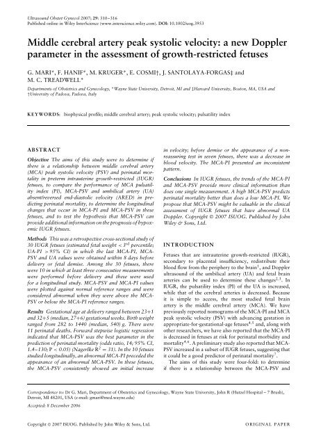
Ultrasound Obstet Gynecol 2007; 29: 310–316<br />
Published online in Wiley InterScience (www.interscience.wiley.com). DOI: 10.1002/uog.3953<br />
<strong>Middle</strong> <strong>cerebral</strong> <strong>artery</strong> <strong>peak</strong> <strong>systolic</strong> <strong>velocity</strong>: a new Doppler<br />
parameter in the assessment of growth-restricted fetuses<br />
G. MARI*, F. HANIF*, M. KRUGER*, E. COSMI†, J. SANTOLAYA-FORGAS‡ and<br />
M. C. TREADWELL*<br />
Departments of Obstetrics and Gynecology, *Wayne State University, Detroit, MI and ‡Harvard University, Boston, MA, USA and<br />
†University of Padova, Padova, Italy<br />
KEYWORDS:<br />
biophysical profile; middle <strong>cerebral</strong> <strong>artery</strong>; <strong>peak</strong> <strong>systolic</strong> <strong>velocity</strong>; pulsatility index<br />
ABSTRACT<br />
Objective The aims of this study were to determine if<br />
there is a relationship between middle <strong>cerebral</strong> <strong>artery</strong><br />
(MCA) <strong>peak</strong> <strong>systolic</strong> <strong>velocity</strong> (PSV) and perinatal mortality<br />
in preterm intrauterine growth-restricted (IUGR)<br />
fetuses, to compare the performance of MCA pulsatility<br />
index (PI), MCA-PSV and umbilical <strong>artery</strong> (UA)<br />
absent/reversed end-diastolic <strong>velocity</strong> (ARED) in predicting<br />
perinatal mortality, to determine the longitudinal<br />
changes that occur in MCA-PI and MCA-PSV in these<br />
fetuses, and to test the hypothesis that MCA-PSV can<br />
provide additional information on the prognosis of hypoxemic<br />
IUGR fetuses.<br />
Methods This was a retrospective cross-sectional study of<br />
30 IUGR fetuses (estimated fetal weight < 3 rd percentile;<br />
UA-PI > 95% CI) in which the last MCA-PI, MCA-<br />
PSV and UA values were obtained within 8 days before<br />
delivery or fetal demise. Among the 30 fetuses, there<br />
were 10 in which at least three consecutive measurements<br />
were performed before delivery and these were used<br />
for a longitudinal study. MCA-PSV and MCA-PI values<br />
were plotted against normal reference ranges and were<br />
considered abnormal when they were above the MCA-<br />
PSV or below the MCA-PI reference ranges.<br />
Results Gestational age at delivery ranged between 23+1<br />
and 32+5(median,27+6) gestational weeks. Birth weight<br />
ranged from 282 to 1440 (median, 540) g. There were<br />
11 perinatal deaths. Forward stepwise logistic regression<br />
indicated that MCA-PSV was the best parameter in the<br />
prediction of perinatal mortality (odds ratio, 14; 95% CI,<br />
1.4–130; P < 0.05) (Nagerlke R 2 = 31). In the 10 fetuses<br />
studied longitudinally, an abnormal MCA-PI preceded the<br />
appearance of an abnormal MCA-PSV. In these fetuses,<br />
the MCA-PSV consistently showed an initial increase<br />
in <strong>velocity</strong>; before demise or the appearance of a nonreassuring<br />
test in seven fetuses, there was a decrease in<br />
blood <strong>velocity</strong>. The MCA-PI presented an inconsistent<br />
pattern.<br />
Conclusions In IUGR fetuses, the trends of the MCA-PI<br />
and MCA-PSV provide more clinical information than<br />
does one single measurement. A high MCA-PSV predicts<br />
perinatal mortality better than does a low MCA-PI. We<br />
propose that MCA-PSV might be valuable in the clinical<br />
assessment of IUGR fetuses that have abnormal UA<br />
Doppler. Copyright © 2007 ISUOG. Published by John<br />
Wiley & Sons, Ltd.<br />
INTRODUCTION<br />
Fetuses that are intrauterine growth-restricted (IUGR),<br />
secondary to placental insufficiency, redistribute their<br />
blood flow from the periphery to the brain 1 , and Doppler<br />
ultrasound of the umbilical <strong>artery</strong> (UA) and fetal brain<br />
arteries can be used to determine these changes 2,3 . In<br />
IUGR, the pulsatility index (PI) of the UA is increased,<br />
while that of the <strong>cerebral</strong> arteries is decreased. Because<br />
it is simple to access, the most studied fetal brain<br />
<strong>artery</strong> is the middle <strong>cerebral</strong> <strong>artery</strong> (MCA). We have<br />
previously reported nomograms of the MCA-PI and MCA<br />
<strong>peak</strong> <strong>systolic</strong> <strong>velocity</strong> (PSV) with advancing gestation in<br />
appropriate-for-gestational-age fetuses 4,5 and, along with<br />
other researchers, we have also reported that the MCA-PI<br />
is decreased in fetuses at risk for perinatal morbidity and<br />
mortality 4,6 . A preliminary study also reported that MCA-<br />
PSV increased in a subset of IUGR fetuses, suggesting that<br />
it could be a good predictor of perinatal mortality 7 .<br />
The aims of this study were four-fold: to determine<br />
if there is a relationship between the MCA-PSV and<br />
Correspondence to: Dr G. Mari, Department of Obstetrics and Gynecology, Wayne State University, John R (Hutzel Hospital – 7 Brush),<br />
Detroit, MI 48201, USA (e-mail: gmari@med.wayne.edu)<br />
Accepted: 8 December 2006<br />
Copyright © 2007 ISUOG. Published by John Wiley & Sons, Ltd.<br />
ORIGINAL PAPER
MCA-PSV and IUGR 311<br />
perinatal mortality in preterm IUGR fetuses that have<br />
an abnormal UA-PI, to compare the performance of<br />
the MCA-PI, MCA-PSV, and UA absent or reversed<br />
end-diastolic flow <strong>velocity</strong> (ARED) in the prediction of<br />
perinatal mortality in these fetuses, to determine the<br />
longitudinal changes that occur in the MCA-PI and MCA-<br />
PSV in these fetuses, and to test the hypothesis that the<br />
MCA-PSV can provide additional information on the<br />
prognosis of hypoxemic IUGR fetuses.<br />
MATERIAL AND METHODS<br />
This study included a retrospective and a prospective<br />
assessment of the MCA-PSV and MCA-PI. All patients<br />
were enrolled into research protocols approved by a<br />
human investigation committee.<br />
Among 55 fetuses diagnosed with IUGR, we selected<br />
singletons that had: (a) an estimated weight < 3 rd<br />
percentile (confirmed at birth), (b) a UA-PI > 95 th<br />
CI, (c) normal anatomy and (d) an MCA Doppler<br />
examination determined with an angle close to 0 ◦ within<br />
8 days before delivery or fetal demise. All were delivered<br />
at < 33 weeks’ gestation.<br />
Thirty fetuses met the entry criteria. The gestational age<br />
at the time of the Doppler study ranged between 23+0and<br />
32+4 (median,27+2) weeks, based on the last menstrual<br />
period for women with a recorded history or on a secondtrimester<br />
scan. Delivery was indicated in the presence<br />
of: (a) non-reassuring fetal testing, (b) fetal demise, or<br />
(c) worsening maternal or fetal conditions as determined<br />
by the managing physician. Non-reassuring fetal testing<br />
was defined as the presence of either continuous late<br />
decelerations or a biophysical profile < 4. When delivery<br />
was indicated and the fetus was < 500 g in weight,<br />
the patient was counseled on the poor prognosis and<br />
offered the option of non-intervention. For fetal weight,<br />
we utilized the normal reference ranges established by<br />
Hadlock et al. 8 .<br />
Doppler studies<br />
Pulsed-wave Doppler ultrasound studies were performed<br />
with four color Doppler systems (Sequoia, Siemens<br />
Medical Solutions, Mountain View, CA, USA; Voluson<br />
530 or 730 Expert, GE Medical Systems, Milwaukee,<br />
WI, USA; HDI 5000, Philips Medical Systems, Bothell,<br />
WA, USA) using 3.5- or 5-MHz probes with spatial <strong>peak</strong><br />
temporal average intensities of < 100 mW/cm 2 in both<br />
imaging and Doppler modes. All recordings were obtained<br />
in the absence of fetal breathing and fetal movements.<br />
For each vessel, an average of three consecutive Doppler<br />
<strong>velocity</strong> waveforms was used for statistical analysis. A<br />
free loop of the UA was sampled, and the PI was used<br />
to analyze the waveforms 9 . The MCA was studied with<br />
an angle close to 0 ◦ between the ultrasound beam and<br />
the direction of blood flow, as reported previously 5 .The<br />
MCA waveforms were quantified using the PI as well<br />
as the PSV. The MCA-PI was considered abnormal if<br />
the measurements were below the lower limit of normal<br />
as reported previously 4 ; the MCA-PSV was considered<br />
abnormal if the measurements were above the upper limit<br />
of normal, as established previously 5 .AUA-PI> 95%<br />
CI for gestational age of our reference range was also<br />
considered abnormal, as was ARED in the UA.<br />
Prospective longitudinal study<br />
Of the 30 IUGR fetuses, we prospectively assessed 10<br />
in which the MCA-PI and the MCA-PSV were recorded<br />
longitudinally from the time the diagnosis was made<br />
until delivery. The number of measurements in these<br />
fetuses ranged from 3 to 8 (median, 5). Gestational ages<br />
at entrance into the study ranged from 20+6 to26+4<br />
(median, 23+5) weeks, and gestational ages at delivery<br />
were between 23+1 and 28+6 (median,26+1) weeks.<br />
Perinatal outcome endpoints included: (a) perinatal<br />
mortality, defined as mortality occurring between<br />
20 weeks’ gestation and 28 days after birth and<br />
Table 1 Demographics of the study population (n = 30)<br />
Characteristic<br />
Median (range)<br />
GA at Doppler study (weeks) 27+2 (23+0–32+4)<br />
Interval between last scan and delivery 1 (0–8)<br />
(days)<br />
GA at delivery (weeks)<br />
Total population 27+6 (23+1–32+5)<br />
Perinatal deaths (n = 11) 25+5 (23+1–28+6)<br />
IUFDs (n = 6) 25+8 (23+1–28+6)<br />
NDs (n = 5) 25+0 (24+6–28+1)<br />
Survivors (n = 19) 29+2 (25+2–32+5)<br />
Birth weight (g)<br />
Total population 540 (282–1440)<br />
Perinatal deaths (n = 11) 440 (282–660)<br />
IUFDs (n = 6) 430 (282–472)<br />
NDs (n = 5) 471 (300–660)<br />
Survivors (n = 19) 760 (360–1440)<br />
GA, gestational age; IUFD, intrauterine fetal demise; ND, neonatal<br />
death.<br />
MCA-PSV (cm/s)<br />
100<br />
90<br />
80<br />
70<br />
60<br />
50<br />
40<br />
30<br />
20<br />
10<br />
0<br />
15 20 25 30 35 40<br />
Gestational age (weeks)<br />
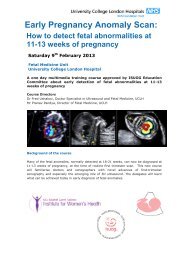
Figure 1 <strong>Middle</strong> <strong>cerebral</strong> <strong>artery</strong> <strong>peak</strong> <strong>systolic</strong> <strong>velocity</strong> (MCA-PSV)<br />
in 30 growth-restricted fetuses plotted on the normal reference<br />
range 5 , showing fetuses which died (ž) and those which<br />
survived (<br />
° ).<br />
Copyright © 2007 ISUOG. Published by John Wiley & Sons, Ltd. Ultrasound Obstet Gynecol 2007; 29: 310–316.
312 Mari et al.<br />
Table 2 Individual characteristics of the study population<br />
Gestational age (weeks) Birth Perinatal<br />
Maternal Indication for weight morbidity<br />
Case pathology at Doppler at delivery MCA-PI MCA-PSV UA-ARED delivery (g) (BPD) Perinatal mortality<br />
1 CHtn 30+0 30+0 A A A NRFT 600 Yes No<br />
2 None 25+1 25+5 A A A IUFD 308 — IUFD<br />
3 None 27+1 27+2 N A A IUFD 472 — IUFD<br />
4 CHtn 24+6 24+6 A N A NRFT 300 — DiedinNICU(1day)<br />
5 CHtn 25+5 25+5 A A A S-PE 360 Yes No<br />
6 CHtn 23+0 23+1 N A A IUFD 282 — IUFD<br />
7 CHtn 25+0 25+0 A A A HELLP 440 — Died in NICU (1 day)<br />
8 None 30+4 30+6 A A N S-PE 1080 No No<br />
9 CHtn 26+0 26+5 A N A NRFT 510 No No<br />
10 None 32+4 32+5 N N N HELLP 1440 No No<br />
11 None 29+2 29+2 A N A NRFT 1000 Yes No<br />
12 CHtn 26+4 26+4 A A A NRFT 460 Yes No<br />
13 None 32+1 32+2 N N N HELLP 1380 No No<br />
14 CHtn 31+3 31+3 A N N S-PE 1240 Yes No<br />
15 None 25+1 25+2 A A A NRFT (PE) 530 Yes No<br />
16 None 27+6 28+1 N A A NRFTS-PE 471 — DiedinNICU(8days)<br />
17 CHtn 26+6 28+0 A N A NRFT 700 Yes No<br />
18 CHtn 26+6 26+6 A A A NRFT 660 — DiedinNICU(3days)<br />
19 CHtn 28+0 28+0 A A A NRFT 467 Yes No<br />
20 None 28+1 28+4 A A A NRFT (S-PIH) 840 Yes No<br />
21 None 28+3 28+6 N A A IUFD 440 — IUFD<br />
22 Hypth 24+0 24+1 N A A IUFD 440 — IUFD<br />
23 Lupus 25+0 26+1 A A A IUFD 420 — IUFD<br />
24 CHtn 31+6 32+0 A N A NRFT 1100 No No<br />
25 SCD 31+1 31+6 A N N S-PIH 1150 No No<br />
26 None 29+1 29+4 A N N S-PIH 820 No No<br />
27 None 27+0 27+1 A A A S-PE 550 Yes No<br />
28 None 29+3 29+4 N N N S-PE 760 No No<br />
29 None 24+6 24+6 A A A S-PE 505 — DiedinNICU(8days)<br />
30 None 27+3 27+3 A N A NRFT (S-PE) 620 Yes No<br />
A, abnormal; BPD, bronchopulmonary dysplasia; CHtn, chronic hypertension; HELLP, hemolysis, elevated liver function tests, low<br />
platelets; Hypth, hyperthyroidism; IUFD, intrauterine fetal demise; MCA-PI, middle <strong>cerebral</strong> <strong>artery</strong> pulsatility index; MCA-PSV, middle<br />
<strong>cerebral</strong> <strong>artery</strong> <strong>peak</strong> <strong>systolic</strong> <strong>velocity</strong>; N, normal; NICU, neonatal intensive care unit; NRFT, non-reassuring fetal testing; PE, pre-eclampsia;<br />
PIH, pregnancy-induced hypertension or gestational hypertension; S, severe; SCD, sickle cell disease; UA-ARED, absent/reversed<br />
end-diastolic flow <strong>velocity</strong>.<br />
MCA-PI<br />
3.5<br />
3.0<br />
2.5<br />
2.0<br />
1.5<br />
1.0<br />
0.5<br />
0.0<br />
15 20 25 30 35 40<br />
Gestational age (weeks)<br />
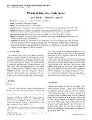
Figure 2 <strong>Middle</strong> <strong>cerebral</strong> <strong>artery</strong> pulsatility index (MCA-PI) in 30<br />
growth-restricted fetuses plotted on the normal reference range 4 ,<br />
showing fetuses which died (ž) and those which survived (<br />
° ).<br />
(b) major neonatal complications, defined as intra<strong>cerebral</strong><br />
hemorrhage (IVH) Grades III or IV according to<br />
the Papile classification 10 or bronchopulmonary dysplasia<br />
(BPD).<br />
Table 3 Predictive value for perinatal mortality of middle <strong>cerebral</strong><br />
<strong>artery</strong> <strong>peak</strong> <strong>systolic</strong> <strong>velocity</strong> (MCA-PSV) above the normal range<br />
for gestational age<br />
Test result<br />
Perinatal<br />
mortality (n)<br />
No perinatal<br />
mortality (n)<br />
Total (n)<br />
MCA-PSV (abnormal) 10 8 18<br />
MCA-PSV (normal) 1 11 12<br />
Total 11 19 30<br />
Sensitivity, 91%; specificity, 58%; positive predictive value, 56%;<br />
negative predictive value, 92% (odds ratio, 14; 95% CI, 1.4–130;<br />
P < 0.05).<br />
Statistical analysis<br />
Statistical analysis was performed using SPSS statistical<br />
software, version 14.0 (SPSS, System for Windows,<br />
Chicago, IL, USA). We evaluated the MCA-PI, MCA-PSV<br />
and UA-ARED obtained at the last examination against<br />
the outcomes of perinatal mortality or major neonatal<br />
complications by forward stepwise logistic regression.<br />
Due to the small number of fetuses that survived,<br />
Copyright © 2007 ISUOG. Published by John Wiley & Sons, Ltd. Ultrasound Obstet Gynecol 2007; 29: 310–316.
MCA-PSV and IUGR 313<br />
(a) 100<br />
MCA-PSV<br />
80<br />
60<br />
40<br />
20<br />
0<br />
15 20 25 30 35 40<br />
(b) 100<br />
MCA-PSV<br />
80<br />
60<br />
40<br />
20<br />
0 15 20 25 30 35 40<br />
IUFD 282 g<br />
Gestational age (weeks)<br />
IUFD 308 g<br />
Gestational age (weeks)<br />
(c) 100<br />
(d) 100<br />
MCA-PSV<br />
80<br />
60<br />
40<br />
20<br />
MCA-PSV<br />
80<br />
60<br />
40<br />
20<br />
0<br />
15 20 25 30 35 40<br />
0 15 20 25 30 35 40<br />
ND 471 g<br />
Gestational age (weeks)<br />
WELL 467 g<br />
Gestational age (weeks)<br />
(e) 100<br />
(f) 100<br />
MCA-PSV<br />
80<br />
60<br />
40<br />
20<br />
MCA-PSV<br />
80<br />
60<br />
40<br />
20<br />
0 15 25 35<br />
0<br />
15 20 25 30 35 40<br />
WELL 360 g<br />
Gestational age (weeks)<br />
ND 440 g<br />
Gestational age (weeks)<br />
(g) 100<br />
(h) 100<br />
MCA-PSV<br />
80<br />
60<br />
40<br />
20<br />
MCA-PSV<br />
80<br />
60<br />
40<br />
20<br />
0 15 20 25 30 35 40<br />
0 15 20 25 30 35 40<br />
ND 505 g<br />
Gestational age (weeks)<br />
IUFD 472 g<br />
Gestational age (weeks)<br />
(i) 100<br />
(j) 100<br />
MCA-PSV<br />
80<br />
60<br />
40<br />
20<br />
MCA-PSV<br />
80<br />
60<br />
40<br />
20<br />
0<br />
15 20 25 30 35 40<br />
0 15 20 25 30 35 40<br />
IUFD 440 g<br />
Gestational age (weeks)<br />
ND 660 g<br />
Gestational age (weeks)<br />
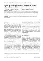
Figure 3 Individual longitudinal values for the middle <strong>cerebral</strong> <strong>artery</strong> <strong>peak</strong> <strong>systolic</strong> <strong>velocity</strong> (MCA-PSV in cm/s) in 10 growth-restricted<br />
fetuses plotted on the reference range 5 . An accurate <strong>velocity</strong> value could not be determined in some studies because an appropriate angle<br />
between the ultrasound beam and the direction of the blood flow could not be obtained. At the bottom left of each graph, the fetal outcome is<br />
indicated: intrauterine fetal demise (IUFD), well at the time of writing (WELL) or neonatal death (ND), with the weight at delivery in grams.<br />
two-tailed chi-square or Fisher’s exact tests were<br />
employed to analyze the relationship between the Doppler<br />
parameters and perinatal morbidity. Student’s t-test for<br />
independent samples was used to compare whether or<br />
not a difference in gestational age existed at delivery<br />
among the babies who survived and those who died,<br />
either in utero or within the first 4 weeks of life. This<br />
test was also used to compare whether a difference in<br />
gestational age existed at delivery among the infants who<br />
developed major neonatal complications and those who<br />
did not. A probability value of P < 0.05 was considered<br />
statistically significant.<br />
We also calculated the sensitivity, specificity, positive<br />
predictive value and negative predictive value for the<br />
parameters selected by the forward stepwise logistic<br />
regression analysis, with the tests’ status being abnormal<br />
Copyright © 2007 ISUOG. Published by John Wiley & Sons, Ltd. Ultrasound Obstet Gynecol 2007; 29: 310–316.
314 Mari et al.<br />
(a) 4<br />
MCA-PI<br />
3<br />
2<br />
1<br />
0 15 20 25 30 35 40<br />
Gestational age (weeks)<br />
(b) 4<br />
MCA-PI<br />
3<br />
2<br />
1<br />
0<br />
15 20 25 30 35 40<br />
Gestational age (weeks)<br />
(c)<br />
MCA-PI<br />
4<br />
3<br />
2<br />
1<br />
(d) 4<br />
MCA-PI<br />
3<br />
2<br />
1<br />
0 15 20 25 30 35 40<br />
Gestational age (weeks)<br />
0 15 20 25 30 35 40<br />
Gestational age (weeks)<br />
(e)<br />
4<br />
(f)<br />
4<br />
MCA-PI<br />
3<br />
2<br />
1<br />
MCA-PI<br />
3<br />
2<br />
1<br />
0 15 20 25 30 35 40<br />
0 15 20 25 30 35 40<br />
Gestational age (weeks)<br />
Gestational age (weeks)<br />
(g) 4<br />
MCA-PI<br />
3<br />
2<br />
1<br />
0 15 20 25 30 35 40<br />
Gestational age (weeks)<br />
(h) 4<br />
MCA-PI<br />
3<br />
2<br />
1<br />
0 15 20 25 30 35 40<br />
Gestational age (weeks)<br />
(i)<br />
4<br />
(j)<br />
4<br />
MCA-PI<br />
3<br />
2<br />
1<br />
MCA-PI<br />
3<br />
2<br />
1<br />
0 15 20 25 30 35 40<br />
Gestational age (weeks)<br />
0<br />
15 20 25 30 35 40<br />
Gestational age (weeks)<br />
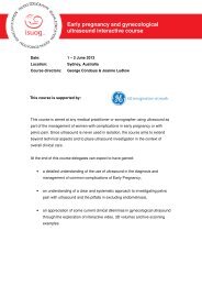
Figure 4 Individual longitudinal values for the middle <strong>cerebral</strong> <strong>artery</strong> pulsatility index (MCA-PI) in the same 10 growth-restricted fetuses as<br />
in Figure 3, plotted on the reference range 4 .<br />
Doppler parameter and the outcome status being presence<br />
or absence of perinatal death.<br />
For the 10 fetuses that were followed longitudinally, we<br />
plotted the MCA-PI and the MCA-PSV values on our reference<br />
ranges to determine the trend of these two parameters<br />
with advancing gestational age in IUGR fetuses.<br />
RESULTS<br />
The characteristics of the study population and the<br />
perinatal morbidity and mortality data are reported in<br />
Tables 1 and 2. There were six fetal demises and in all<br />
cases the mothers opted for no intervention following<br />
counseling due to extreme prematurity and low birth<br />
weight. With the exception of those that died in utero, all<br />
but one of the fetuses were delivered by Cesarean section.<br />
The forward stepwise logistic regression indicated that the<br />
MCA-PSV predicted the perinatal mortality (odds ratio,<br />
14; 95% CI, 1.4–130; P < 0.05) (Nagerlke R 2 = 31).<br />
The fetuses that died were at a significantly lower<br />
gestational age at the time of delivery than were those that<br />
survived (25+6 ± 1+6 weeks vs. 29+0 ± 2+3 weeks;<br />
Copyright © 2007 ISUOG. Published by John Wiley & Sons, Ltd. Ultrasound Obstet Gynecol 2007; 29: 310–316.
MCA-PSV and IUGR 315<br />
P = 0.001). There were no survivors at ≤ 25+0 weeks’<br />
gestation (two intrauterine (IUFD) and three neonatal<br />
deaths), whereas at > 29 weeks’ gestation all the fetuses<br />
survived (n = 10), and between 25 and 29 weeks six died<br />
(four fetal demises and two neonatal deaths).<br />
Figures 1 and 2 show the MCA-PSV and MCA-PI,<br />
respectively, plotted on our reference ranges 4,5 . Table 3<br />
gives the sensitivity, specificity, and positive and negative<br />
predictive values of MCA-PSV above the normal range<br />
for perinatal mortality.<br />
Among the infants who survived, none developed<br />
IVH Grade III or IV, whereas 11 developed BPD.<br />
The gestational age at delivery was lower in the<br />
infants who developed BPD (27+6 ± 1+6 weeks vs.<br />
30+3 ± 2+1 weeks; P < 0.05). BPD was significantly<br />
related to an abnormal MCA-PSV (odds ratio, 12; 95%<br />
CI, 1.1–139; P = 0.037), and UA-ARED (odds ratio, 30;<br />
95% CI, 2.2–405; P = 0.006). The MCA-PI odds ratio<br />
was 0.6 (95% CI, 0.4–1.1; P = 0.058).<br />
In the 10 fetuses followed longitudinally, the abnormal<br />
MCA-PI preceded the appearance of an abnormal<br />
MCA-PSV. In six of these 10 fetuses, the last MCA<br />
measurements were obtained within 12 h of delivery or<br />
fetal demise. In two other fetuses, measurements were<br />
performed within 2 days of delivery; one of these two<br />
fetuses died in utero.Inthelasttwofetuses,measurements<br />
were performed within 4 days of delivery; both fetuses<br />
died in utero. In all 10 fetuses, the MCA-PSV showed<br />
an initial increase in <strong>velocity</strong> and it decreased before<br />
demise or the appearance of non-reassuring testing in<br />
seven fetuses (Figure 3). In the other three fetuses, the<br />
MCA-PSV had the highest <strong>velocity</strong> value before birth<br />
and a normal arterial pH value was recorded at birth 11 ,<br />
with Apgar scores at 5 min of 7, 8 and 9. These three<br />
infants did well initially, but two died within 8 days of<br />
delivery; the third infant, who had a birth weight of<br />
360 g, did well. The MCA-PI did not have a consistent<br />
pattern: initially, it was abnormal in most of the fetuses;<br />
in three fetuses it decreased progressively with advancing<br />
gestation, in another six fetuses, it increased towards<br />
normal before delivery and in one fetus, it remained more<br />
or less unchanged (Figure 4).<br />
DISCUSSION<br />
In this cross-sectional and longitudinal assessment of the<br />
MCA-PI and MCA-PSV in IUGR fetuses, our data have<br />
shown that, although the MCA waveforms change in<br />
IUGR fetuses, the MCA-PSV predicts perinatal mortality<br />
more accurately than does the MCA-PI. The reason for<br />
this difference can be found in the evaluation of our longitudinal<br />
data. Initially, the MCA-PI was abnormal in most<br />
of the fetuses, but in six an increased MCA-PI with a tendency<br />
towards normalization occurred before delivery or<br />
fetal demise. In three other fetuses, the MCA-PI progressively<br />
decreased up to delivery, and in one fetus it changed<br />
little. The MCA-PSV, on the other hand, exhibited a welldefined<br />
pattern, progressively increasing with advancing<br />
gestation in all fetuses, with a tendency to slightly<br />
decrease, just before delivery or the occurrence of fetal<br />
demise, in seven fetuses. Despite this decrease, however,<br />
the MCA-PSV value remained above the upper limit of<br />
normal until a few hours prior to delivery or fetal demise.<br />
The MCA-PI together with the UA-PI, has long been<br />
considered the gold standard for the assessment of fetuses<br />
with IUGR. Our findings, though, suggest that not<br />
only does the MCA-PSV complement the information<br />
provided by the MCA-PI, but that it actually provides<br />
clearer information than does the MCA-PI. In IUGR<br />
fetuses with an abnormal MCA-PI and a normal MCA-<br />
PSV, the IUGR condition is less severe than in cases in<br />
which both parameters are abnormal. However, when<br />
the IUGR condition intensifies, the MCA-PSV increases<br />
and becomes abnormal. Our data also suggest that when<br />
an MCA-PI value is increasing or an MCA-PSV value is<br />
decreasing, the fetus starts to decompensate. For example,<br />
four fetuses died following a drop in MCA-PSV value, and<br />
in each case the MCA-PI increased before the fetal demise<br />
occurred.<br />
Most of our data were collected within 48 h before<br />
delivery or fetal demise, when the MCA-PI had normalized<br />
in some fetuses. This might explain why the MCA-PI<br />
predicted the perinatal mortality less well than did the<br />
MCA-PSV. UA-ARED, on the other hand, was present in<br />
most IUGR fetuses and might explain why the MCA-PSV<br />
that was normal in most of the fetuses that survived and<br />
abnormal in most of the fetuses that died predicted the<br />
perinatal mortality better than did the UA-ARED.<br />
Although we confirmed that gestational age plays<br />
an important role in the perinatal mortality of IUGR<br />
fetuses, we found that among IUGR fetuses delivered at<br />
≤ 25+0 weeks’ gestation, there were no survivors, while<br />
all IUGR fetuses survived when they were delivered at<br />
> 29 weeks’ gestation. In terms of survival rate, the IUGR<br />
group delivered at > 25 and < 29 weeks’ gestation was the<br />
most uncertain group. Interestingly, although the MCA-<br />
PI at this gestational age was abnormal in 12/15 fetuses,<br />
none of the fetuses died when the MCA-PSV was normal.<br />
These outcomes reinforce the concept that the MCA-PSV<br />
is a better predictor of perinatal mortality than is the<br />
MCA-PI.<br />
We have reported previously that the MCA-PSV<br />
is a reliable predictor of moderate and severe fetal<br />
anemia, whereas it is less sensitive in predicting mild<br />
anemia 12 . While IUGR fetuses can be anemic at birth,<br />
they are rarely severely anemic, although they can also<br />
be polycythemic 13 . This is probably the reason why a<br />
previous study on mild and non-anemic IUGR fetuses<br />
was unable to find a correlation between anemia and<br />
MCA-PSV 14 .<br />
We would like to propose here that the pathophysiological<br />
explanation for the observed high MCA-PSV in IUGR<br />
fetuses and anemic fetuses is different. In anemic fetuses,<br />
as reported previously, the high MCA-PSV is most likely<br />
secondary to a raised cardiac output and decreased blood<br />
viscosity 12 , whereas in IUGR fetuses, the increased MCA-<br />
PSV reflects increased blood flow to the brain through an<br />
Copyright © 2007 ISUOG. Published by John Wiley & Sons, Ltd. Ultrasound Obstet Gynecol 2007; 29: 310–316.
316 Mari et al.<br />
elevated left cardiac output 15 and an increased placental<br />
vascular resistance.<br />
Although betamethasone can affect blood <strong>velocity</strong> 16 ,it<br />
is unlikely that this was the cause of high MCA-PSV in<br />
our study because betamethasone was administered to all<br />
our patients.<br />
Our data, combined with previously reported data,<br />
allow us to postulate that in fetuses with IUGR<br />
secondary to placental insufficiency, the following occurs.<br />
Initially, a low MCA-PI might reflect decreased brain<br />
vascular resistance and a slight increase in blood flow.<br />
With increased severity, the MCA-PSV increases, as a<br />
consequence of increased left cardiac output 15 . When the<br />
process becomes more severe, a portion of the blood<br />
ejected from the right ventricle is shifted to the brain<br />
through the aortic isthmus because of a high vascular<br />
resistance in the descending aorta 17 . Although the MCA-<br />
PSV decreased before IUFD in four of the fetuses we<br />
studied, it remained above the normal range. This might<br />
have been a consequence of the exaggerated vasodilatation<br />
or vasoconstriction that occurs in the overstressed fetus.<br />
We cannot currently conclude if blood pressure also plays<br />
a role in these <strong>cerebral</strong> blood flow changes.<br />
The gestational age was lower in the group that<br />
developed BPD. Because of the small number of neonates<br />
that survived (n = 19), we did not perform logistic<br />
regression analysis, and we were not able to subdivide<br />
these IUGR fetuses based on gestational age. We believe<br />
that studies with a larger number of fetuses should be<br />
undertaken to further explore whether the MCA-PSV<br />
and the UA-ARED are better correlated with perinatal<br />
morbidity than is the MCA-PI.<br />
It is important to emphasize that the IUGR group<br />
reported here represented a heterogeneous group because<br />
the maternal pathology present before the pregnancy and<br />
the pathology occurring during the pregnancy influenced<br />
the time of delivery in many of our fetuses. Therefore, a<br />
study that includes only idiopathic IUGR fetuses (cases<br />
in which no cause can be found for the placental<br />
insufficiency), as reported previously for other Doppler<br />
parameters 18 , is warranted.<br />
In summary, our data indicate that: (a) the trends of<br />
the MCA-PSV and MCA-PI provide more information<br />
than does one single measurement of either, and (b) the<br />
MCA-PSV might be clinically more informative than is the<br />
MCA-PI in the management of IUGR fetuses. We believe<br />
that prospective longitudinal studies in IUGR fetuses with<br />
documented long-term outcome and aimed at assessing<br />
the correlation of the longitudinal changes of the MCA-<br />
PSV with those of other vessels, such as the aortic isthmus<br />
and ductus venosus, could be profitable.<br />
ACKNOWLEDGMENTS<br />
We would like to thank the staff, technicians, nurses and<br />
faculty of the PRB and the Department of Obstetrics<br />
and Gynecology at Wayne State University and Mamtha<br />
Balasumbramian for their support. This work was<br />
supported in part by the Intramural Research Program<br />
of the National Institute of Child Health and Human<br />
Development, NIH, DHHS.<br />
REFERENCES<br />
1. Cohn HE, Sacks EJ, Heymann MA, Rudolph AM. Cardiovascular<br />
responses to hypoxemia and acidemia in fetal lambs. Am<br />
J Obstet Gynecol 1974; 120: 817–824.<br />
2. Trudinger BJ, Giles WB, Cook CM, Bombardieri J, Collins L.<br />
Fetal umbilical <strong>artery</strong> flow <strong>velocity</strong> waveforms and placental<br />
resistance: clinical significance. BrJObstetGynaecol1985; 92:<br />
23–30.<br />
3. Wladimiroff JW, Tonge HM, Stewart PA. Doppler ultrasound<br />
assessment of the <strong>cerebral</strong> blood flow in the human fetus. Br J<br />
Obstet Gynaecol 1986; 93: 471–475.<br />
4. Mari G, Deter RL. <strong>Middle</strong> <strong>cerebral</strong> <strong>artery</strong> flow <strong>velocity</strong><br />
waveforms in normal and small-for-gestational-age fetuses. Am<br />
J Obstet Gynecol 1992; 166: 1262–1270.<br />
5. Mari G, Adrignolo A, Abuhamad A, Pirhonen J, Jones DC,<br />
Ludomirsky A, Copel JA. Diagnosis of fetal anemia with<br />
Doppler ultrasound in the pregnancy complicated by maternal<br />
blood group immunization. Ultrasound Obstet Gynecol 1995;<br />
5: 400–405.<br />
6. Vyas S, Nicolaides KH, Bower S, Campbell S. <strong>Middle</strong> <strong>cerebral</strong><br />
<strong>artery</strong> flow <strong>velocity</strong> waveforms in fetal hypoxaemia. BrJObstet<br />
Gynaecol 1990; 97: 797–803.<br />
7. Ozcan T, Sbracia M, d’Ancona RL, Copel JA, Mari G. Arterial<br />
and venous Doppler velocimetry in the severely growthrestricted<br />
fetus and associations with adverse perinatal outcome.<br />
Ultrasound Obstet Gynecol 1998; 12: 39–44.<br />
8. Hadlock FP, Harrist RB, Martinez-Poyer J. In utero analsysis of<br />
fetal growth: a sonographic weight standard. Radiology 1991;<br />
181: 129–133.<br />
9. Gosling RG, King DH. Ultrasound Angiology. Churchill-<br />
Livingstone: New York, 1975; 61–98.<br />
10. Papile LA, Burstein J, Burstein R, Koffler H. Incidence and<br />
evolution of subependymal and intraventricular hemorrhage: a<br />
study of infants with birth weight less than 1,500 gm. JPediatr<br />
1978; 92: 529–534.<br />
11. Ramin SM, Gilstrap LC, III, Leveno KJ, Burris J, Little BB.<br />
Umbilical <strong>artery</strong> acid-base status in the preterm infant. Obstet<br />
Gynecol 1989; 74: 256–258.<br />
12. Mari G, Deter RL, Carpenter RL, Rahman F, Zimmerman<br />
R, Moise KJ, Jr. Dorman KF, Ludomirsky A, Gonzalez R,<br />
Gomez R, Oz U, Detti L, Copel JA, Bahado-Singh R, Berry S,<br />
Martinez-Poyer J, Blackwell SC. Noninvasive diagnosis by<br />
Doppler ultrasonography of fetal anemia due to maternal<br />
red-cell alloimmunization. Collaborative Group for Doppler<br />
Assessment of the Blood Velocity in Anemic Fetuses. NEnglJ<br />
Med 2000; 342: 9–14.<br />
13. Resnik R. Intrauterine growth restriction. Obstet Gynecol 2002;<br />
99: 490–496.<br />
14. Makh DS, Harman CR, Baschat AA. Is Doppler prediction of<br />
anemia effective in the growth-restricted fetus? Ultrasound<br />
Obstet Gynecol 2003; 22: 489–492.<br />
15. al Ghazali W, Chita SK, Chapman MG, Allan LD. Evidence<br />
of redistribution of cardiac output in asymmetrical growth<br />
retardation. BrJObstetGynaecol1989; 96: 697–704.<br />
16. Simchen MJ, Alkazaleh F, Adamson SL, Windrim R, Telford J,<br />
Beyene J, Kingdom J. The fetal cardiovascular response to<br />
antenatal steroids in severe early-onset intrauterine growth<br />
restriction. Am J Obstet Gynecol 2004; 190: 296–304.<br />
17. Fouron JC. The unrecognized physiological and clinical significance<br />
of the fetal aortic isthmus. Ultrasound Obstet Gynecol<br />
2003; 22: 441–447.<br />
18. Cosmi E, Ambrosini G, D’Antona D, Saccardi C, Mari G.<br />
Doppler, cardiotocography, and biophysical profile changes<br />
in growth-restricted fetuses. Obstet Gynecol 2005; 106:<br />
1240–1245.<br />
Copyright © 2007 ISUOG. Published by John Wiley & Sons, Ltd. Ultrasound Obstet Gynecol 2007; 29: 310–316.