Cloning and Expression of a Burkholderia pseudomallei ... - EJUM
Cloning and Expression of a Burkholderia pseudomallei ... - EJUM
Cloning and Expression of a Burkholderia pseudomallei ... - EJUM

You also want an ePaper? Increase the reach of your titles
YUMPU automatically turns print PDFs into web optimized ePapers that Google loves.
<strong>Cloning</strong> <strong>and</strong> expression <strong>of</strong> B. <strong>pseudomallei</strong> peptidase<br />
36<br />
<strong>Expression</strong> <strong>and</strong> characterization <strong>of</strong> recombinant peptidase<br />
M23B<br />
<strong>Expression</strong> <strong>of</strong> the B. <strong>pseudomallei</strong> peptidase M23B<br />
gene was performed in E. coli BL21 Star (DE3) at<br />
30ºC with 1 mM IPTG induction. <strong>Expression</strong> was carried<br />
out at a lower temperature to avoid formation <strong>of</strong> inclusion<br />
bodies [17]. The growth <strong>of</strong> E. coli is slower at<br />
temperatures below 37ºC [18], thus, an induction time <strong>of</strong><br />
3 <strong>and</strong> 5 hours was performed to determine the optimal<br />
induction period. <strong>Expression</strong> <strong>of</strong> the peptidase M23B gene<br />
was expected to produce a protein <strong>of</strong> 36 kDa.<br />
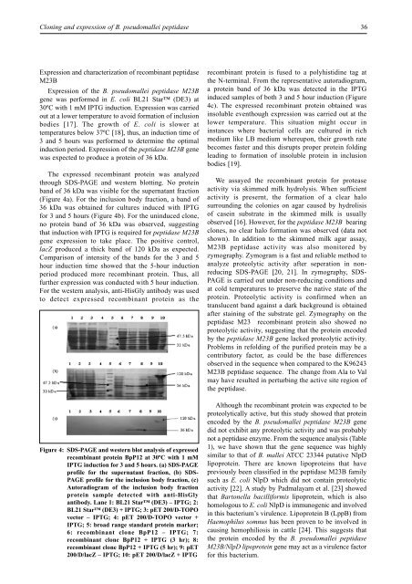
The expressed recombinant protein was analyzed<br />
through SDS-PAGE <strong>and</strong> western blotting. No protein<br />
b<strong>and</strong> <strong>of</strong> 36 kDa was visible for the supernatant fraction<br />
(Figure 4a). For the inclusion body fraction, a b<strong>and</strong> <strong>of</strong><br />
36 kDa was obtained for cultures induced with IPTG<br />
for 3 <strong>and</strong> 5 hours (Figure 4b). For the uninduced clone,<br />
no protein b<strong>and</strong> <strong>of</strong> 36 kDa was observed, suggesting<br />
that induction with IPTG is required for peptidase M23B<br />
gene expression to take place. The positive control,<br />
lacZ produced a thick b<strong>and</strong> <strong>of</strong> 120 kDa as expected.<br />
Comparison <strong>of</strong> intensity <strong>of</strong> the b<strong>and</strong>s for the 3 <strong>and</strong> 5<br />
hour induction time showed that the 5-hour induction<br />
period produced more recombinant protein. Thus, all<br />
further expression was conducted with 5 hour induction.<br />
For the western analysis, anti-HisGly antibody was used<br />
to detect expressed recombinant protein as the<br />
Figure 4: SDS-PAGE <strong>and</strong> western blot analysis <strong>of</strong> expressed<br />
recombinant protein BpP12 at 30ºC with 1 mM<br />
IPTG induction for 3 <strong>and</strong> 5 hours. (a) SDS-PAGE<br />
pr<strong>of</strong>ile for the supernatant fraction, (b) SDS-<br />
PAGE pr<strong>of</strong>ile for the inclusion body fraction, (c)<br />
Autoradiogram <strong>of</strong> the inclusion body fraction<br />
protein sample detected with anti-HisGly<br />
antibody. Lane 1: BL21 Star (DE3) – IPTG; 2:<br />
BL21 Star (DE3) + IPTG; 3: pET 200/D-TOPO<br />
vector – IPTG; 4: pET 200/D-TOPO vector +<br />
IPTG; 5: broad range st<strong>and</strong>ard protein marker;<br />
6: recombinant clone BpP12 – IPTG; 7:<br />
recombinant clone BpP12 + IPTG (3 hr); 8:<br />
recombinant clone BpP12 + IPTG (5 hr); 9: pET<br />
200/D/lacZ – IPTG; 10: pET 200/D/lacZ + IPTG<br />
recombinant protein is fused to a polyhistidine tag at<br />
the N-terminal. From the representative autoradiogram,<br />
a protein b<strong>and</strong> <strong>of</strong> 36 kDa was detected in the IPTG<br />
induced samples <strong>of</strong> both 3 <strong>and</strong> 5 hour induction (Figure<br />
4c). The expressed recombinant protein obtained was<br />
insoluble eventhough expression was carried out at the<br />
lower temperature. This situation might occur in<br />
instances where bacterial cells are cultured in rich<br />
medium like LB medium whereupon, their growth rate<br />
becomes faster <strong>and</strong> this disrupts proper protein folding<br />
leading to formation <strong>of</strong> insoluble protein in inclusion<br />
bodies [19].<br />
We assayed the recombinant protein for protease<br />
activity via skimmed milk hydrolysis. When sufficient<br />
activity is presernt, the formation <strong>of</strong> a clear halo<br />
surrounding the colonies on agar caused by hydrolisis<br />
<strong>of</strong> casein substrate in the skimmed milk is usually<br />
observed [16]. However, for the peptidase M23B bearing<br />
clones, no clear halo formation was observed (data not<br />
shown). In addition to the skimmed milk agar assay,<br />
M23B peptidase activity was also monitored by<br />
zymography. Zymogram is a fast <strong>and</strong> reliable method to<br />
analyze proteolytic activity after seperation in nonreducing<br />
SDS-PAGE [20, 21]. In zymography, SDS-<br />
PAGE is carried out under non-reducing conditions <strong>and</strong><br />
at cold temperatures to preserve the native state <strong>of</strong> the<br />
protein. Proteolytic activity is confirmed when an<br />
translucent b<strong>and</strong> against a dark background is obtained<br />
after staining <strong>of</strong> the substrate gel. Zymography on the<br />
peptidase M23 recombinant protein also showed no<br />
proteolytic activity, suggesting that the protein encoded<br />
by the peptidase M23B gene lacked proteolytic activity.<br />
Problems in refolding <strong>of</strong> the purified protein may be a<br />
contributory factor, as could be the base differences<br />
observed in the sequence when compared to the K96243<br />
M23B peptidase sequence. The change from Ala to Val<br />
may have resulted in perturbing the active site region <strong>of</strong><br />
the peptidase.<br />
Although the recombinant protein was expected to be<br />
proteolytically active, but this study showed that protein<br />
encoded by the B. <strong>pseudomallei</strong> peptidase M23B gene<br />
did not exhibit any proteolytic activity <strong>and</strong> was probably<br />
not a peptidase enzyme. From the sequence analysis (Table<br />
1), we have shown that the gene sequence was highly<br />
similar to that <strong>of</strong> B. mallei ATCC 23344 putative NlpD<br />
lipoprotein. There are known lipoproteins that have<br />
previously been classified in the peptidase M23B family<br />
such as E. coli NlpD which did not contain proteolytic<br />
activity [22]. A study by Padmalayam et al. [23] showed<br />
that Bartonella bacilliformis lipoprotein, which is also<br />
homologous to E. coli NlpD is immunogenic <strong>and</strong> involved<br />
in this bacterium’s virulence. Lipoprotein B (LppB) from<br />
Haemophilus somnus has been proven to be involved in<br />
causing hemophiliosis in cattle [24]. This suggests that<br />
the protein encoded by the B. <strong>pseudomallei</strong> peptidase<br />
M23B/NlpD lipoprotein gene may act as a virulence factor<br />
for this bacterium.