Trypanosoma cruzi: Amastigotes and ... - ResearchGate
Trypanosoma cruzi: Amastigotes and ... - ResearchGate
Trypanosoma cruzi: Amastigotes and ... - ResearchGate

Create successful ePaper yourself
Turn your PDF publications into a flip-book with our unique Google optimized e-Paper software.
EXPERIMENTAL PARASITOLOGY 73, 1-14 (191)<br />
<strong>Trypanosoma</strong> <strong>cruzi</strong>: <strong>Amastigotes</strong> <strong>and</strong> Trypomastigotes Interact with<br />
Different Structures on the Surface of HeLa Cells<br />
RENATO A. MORTARA<br />
Department of Microbiology, Immunology, <strong>and</strong> Parasitology, Escola Paulista de Medicina, Rua Botucatu,<br />
862 6” <strong>and</strong>ar, 04023, Scio Pa&o, SP, Brazil<br />
MORTARA, RENATO A. 1991. <strong>Trypanosoma</strong> <strong>cruzi</strong>: <strong>Amastigotes</strong> <strong>and</strong> trypomastigotes interact<br />
with different structures on the surface of HeLa cells. Experimental Parasitology<br />
73, 1-14. It is generally accepted that <strong>Trypanosoma</strong> <strong>cruzi</strong> trypomastigotes represent the<br />
infective forms of the etiological agent of Chagas’ disease. However, the invasive capacity<br />
of amastigotes <strong>and</strong> their ability to sustain a complete infective cycle in mammalian cultured<br />
cells <strong>and</strong> hosts has been recently demonstrated. In order to compare the process of cell<br />
invasion by these different infective forms, I examined the interactions of trypomastigotes<br />
<strong>and</strong> amastigotes with HeLa cells using a new <strong>and</strong> simple method that improves parasitecell<br />
interactions <strong>and</strong> significantly reduces incubation periods. T. <strong>cruzi</strong> forms were centrifuged<br />
onto HeLa cells grown on coverslips <strong>and</strong> parasite-cell interactions were examined by<br />
fluorescence <strong>and</strong> scanning electron microscopy. As expected, it was observed that all parasite<br />
forms attach <strong>and</strong> eventually enter the cells. However, whereas trypomastigotes preferentially<br />
invade HeLa cells at the edges, as has recently been demonstrated for other cell<br />
types, the initial steps of amastigote-HeLa cell interaction involve binding <strong>and</strong> entangling of<br />
the parasite to surface microvilli. Thus, different T. <strong>cruzi</strong> infective forms interact with<br />
different cell surface structures that could express different receptors at the HeLa cell<br />
membrane. 0 1991 Academic Press, ICC<br />
INDEX DESCRIPTORS AND ABBREVIATIONS: <strong>Trypanosoma</strong> <strong>cruzi</strong>; Amastigote; Metacyclic<br />
trypomastigote; HeLa cell; Parasite-mammalian cell interactions; Actin; Cytoskeleton; Microvillus;<br />
RPM1 1640 containing 10% fetal bovine serum (R-10); Cytochalasin D (CD);<br />
Scanning electron microscopy &EM).<br />
In order to establish a complete life cycle<br />
within the mammalian host, Trypan~~rna<br />
<strong>cruzi</strong>, the ethiological agent of Chagas’ disease,<br />
must invade cells, divide intracellularly<br />
as amastigotes, <strong>and</strong>, finally, transform<br />
into trypomastigotes that may eventually<br />
be ingested by triatomine vectors. It has<br />
been known for a while that trypomastigotes,<br />
derived from blood of infected mammalian<br />
hosts or mammalian cultured cells,<br />
<strong>and</strong> metacyclic trypomastigotes, obtained<br />
from axenic cultures or urine from infected<br />
vectors, represent the classic infective<br />
forms of the parasite (Brener 1973; De<br />
Souza 1984). More recent work, however,<br />
has also demonstrated that amastigotes, derived<br />
from infected cells or organs, can also<br />
infect <strong>and</strong> multiply within mammalian cells<br />
<strong>and</strong> hosts (Pan 1978; Umezawa et al. 1985;<br />
Ley et al. 1988). In an elegant piece of<br />
work, Schenkman et al. (1988) demonstrated<br />
that T. <strong>cruzi</strong> trypomastigotes invade<br />
mammalian cells in a polarized fashion <strong>and</strong><br />
preferentially penetrate fibroblasts at the<br />
cell periphery. By contrast, the mode of interaction<br />
of amastigotes with mammalian<br />
cells has not yet been carefully examined.<br />
In spite of extensive studies on this subject,<br />
the molecular nature of the membrane<br />
elements involved in the initial steps of parasite<br />
attachment is only beginning to<br />
emerge (Quaissi et al. 1984; Kierszenbaum<br />
<strong>and</strong> Stiles 1985; Zingales <strong>and</strong> Colli 1985).<br />
Since actin microtilaments are the major<br />
contractile elements of the mammalian cell<br />
cytoskeleton associated with the plasma<br />
membrane (Alberts et al. 1983), their involvement<br />
in the uptake by phagocytic <strong>and</strong><br />
1<br />
0014-4894/91 $3.00<br />
Copyright 0 1991 by Academic Press, Inc.<br />
All rights of reproduction in any form reserved.
2 RENATOA.MORTARA<br />
nonphagocytic cells of the various parasite previously infected cells. Supematants were centristages<br />
has been examined by several au- fuged at 200s for 2 min to eliminate most cellular debris<br />
<strong>and</strong> parasites were subsequently collected at<br />
thors. The evidences are somewhat con-<br />
25OOg for 15 min. During the isolation procedure, tryflicting<br />
<strong>and</strong> mostly based on the analysis of pomastigotes spontaneously differentiated into<br />
the effect of cytochalasin B on the host cell amastigotes (Andrews et al. 1987) <strong>and</strong> mixtures conbut,<br />
nevertheless, favor a possible partici- taining from 5 to 50% trypomastigotes were usually<br />
pation of actin filaments in a classical en- obtained in this way. After purification, metacyclic<br />
trypomastigotes (7 x 10’ parasites ml-‘) <strong>and</strong> trypodocytic<br />
interiorization, particularly of trymastigote-amastigote<br />
mixtures (2-4 X 10’ parasites<br />
pomastigotes (Alex<strong>and</strong>er 1975; Nogueira ml-‘) were resuspended in R-10 medium <strong>and</strong> incu<strong>and</strong><br />
Cohn 1976; Kipnis et al. 1979; Hen- bated at 37°C for 30 min before the centrifugation proriquez<br />
et al. 1981; Meirelles et al. 1982). cedure. Parasite/cell ratios usually ranged between 5: 1<br />
In a series of studies on the interaction <strong>and</strong> 1511.<br />
The centrifugation of parasites onto HeLa cells was<br />
between enteropathogenic Escherichia coli<br />
carried out essentially as described earlier (Silva et (I/.<br />
<strong>and</strong> HeLa cells, it was demonstrated that 1989). Briefly, HeLa cells were grown for at least 24 hr<br />
localized adherence of the bacteria caused onto glass coverslips in 2-cm-diameter petri dishes<br />
aggregation of actin filaments due to bacte- containing 1.5 ml R-IO medium. The medium was asrial<br />
clumping of surface microvilli (Silva et pirated <strong>and</strong> after addition of the parasite suspensions<br />
the dishes were immediately centrifuged in a swing-out<br />
al. 1989). To study the interactions between<br />
rotor at 2000~ for 15 min at 30°C. After centrifugation,<br />
T. <strong>cruzi</strong> <strong>and</strong> a mammalian cultured cell line, unattached parasites were removed by washing the<br />
I have adapted the centrifugation method coverslips 10 times with phosphate-buffered saline<br />
used in that work (Silva et al. 1989) which (PBS). Associated parasites produced normal infecimproves<br />
parasite-cell interactions <strong>and</strong> re- tions as determined by the observation of amastigotebearing<br />
cells after 2448 hr <strong>and</strong> by the presence of<br />
duces the time of manipulations. Followed<br />
trypomastigotes in culture supernatants after 4-5 days.<br />
by a combination of fluorescence <strong>and</strong> scan- One set of coverslips was fixed with methanol <strong>and</strong><br />
ning electron microscopy, the mode of as- stained with the Giemsa reagent. Duplicate sets of insociation<br />
between different T. <strong>cruzi</strong> forms fected <strong>and</strong> control coverslips were immediately fixed<br />
<strong>and</strong> HeLa cells was examined. It was ob- in 3.7% formaldehyde/PBS for 30 min at room temperature<br />
for immunofluorescence observations or proserved<br />
that trypomastigotes tend to invade<br />
cessed for scanning electron microscopy (SEM).<br />
HeLa cells at the periphery, as expected The formaldehyde-fixed coverslips were washed<br />
from previous studies (Schenkman et al.<br />
1988). By contrast, amastigotes, which can<br />
induce the formation of actin aggregates,<br />
detected at the optical microscopy level,<br />
bind to microvilli forming small aggregates<br />
at the cell surface before entering the HeLa<br />
cells.<br />
MATERIALSAND<br />
METHODS<br />
The T. <strong>cruzi</strong> G strain used throughout this study was<br />
isolated from an infected opossum by Mena-Barreto in<br />
the Amazon (Yoshida 1983). Epimastigotes were<br />
grown in LIT medium (Camargo 1964) <strong>and</strong> metacyclic<br />
trypomastigotes were isolated from late stationary LIT<br />
cultures as previously described (Mortara ef al. 1988).<br />
HeLa cells were grown at 37°C in RPM1 1640 medium<br />
supplemented with 10% fetal bovine serum <strong>and</strong> 40 ug<br />
ml-’ gentamycin (R-10 medium) in a 5% CO, humid<br />
atmosphere. HeLa cell-derived trypomastigotes <strong>and</strong><br />
three times with PBS <strong>and</strong> after a 15-min incubation in<br />
50 mM NH&l/PBS to quench excess aldehyde groups,<br />
the coverslips were treated with PBS containing 0.25%<br />
gelatin, 0.05% NaN, (PGN), <strong>and</strong> 0.1% saponin to improve<br />
permeabilization <strong>and</strong> prevent nonspecific antibody<br />
binding during the subsequent incubations. In<br />
the series of experiments carried out to determine parasite<br />
interiorization, coverslips were fixed in 2% glutaraldehyde<br />
(Sigma, EM grade) in PBS for 1 hr at room<br />
temperature. After three washings in PBS, the coverslips<br />
were incubated with 0.138 M ethanolamine, pH<br />
8.3, for 30 min at room temperature to block excess<br />
aldehyde groups, washed with PBS, <strong>and</strong> soaked in<br />
PGN. Permeabilization of glutaraldehyde-fixed samples<br />
was achieved by soaking the coverslips in PGN<br />
containing 1% Nonidet-P40 (NP-40) before antibody<br />
incubation as described below.<br />
After 15-30 min, excess liquid was blotted with filter<br />
paper <strong>and</strong> the coverslips were inverted for 45 min onto<br />
25-pl drops of 2C2 (Andrews et al. 1987) or 3F5 antiT.<br />
<strong>cruzi</strong> monoclonal antibodies (3F5 reacts with a surface<br />
glycoconjugate expressed on the cell surface of metaamastigotes<br />
were isolated from culture supernatants of cyclic trypomastigotes <strong>and</strong> epimastigotes-R. A. Mor-
T. <strong>cruzi</strong> INFECTIVE FORMS 3<br />
tara <strong>and</strong> S. da Silva, manuscript in preparation)-both<br />
ascitic fluids diluted 1: 100 in PGN-in a humid chamber.<br />
Subsequently, samples were washed five times<br />
with PBS <strong>and</strong> treated in the same way with a solution<br />
containing the appropriate FITC conjugate (Dako<br />
Corp.) <strong>and</strong> phalloidin coupled to rhodamine (Faulstich<br />
ef al. 1983), a kind gift of H. Faulstich. T. <strong>cruzi</strong> actin<br />
was not labeled with the concentrations of phalloidinrhodamine<br />
used in this study <strong>and</strong> required to 30- to<br />
40-fold increase to be visualized (Mortara 1989; R. A.<br />
Mortara, unpublished observations). After washing<br />
the conjugate/phalloidin mixture as above, coverslips<br />
were mounted in 90% glyceroli0.2M glycine, pH 9.2.<br />
Photographs were taken using black <strong>and</strong> white Ilford<br />
HP5 400 ASA films on a Leitz Laborlux D microscope<br />
equipped for epifluorescence with selective FITC <strong>and</strong><br />
rhodamine filters using 40 <strong>and</strong> 100x phase-contrast objectives<br />
(Silva et al. 1989).<br />
For experiments with cytochalasin D (CD, obtained<br />
from Sigma Chemical Co.), appropriate amounts (OS-<br />
5 (~1) of a 5 mg ml-’ stock solution in dimethylsulfoxide<br />
were added to the HeLa cell supematants <strong>and</strong> samples<br />
incubated for 15 min (final CD concentration<br />
ranged between 1 <strong>and</strong> 5 pg ml-‘). After this period,<br />
coverslips were washed three times with R-10 medium<br />
before parasite centrifugations. The parasite suspensions<br />
were then added <strong>and</strong> the dishes subjected to the<br />
centrifugation procedure.<br />
For SEM cells grown on glass coverslips were<br />
washed 10 times in PBS after parasite centrifugation,<br />
fixed with 2.5% glutaraldehyde (Sigma, EM Grade) in<br />
0.1 M phosphate buffer, pH 7.2, postfixed with 1%<br />
osmium tetroxide in 0.1 M phosphate buffer, pH 7.2,<br />
dehydrated in an ethanol series, <strong>and</strong> critical-point<br />
dried with CO,. The coverslips were mounted on appropriate<br />
stubs, coated with gold, <strong>and</strong> examined on a<br />
JEOL JSM 25 SII scanning electron microscope operated<br />
at 25 kV.<br />
RESULTS<br />
The interaction of different T. <strong>cruzi</strong> developmental<br />
forms with HeLa cells was examined<br />
in this study. To improve the association<br />
with HeLa cells <strong>and</strong> reduce the manipulation<br />
time, parasites were centrifuged<br />
onto HeLa cells grown on coverslips <strong>and</strong><br />
after washing unattached parasites, samples<br />
were processed for immunofluorescence<br />
<strong>and</strong> scanning electron microscopy.<br />
On average, these experiments were performed<br />
within 18 min from the addition of<br />
the parasites to the cells until sample fixation.<br />
Under such conditions, metacyclic<br />
trypomastigotes readily attached to HeLa<br />
cells <strong>and</strong> images suggesting parasite invasion<br />
were frequently observed under phasecontrast<br />
microscopy <strong>and</strong> immunofluorescence<br />
(Figs. IA <strong>and</strong> IB). Following the attachment<br />
of metacyclic trypomastigotes,<br />
rearrangements of actin microfilaments<br />
from HeLa cells were not frequently detected,<br />
as judged by the analysis of phalloidin-rhodamine<br />
distribution under conventional<br />
fluorescence microscopy (Fig. IC).<br />
Further observation of attached metacyclic<br />
trypomastigotes showed that after 24 hr all<br />
parasites had entered the cells leading to<br />
normal infections of more than 60% of the<br />
cells, as judged by the presence of intracellular<br />
amastigote forms (result not shown).<br />
The observation of corresponding samples<br />
at the scanning electron microscope<br />
showed that the dorsal surface of the HeLa<br />
cells used in this study (see also Silva et al.<br />
1989) was covered with microvilli (Fig. 2).<br />
A substantial proportion (94%, II = 254) of<br />
the metacyclic trypomastigotes observed<br />
were end-on associated with their posterior<br />
(kinetoplast) portion toward the cells <strong>and</strong><br />
had part of their cell body already inside the<br />
HeLa cells, suggesting that they were engaged<br />
in the process of invasion (e.g., Fig.<br />
2B). The trypomastigotes were usually<br />
(89% of parasites scored, IZ = 224) observed<br />
at cellular edges (Fig. 2A) <strong>and</strong> intercellular<br />
contacts (Fig. 2B) not more than 1<br />
km away from the cell borders <strong>and</strong> did not<br />
appear to associate with surface microvilli<br />
(Figs. 2B-2D). Interestingly, in some cases<br />
more than one metacyclic trypomastigote<br />
was found quite close to others(s) parasite(s)<br />
penetrating a cell. Under identical<br />
experimental conditions, epimastigotes did<br />
not attach to HeLa cells (see Table I).<br />
When mixtures of tissue culture-derived<br />
amastigotes <strong>and</strong> trypomastigotes were centrifuged<br />
onto HeLa cells, actin aggregation<br />
underneath the sites of amastigote adhesion<br />
was observed (Figs. 3A-3D). At the optical<br />
microscopy level it was observed that, using<br />
the centrifugation protocol, amastigotes<br />
usually displayed a greater tendency to as-
RENATO<br />
A. MORTARA<br />
sociate with HeLa cells than tissue culturederived<br />
trypomastigotes (see Table I). Using<br />
glutaraldehyde-fixed <strong>and</strong> unpermeabilized<br />
samples, it could be determined that<br />
amastigotes were already inside the cell after<br />
the centrifugation step since parasites<br />
visible by phase contrast were not labeled<br />
with the monoclonal antibody (Figs. 3A-<br />
3D). By contrast, when samples were fixed<br />
in glutaraldehyde <strong>and</strong> subsequently permeabilized<br />
with 1% NP-40, all amastigotes associated<br />
with the HeLa cells were labeled<br />
with monoclonal antibody 2C2 (Figs. 3D-<br />
3F). Actin aggregation was evident at the<br />
sites of amastigote attachment <strong>and</strong> the<br />
amastigotes without the corresponding microfilament<br />
aggregate were usually those<br />
that did not label with the antibody <strong>and</strong><br />
were considered to be already within the<br />
cell (Figs. 3A-3D). Quantitation of these<br />
samples indicated that after 15 min of centrifugation,<br />
41% of the attached amastigotes<br />
were already inside the HeLa cells<br />
(Table II). Since all changes in actin distribution<br />
after amastigote attachment to HeLa<br />
cells were monitored at the fluorescence<br />
microscope by double-labeling samples<br />
with phalloidin-rhodamine <strong>and</strong> monoclonal<br />
antibody 2C2, controls were performed to<br />
ensure that 2C2 (raised against the major<br />
surface glycoprotein of Y strain amastigotes-Andrews<br />
et al. 1987, 1988) gave a<br />
positive reaction with 100% of the amastigotes<br />
of the G strain <strong>and</strong> that phalloidinrhodamine,<br />
at the concentration used in<br />
this study, did not label parasite actin (Fig.<br />
4). Attachment <strong>and</strong> entry of amastigotes<br />
also led to successful infections <strong>and</strong> cells<br />
displaying several perinuclearly arranged<br />
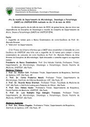
FIG. 1. HeLa cell actin distribution following T.<br />
<strong>cruzi</strong> metacyclic trypomastigote centrifugation. (A)<br />
Phase-contrast micrograph of cells with associated<br />
parasites (arrows); (B) immunofluorescence image of<br />
metacyclics stained with monoclonal antibody 3F5<br />
with suggestive images (arrows) of cell invasion; (C)<br />
same fields labeled with phalloidin-rhodamine, with<br />
typical actin filament bundles. Magnification: x685.<br />
Bar = 10 (*m.
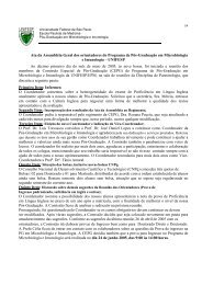
FIG. 2. Scanning electron microscopy of HeLa cells after centrifugation of T. <strong>cruzi</strong> metacyclic<br />
trypomastigotes. (A-D) Images of parasites invading HeLa cells at the edges. (C <strong>and</strong> D) Metacyclic<br />
trypomastigotes penetrating a cell close to each other. Magnifications: (A) X 1000; (B <strong>and</strong> C) x4500;<br />
(D) x3000. Bars = 5 km.<br />
amastigotes were already detectable 24 hr<br />
postinfection (Table II <strong>and</strong> Fig. 5).<br />
Examination by SEM of samples fixed<br />
immediately after the centrifugation step<br />
revealed that amastigotes adhered to the<br />
dorsal surface of HeLa cells (Fig. 6A). At<br />
higher magnifications, the adherence sites<br />
consistently displayed microvilli attached<br />
to the parasite surface forming a small<br />
clump (Figs. 6B-6F). Quantitative studies<br />
carried out at high magnifications (X 10,000)<br />
indicated that 95% of the attached amastigotes<br />
were actually entangled with surface<br />
microvilli whereas the number of contaminating<br />
trypomastigotes associated with the<br />
cells was comparatively small. Up to 10<br />
amastigotes could associate with a single<br />
HeLa cell but the majority of infected cells<br />
contained between one <strong>and</strong> three parasites<br />
on their dorsal surface (Fig. 7).<br />
The role of microtilaments on the attachment<br />
<strong>and</strong> interiorization of T. <strong>cruzi</strong> into<br />
mammalian cells has been probed with cytochalasin<br />
B. Cytochalasin D was used in<br />
this study because it has a higher affinity for<br />
actin filaments <strong>and</strong> its effects on mammalian<br />
cells is also more specific than those of<br />
cytochalasin B (Cooper 1987). Using the<br />
glutaraldehyde fixation approach it was<br />
possible to distinguish between attached<br />
<strong>and</strong> internalized amastigotes in cytochalasin<br />
D-treated HeLa cells. Cytochalasin D<br />
treatment as performed here (5 p.g ml - ’ for<br />
15 min) caused dramatic changes in cell<br />
morphology (see below) <strong>and</strong> despite a significant<br />
reduction in the proportion of intracellular<br />
parasites, it did not block the entry<br />
of amastigotes (Table II). Figure 8 shows<br />
one of such samples in which attached (labeled)<br />
<strong>and</strong> internalized (unlabeled) amastigotes<br />
can be distinguished. The distribution<br />
of actin in CD-treated cells was also affected<br />
<strong>and</strong> microfilament aggregates were<br />
visible throughout the cytoplasm (Fig. 8).
6 RENATO A. MORTARA<br />
TABLE I<br />
Treatment of HeLa Cells with Cytochalasin D:<br />
Effect on Ttypanosoma <strong>cruzi</strong> Association*<br />
No. of<br />
parasites<br />
Cytochalasin D associated<br />
T. <strong>cruzi</strong> form (wg ml-‘) per cell<br />
Metacyclic<br />
trypomastigote@ 0 3.18 k 0.36<br />
2 2.65 -r- 0.32<br />
5 2.48 2 0.40<br />
<strong>Amastigotes</strong>l<br />
trypomastigotes<br />
(70:30 ratio) 0 Ad 3.01 2 0.86<br />
T 0.24 2 0.03<br />
2 A 3.48 4 0.79<br />
T 0.33 ? 0.13<br />
5 A 2.93 t 0.93<br />
T 0.34 2 0.11<br />
Epimastigotes 0 0<br />
0 Parasites scored on at least 400 cells on duplicate<br />
cover slips after Giemsa staining.<br />
b Figures represent means 5 st<strong>and</strong>ard error of two<br />
independent experiments.<br />
c Figures represent means k st<strong>and</strong>ard error of three<br />
independent experiments.<br />
d A, amastigotes; T, trypomastigotes.<br />
Phalloidin staining also revealed that at<br />
some sites of amastigote attachment, cellular<br />
actin appeared to surround the entire<br />
parasite, suggesting that a membrane process<br />
is preventing the binding of monoclonal<br />
2C2 to the parasites (vertical arrows in<br />
Figs. 8A <strong>and</strong> SC).<br />
Cells treated with cytochalasin D displayed<br />
profound morphological changes.<br />
They tended to round up <strong>and</strong> their attachments<br />
to the substratum appeared to retract;<br />
moreover, their surface presented<br />
few microvilli <strong>and</strong> became covered with<br />
blebs (Fig. 9A). Quantitation of amastigote<br />
binding to CD-treated HeLa cells at the<br />
SEM level (Figs. 9C <strong>and</strong> 9D) was not possible<br />
since the abundance of surface blebs<br />
masked the attached parasites (see, for instance,<br />
Fig. 90). On the other h<strong>and</strong>, metacyclic<br />
trypomastigotes are frequently seen<br />
invading CD-treated HeLa cells, preserving<br />
their preference for the cell periphery (Figs.<br />
9E <strong>and</strong> 9F).<br />
Another quantitative evaluation of the effect<br />
of treating HeLa cells with cytochalasin<br />
D on T. <strong>cruzi</strong> association was obtained<br />
with samples stained with Giemsa reagent.<br />
Quantitation was expressed in terms of parasites<br />
associated per cell, since this staining<br />
procedure does not allow the clear distinction<br />
between interiorized <strong>and</strong> attached<br />
forms (Table I). Metacyclic trypomastigote<br />
association with HeLa cells was hardly affected<br />
by CD treatment <strong>and</strong> also no significant<br />
differences were observed in the attachment<br />
of amastigotes, both to control<br />
<strong>and</strong> to CD-treated cells (Table I). Similarly,<br />
tissue culture-derived trypomastigotes associated<br />
with CD-treated HeLa cells in levels<br />
comparable to that of the control samples<br />
(Table I). Incubation of cytochalasin<br />
D-treated samples for 24 hr (in medium<br />
without the drug) resulted in infections<br />
comparable to that of the controls, both for<br />
amastigotes (Table II) <strong>and</strong> metacyclic trypomastigotes<br />
(not shown).<br />
DISCUSSION<br />
Based on previous observations that enteropathogenic<br />
E. coli cause actin aggregation<br />
after localized adhesion of the bacteria<br />
to HeLa cells (Silva et al. 1989) <strong>and</strong> considering<br />
the evidences of a possible involvement<br />
of actin filaments in the interiorization<br />
process of T. <strong>cruzi</strong> in mammalian<br />
cells (Nogueira <strong>and</strong> Cohn 1976; Kipnis et<br />
al. 1979; Henriquez et al. 1981; Meirelles et<br />
al. 1982), the interaction of T. <strong>cruzi</strong> with<br />
HeLa cells was studied in order to examine<br />
possible effects of different developmental<br />
forms of the parasite on the host cell microfilament<br />
system. For such studies a centrifugation<br />
procedure previously described in<br />
our laboratory (Silva et al. 1989) was employed<br />
to maximize parasite-HeLa cell interactions<br />
<strong>and</strong> to reduce manipulation time.<br />
Results with metacyclic trypomastigotes<br />
confirmed previous observations (Schenkman<br />
et al. 1988) that these forms tend to<br />
invade cells at the periphery (Fig. 2). By<br />
contrast, amastigotes attach preferentially
T. Cruzi INFECTIVE FORMS 7<br />
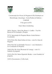
FIG. 3. Centrifugation of T. <strong>cruzi</strong> amastigotes onto HeLa cells induces aggregation of actin filaments<br />
<strong>and</strong> leads to parasite entry. (A-C) Phase-contrast microscopy, 2C2 staining, <strong>and</strong> actin distribution in<br />
glutaraldehyde-fixed samples. (D-F) Equivalent samples stained as before after permeabilization with<br />
1% Nonidet-P40-note that all parasites are labeled with the antibody. Black arrows in (A): intemalized<br />
amastigotes; black arrowheads in (A) indicate attached parasites, also labeled with 2C2 (see B);<br />
white arrows in (C): actin aggregates corresponding to attached amastigotes. Magnification: x710. Bar<br />
= 10 pm.<br />
to the dorsal surface of HeLa cells where<br />
they induce actin filament aggregation that<br />
corresponds to clumps of surface microvilli<br />
<strong>and</strong> parasites (Figs. 4 <strong>and</strong> 6). Attached <strong>and</strong><br />
internalized amastigotes could be distinguished<br />
using differential antibody accessibility<br />
after glutaraldehyde fixation (Fig. 4<br />
<strong>and</strong> Table II). The finding that 41% of the<br />
amastigotes were localized within the cells<br />
after 15 min required for the centrifugation<br />
step supports the notion that this procedure<br />
promotes infection by amastigote forms.<br />
Under the experimental conditions described<br />
here, trypomastigotes derived from<br />
infected cells displayed a lower degree of<br />
association with HeLa than amastigotes. It
8 RENATO A. MORTARA<br />
TABLE II<br />
Entry of <strong>Trypanosoma</strong> <strong>cruzi</strong> <strong>Amastigotes</strong>” into HeLa<br />
Cellsb Effect of Cytochalasin D<br />
Percentage of<br />
Cytochalasin DC parasitesd inside No. amastigotes’<br />
(kg mlm’) the cells inside 100 cells<br />
0 41.3 k 6.8 155 t 13<br />
5 11.2 2 0.9 422 10<br />
0 97.1 2 l.lf 255 ? 35<br />
5 95.4 k 2.P 247 2 42<br />
a These parasite preparations contained more than<br />
95% amastigotes.<br />
b Parasites were centrifuged onto HeLa cells <strong>and</strong><br />
after glutaraldehyde fixation, samples were stained<br />
with monoclonal antibody 2C2.<br />
c Cells were incubated for 15 min with 5 ug ml-’<br />
CD.<br />
d Percentage refers to the ratio of parasites not labeled/total<br />
parasites on at least 200 cells on duplicate<br />
cover slips. Figures represent means rt st<strong>and</strong>ard error<br />
of two independent experiments.<br />
p Intracellular amastigotes scored on 200 cells on<br />
duplicate coverslips. Figures represent means 2 st<strong>and</strong>ard<br />
error of two independent experiments.<br />
f Figures obtained following incubation in drug-free<br />
medium for 24 hr, after centrifugation.<br />
should be pointed out, however, that these<br />
associations do not reflect the relative infectivities<br />
under conventional conditions<br />
where trypomastigotes clearly display a<br />
much greater capacity to infect mammalian<br />
cells (e.g., Schenkman et al. 1988).<br />
Surface projections like microvilli <strong>and</strong><br />
filopodia are probing organelles (Vasiliev<br />
1981) thought to be involved in initial steps<br />
of parasitexell interactions (McGee et al.<br />
1988). The interaction of amastigotes with<br />
microvilli on the dorsal surface of HeLa<br />
cells forming small clumps (Fig. 6) confirms<br />
that these membrane projections act as<br />
docking elements to the spheroid amastigotes.<br />
These interactions could involve<br />
specific parasite <strong>and</strong> microvillus membrane<br />
components. This apparently specific association<br />
with microvilli could imply that<br />
these surface protrusions concentrate specific<br />
membrane receptor molecules for<br />
amastigotes, as seems to occur in other systems<br />
(e.g., Koch <strong>and</strong> Smith, 1978). Inter-<br />
FIG. 4. Control of T. <strong>cruzi</strong> amastigote staining with<br />
monoclonal antibody 2C2 <strong>and</strong> phalloidin-rhodamine.<br />
(A) Phase-contrast micrograph of parasites; (B) 2C2<br />
monoclonal antibody staining, <strong>and</strong> (C) phalloidinrhodamine<br />
staining of actin using the same concentrations<br />
of the reagent as in Figs. 1, 3, <strong>and</strong> 8.<br />
estingly, when HeLa cells are treated with<br />
cytochalasin D at concentrations that dramatically<br />
altered their surface morphology<br />
(Fig. 9) <strong>and</strong> completely blocked localized<br />
adherence of enteropathogenic E. coli<br />
(Silva et al. 1989), amastigote (<strong>and</strong> trypomastigote)<br />
adhesion was still observed in<br />
levels comparable to those of the controls<br />
(Table I). Since cytochalasin D-treated<br />
cells express fewer microvilli while retaining<br />
their capacity to physically attach to<br />
amastigotes, it could be tentatively assumed<br />
that the putative receptor molecule(s)<br />
remains functionally expressed at<br />
the surface <strong>and</strong> that the microvillar structure<br />
is not required for parasite adhesion.
T. <strong>cruzi</strong> INFECTIVE FORMS 9<br />
FIG. 5. Amastigote attachment <strong>and</strong> invasion causes<br />
normal infection. <strong>Amastigotes</strong> were centrifuged onto<br />
HeLa cells, washed, <strong>and</strong> incubated for 24 hr before<br />
fixation <strong>and</strong> 2C2 staining. (A) Phase-contrast <strong>and</strong> (B)<br />
2C2 labeling of amastigotes. Magnification: X260. Bar<br />
= 10 pm.<br />
This is again in contrast to the effect observed<br />
with the adhesion of enteropathogenie<br />
E. cofi that is almost abolished when<br />
HeLa cells are exposed to conditions that<br />
reduce the expression of microvilli (Silva ef<br />
al. 1989). Although cytochalasin D did not<br />
significantly affect parasite attachment (Table<br />
I), the disruption of cellular microfila-<br />
ments with CD greatly inhibited amastigote<br />
entry after the centrifugation procedure<br />
(Table II).<br />
At this point it would be interesting to<br />
consider the role of host cell actin microtilaments<br />
on T. <strong>cruzi</strong> attachment <strong>and</strong> invasion.<br />
Previous studies based mainly on the<br />
inhibitory effect of cytochalasin B were<br />
taken as indications that microfilaments<br />
participate in the uptake of T. <strong>cruzi</strong> by cells<br />
(Alex<strong>and</strong>er 1975; Nogueira <strong>and</strong> Cohn 1976;<br />
Henriquez et al. 1981; Meirelles et al.<br />
1982). In these studies it was shown that<br />
severe disruption of the cortical microtilament<br />
system with cytochalasin D did not<br />
cause a significant effect on the degree of<br />
association of different T. <strong>cruzi</strong> infective<br />
forms with HeLa cells (Table I) <strong>and</strong> subsequent<br />
incubations in medium without the<br />
drug resulted in normal infections (Table<br />
II). It is noteworthy that metacyclic trypomastigotes<br />
retain their preference to invade<br />
the cells at the periphery, even in CDtreated<br />
cells (see Figs. 2 <strong>and</strong> 9). In a recent<br />
study, Schenkman et al. (1991) observed<br />
that CD, at the same concentration used<br />
here, did not affect the entry of Y-strain<br />
trypomastigotes into HeLa cells. These<br />
findings support the notion that (1) attachment<br />
is required for invasion <strong>and</strong> (2) attached<br />
parasites will eventually enter (even<br />
CD-treated) cells. This is intriguing in view<br />
of the accepted notion that a classical receptor-mediated<br />
endocytosis mechanism is<br />
believed to operate in the uptake of the parasite<br />
(Kierszenbaum <strong>and</strong> Stiles, 1985;<br />
Schenkman et al. 1988; Zingales <strong>and</strong> Colli<br />
1985). In such a mechanism, motility of<br />
membrane receptors tethered to the cytoskeleton<br />
leading to the uptake of surface<br />
lig<strong>and</strong>s is thought to require a functional<br />
cortical meshwork of actin microfilaments,<br />
since such processes are promptly inhibited<br />
by cytochalasins (e.g., Cooper 1985). The<br />
observations that the infective forms of T.<br />
<strong>cruzi</strong> keep their capacity to associate with<br />
<strong>and</strong> invade (albeit to a lower degree in the<br />
case of the amastigotes) cells bearing dis-
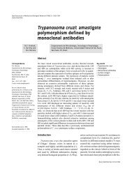
FIG. 6. T. <strong>cruzi</strong> amastigote centrifuged onto HeLa cells entangle to microvilli. (A-H) Scanning<br />
electron micrographs showing amastigotes attached to the dorsal surface of HeLa cells. Images in<br />
(B-H) show details of clumps formed with microvilh bound to amastigotes. Magnifications: (A) X 1500;<br />
(B) x4500; (C <strong>and</strong> D) x7000; (E) x4500; (F) x7000; (G <strong>and</strong> H) X10,000. Bars in A, B, C, <strong>and</strong> H =<br />
5 pm.
T. <strong>cruzi</strong> INFECTIVE FORMS 11<br />
123456769<br />
AMASTIGOTES PER CELL<br />
FIG. 7. Frequency of T. <strong>cruzi</strong> amastigote attachment<br />
per HeLa cells after parasite centrifugation.<br />
Black bars: determined by SEM; hatched bars: determined<br />
after Giemsa staining.<br />
rupted microfilaments <strong>and</strong> altered surface<br />
morphology is consistent with the notion<br />
that the interaction of parasite lig<strong>and</strong>s with<br />
appropriate functional receptor molecules<br />
at specific surface sites, as envisaged by<br />
King (1988), could be sufficient to trigger a<br />
parasite-directed endocytosis (McGee ef al.<br />
1988) efficient enough to overcome the<br />
damage caused to the host cell cytoskeleton<br />
by cytochalasin D. The highly motile trypomastigotes<br />
would be more efficient than<br />
amastigotes in such an internalization<br />
mechanism involving a positive participation<br />
of T. <strong>cruzi</strong>, which could be envisaged<br />
as an active entry process as previously<br />
suggested by Kipnis et al. (1979).<br />
Finally, the preference of trypomastigotes<br />
for the cell periphery <strong>and</strong> the association<br />
of amastigotes with surface microvilli<br />
FIG. 8. Cytochalasin D inhibits but does not block<br />
amastigote entry into HeLa cells. Cells were fixed<br />
with glutaraldehyde after treatment with 5 ug ml - t CD<br />
for 15 min <strong>and</strong> parasite centrifugation. (A) Phasecontrast<br />
micrograph, (B) 2C2 staining, <strong>and</strong> (C) actin<br />
distribution. Black arrows in (A), internalized amastigotes;<br />
arrowheads in (A), attached amastigotes (see<br />
B); white arrows in (C), actin aggregates at amastigote<br />
attachment (horizontal) <strong>and</strong> internalization (vertical)<br />
sites. Vertical arrows at top right in (A) <strong>and</strong> (C) indicate<br />
internalized amastigote tightly covered with cellular<br />
actin. Magnification x800. Bar = 10 urn.
12 RENATO A.MORTARA<br />
FIG. 9. T. <strong>cruzi</strong> forms attach to cytochalasin D-treated HeLa cells. Scanning electron microscopy<br />
of different T. <strong>cruzi</strong> infective forms centrifuged onto HeLa cells after incubation for 15 min with 5 p.g<br />
ml-’ CD. (A) General aspect of treated cells. (B-D) Cells after amastigote:trypoamstigote mixture<br />
(70:30 ratio) centrifugation (arrow in D points to an attached amastigote); (E <strong>and</strong> F) cells after metacyclic<br />
trypomastigote centrifugation; note trypomastigotes penetrating the cells at the edges. Magnifications:<br />
(A) x1000; (B) x2000; (C-E) x4500; (F) x4000. Bars in A, B, D, <strong>and</strong> F = 5 pm.<br />
shows that different T. <strong>cruzi</strong> developmental<br />
forms use or mobilize distinct structures<br />
<strong>and</strong>/or receptors to attach <strong>and</strong> invade HeLa<br />
cells.<br />
ACKNOWLEDGMENTS<br />
I thank Sebastiio Cruz, Helena C. Barros, <strong>and</strong> W<strong>and</strong>erley<br />
de Souza for their help with the scanning microscopy.<br />
I am grateful to my colleagues Nobuko<br />
Yoshida <strong>and</strong> Jose Franc0 da Silveira for their comments<br />
<strong>and</strong> suggestions on the manuscript. I am particularly<br />
grateful to Sergio Schenkman for helpful discussions<br />
<strong>and</strong> communication of his results prior to publication.<br />
This work was supported by grants from the<br />
Fundacao de Amparo a Pesquisa do Estado de Slo<br />
Paulo-FAPESP <strong>and</strong> Conselho National de desenvolvimento<br />
Cientitico e Tecnologico-CNPq, <strong>and</strong> UNDP/<br />
World Bank/WHO Special Programme for Research<br />
<strong>and</strong> Training in Tropical Diseases <strong>and</strong> the Rockefeller<br />
Foundation.
T. <strong>cruzi</strong> INFECTIVE FORMS 13<br />
REFERENCES<br />
ALBERTS, B., BRAY, D., LEWIS, J., RAFF, M., ROB-<br />
ERTS, K., AND WATSON, J. D. 1983. The cytoskeleton.<br />
In “The Molecular Biology of the Cell,” pp.<br />
549-609. Garl<strong>and</strong>, New York/London.<br />
ALEXANDER, J. 1975. Effect of the antiphagocytic<br />
agent cytochalasin B on macrophage invasion by<br />
Leishmaniu mexicana promastigotes <strong>and</strong> <strong>Trypanosoma</strong><br />
<strong>cruzi</strong> epimastigotes. Journal of Protozoology<br />
22, 237-240.<br />
ANDREWS, N. W., HONG, K. S., ROBBINS, E. S.,<br />
AND NUSSENZWEIG, V. 1987. Stage-specific surface<br />
antigens expressed during the morphogenesis of vertebrate<br />
forms of <strong>Trypanosoma</strong> <strong>cruzi</strong>. Experimental<br />
Parasitology 64, 474-484.<br />
ANDREWS, N. W., ROBBINS, E. S., LEY, V., HONG,<br />
K. S., AND NUSSENZWEIG, V. 1988. Developmentally<br />
regulated, phospholipase C-mediated release of<br />
the major surface glycoprotein of amastigotes of<br />
<strong>Trypanosoma</strong> <strong>cruzi</strong>. The Journal of Experimental<br />
Medicine 167, 300-3 14.<br />
BRENER, Z. 1973. Biology of <strong>Trypanosoma</strong> <strong>cruzi</strong>. Annual<br />
Reviews of Microbiology 27, 347-554.<br />
CAMARGO, E. P. 1964. Growth <strong>and</strong> differentiation in<br />
<strong>Trypanosoma</strong> <strong>cruzi</strong>: Origin of metacyclic trypomastigotes<br />
in liquid media. Revista do Instituto de<br />
Medicina Tropical de Sdo Paul0 6, 93-100.<br />
COOPER, J. A. 1987. Effects of cytochalasins <strong>and</strong> phalloidin<br />
on actin. Journal of Cell Biology 105, 1473-<br />
1478.<br />
DE SOUZA, W. 1984. Cell Biology of <strong>Trypanosoma</strong><br />
<strong>cruzi</strong>. international Reviews of Cytology 86, 197-<br />
283.<br />
FAULSTICH, H., TRISCHMAN, H., AND MAYER, D.<br />
1983. Preparation of tetramethyl rhodaminylphalloidin<br />
<strong>and</strong> uptake of the toxin into short-term<br />
cultured hepatocytes. Experimental Cell Research<br />
144, 63-72.<br />
HENRIQUEZ, D., PIUS, R., AND PIUS, M. M. 1981.<br />
The effect of surface membrane modifications of tibroblastic<br />
cells on the entry process of <strong>Trypanosoma</strong><br />
<strong>cruzi</strong> trypomastigotes. Molecular <strong>and</strong> Biochemical<br />
Parasitology 2, 35%366.<br />
KIERSZENBAUM, R., AND STILES, B. 1985. Evidence<br />
supporting the existence of a host cell surface receptor<br />
for Ttypanosoma <strong>cruzi</strong>. Journal of Protozoology<br />
32, 364-366.<br />
KING, C. A. 1988. Cell motility of sporozoan protozoa.<br />
Parasitology Today 4( 1 I), 3 15-3 19.<br />
KIPNIS, T. L., CALICH, V. L. G., AND DIAS DA<br />
SILVA, W. 1979. Active entry of bloodstream forms<br />
of <strong>Trypanosoma</strong> <strong>cruzi</strong> into macrophages. Parasitology<br />
78, 89-98.<br />
KOCH, G. L. E., AND SMITH, M. J. 1978. An association<br />
between actin <strong>and</strong> the major histocompatibility<br />
antigen H-2. Nature (London) 273, 274-278.<br />
LEY, V., ANDREWS, N. W., ROBBINS, E. S., AND<br />
NUSSENZWEIG, V. 1988. <strong>Amastigotes</strong> of <strong>Trypanosoma</strong><br />
<strong>cruzi</strong> sustain an infective cycle in mammalian<br />
cells. Journal of Experimental Medicine 168, 649-<br />
659.<br />
MCGEE, Z., GORBY, G. L., WYRICK, P. B., HODINKA,<br />
R., AND HOFFMAN, L. H. 1988. Parasite-directed<br />
endocytosis. Reviews on Infectious Diseases 10,<br />
S311-S316.<br />
MEIRELLES, M. N. L., ARA~JO JORGE, T. C., AND DE<br />
SOUZA, W. 1982. Interaction of <strong>Trypanosoma</strong> <strong>cruzi</strong><br />
with macrophages in vitro: Dissociation of the attachment<br />
<strong>and</strong> internalization phases by low temperature<br />
<strong>and</strong> cytochalasin B. Zeitschrift fir Parasitenkunde<br />
68, 7-14.<br />
MORTARA, R. A., ARAGUTH, M. F., AND YOSHIDA,<br />
N. 1988. Reactivity of stage-specific monoclonal antibody<br />
lG7 with metacyclic trypomastigotes of Try-<br />
panosoma <strong>cruzi</strong> strains: Lytic property <strong>and</strong> antigen<br />
polymorphism. Parasite Immunology 10, 369-378.<br />
MORTARA, R. A. 1989. Studies on trypanosomatid actin.<br />
I. lmmunochemical <strong>and</strong> biochemical identification.<br />
Journal of Protozoology 36, 8-13.<br />
NOGUEIRA, N., AND COHN, Z. 1976. <strong>Trypanosoma</strong><br />
<strong>cruzi</strong>: Mechanism of entry <strong>and</strong> intracellular fate in<br />
mammalian cells. Journal of Experimental Medicine<br />
143, 1402-1420.<br />
NOISIN, E. L., AND VILLALTA, F. 1989. Fibronectin<br />
increases <strong>Trypanosoma</strong> <strong>cruzi</strong> amastigote binding to<br />
<strong>and</strong> uptake by murine macrophages <strong>and</strong> human<br />
monocytes. Infection <strong>and</strong> Immunity 57, 1030-1034.<br />
OUAISSI, M. A., AFCHAIN, A., CAPRON, A., AND GRI-<br />
MAUD, J. A. 1984. Fibronectin receptors on Try-<br />
panosoma <strong>cruzi</strong> trypomastigotes <strong>and</strong> their biological<br />
role. Nature (London) 308, 380-382.<br />
PAN, S. C.-T. 1978. <strong>Trypanosoma</strong> <strong>cruzi</strong>: In virro interactions<br />
between cultured amastigotes <strong>and</strong> human<br />
skin-muscle cells. Experimental Parasitology 45,<br />
274-286.<br />
SCHENKMAN, S., ANDREWS, N. W., NUSSENZWEIG,<br />
V., AND ROBBINS, E. S. 1988. <strong>Trypanosoma</strong> <strong>cruzi</strong><br />
invade a mammalian epithelial cell in a polarized<br />
manner. Cell 55, 157-165.<br />
SCHENKMAN, S., ROBINS, E. S., AND NUSSENZWEIG,<br />
V. 1991. The attachment <strong>and</strong> penetration of <strong>Trypanosoma</strong><br />
<strong>cruzi</strong> into mammalian cells require<br />
parasite energy <strong>and</strong> can be independent of the target<br />
cell cytoskeleton. Infection <strong>and</strong> Immunity, 59,<br />
645-654.<br />
SILVA, M. L. M., MORTARA, R. A., BARROS, H. C.,<br />
DE SOUZA, W., AND TRABULSI, L. R. 1989. Aggre-
14 RENATO A. MORTARA<br />
gation of membrane associated actin filaments following<br />
localized adherence of enteropathogenic<br />
Escherichia coli to HeLa cells. Journa/ of Cell Science<br />
93, 4394l6.<br />
UMEZAWA, E. S., MILDER, R. V., AND ABRAHAM-<br />
SON, 1. A. 1985. <strong>Trypanosoma</strong> <strong>cruzi</strong> amastigotes:<br />
Development in vitro <strong>and</strong> infectivity in vivo of the<br />
forms isolated from spleen <strong>and</strong> liver. Acta Tropica<br />
42, 25-32.<br />
VASILIEV, J. M. 1981. The role of pseudopodial reactions<br />
in the attachment of normal <strong>and</strong> transformed<br />
cells. In “International Cell Biology” (H. G. Schweiger,<br />
Ed.). Springer-Verlag, Berlin.<br />
YOSHIDA, N. 1983. Surface antigens of metacyclic trypomastigotes<br />
of <strong>Trypanosoma</strong> <strong>cruzi</strong>. Infection <strong>and</strong><br />
Immunity 40, 836-839.<br />
ZINGALES, B., AND COLLI, W. 1985. <strong>Trypanosoma</strong><br />
<strong>cruzi</strong>: Interaction with host cells. Current Topics in<br />
Microbiology <strong>and</strong> Immunology 117, 129-152.<br />
Received 24 July 1990; accepted with revision 22 January<br />
1991