Axio Imager 2
Axio Imager 2
Axio Imager 2

You also want an ePaper? Increase the reach of your titles
YUMPU automatically turns print PDFs into web optimized ePapers that Google loves.
Microscopy from Carl Zeiss<br />
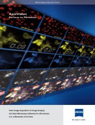
<strong>Axio</strong> <strong>Imager</strong> 2<br />
Progress Meets Performance<br />
Trend-setting technology for brilliant results<br />
in all life science research applications
<strong>Axio</strong> <strong>Imager</strong> 2 from Carl Zeiss.<br />
Success in Series.<br />
Always provide the best tools for the study of life – with this objective in mind<br />
Carl Zeiss introduced <strong>Axio</strong> <strong>Imager</strong> in 2004. This objective still applies. The result: the<br />
new <strong>Axio</strong> <strong>Imager</strong> product generation. With outstanding performance. With unrivaled<br />
optics. With an unmatched range of application. And with maximum ease of use.<br />
<strong>Axio</strong> <strong>Imager</strong>: Trailblazer in Terms of Performance<br />
More flexibility for more performance: from simple observation<br />
and image acquisition to highly complex analyses,<br />
there are six different stands available, which allow you<br />
to adapt the system exactly to your individual application<br />
by providing many different system components. Taken<br />
together these are trend-setting performance characteristics<br />
and technical innovations for outstanding research<br />
results.<br />
Inhalt<br />
<strong>Axio</strong> <strong>Imager</strong> 2 from Carl Zeiss 2-3<br />
Optics 4-5<br />
Fluorescence 6-7<br />
Applications 8-10<br />
Imaging Systems and System Tables 11-13<br />
Ergonomy and Ease of Operation 14<br />
Stand Design 16-18<br />
System Overview 19-25<br />
2
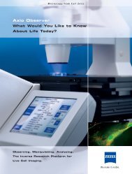
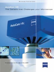
Respiratory epithelium cells<br />
COS cell culture<br />
• Encoding: Readout of magnification, illumination<br />
or contrast settings, respectively, and transfer to the<br />
<strong>Axio</strong>Vision image processing software.<br />
• Motorization for reproducible settings and automatic<br />
procedures.<br />
• Excellent optics and uniform illumination in transmitted<br />
light and fluorescence applications.<br />
• Highest precision due to new high-performance focus,<br />
even in cases of constant load and heavy stages.<br />
• Intelligent control concept for ergonomic work and<br />
multi-user operation<br />
• Preconfigured stand configurations for a broad<br />
application spectrum<br />
• Assured future use supported by modular system<br />
architecture<br />
3
Optics.<br />
Brilliant Performance.<br />
Excellent optical quality: That is what the Carl Zeiss research class stands for.<br />
<strong>Axio</strong> <strong>Imager</strong> 2 boosts this performance even further. From the transmitted light<br />
beam path to the new motorized DIC turret to the high-performance objectives<br />
<strong>Axio</strong> <strong>Imager</strong> provides excellent results even with extremely weak signals.<br />
Visibly more information: the IC²S beam path<br />
IC²S stands for Infinity Contrast & Color Corrected System.<br />
This patented beam path is based on the optimization<br />
of the proven Carl Zeiss ICS Infinity Optics (ICS).<br />
New: the transmitted light beam path for uniform illumination.<br />
The optics of the universal and long-distance<br />
condensers have been adapted to all applications. Even<br />
at low magnifications and large working distances considerably<br />
better resolution and contrast is achieved.<br />
<strong>Axio</strong> <strong>Imager</strong>’s optical system provides you with remarkable<br />
performance: higher image contrast, perfect uniformity<br />
and unrivalled resolution in every contrast technique.<br />
Simple upgrading: the freely accessible infinite space<br />
With its freely accessible infinite space, <strong>Axio</strong> <strong>Imager</strong> allows<br />
additional components such as light sources and<br />
detectors to be added as needed. An individual system<br />
solution that is tuned to the respective application can be<br />
simply and rapidly configured.<br />
Unrivalled in every respect: the objectives<br />
For the new <strong>Axio</strong> <strong>Imager</strong> 2 product line Carl Zeiss has<br />
extended the high-performance objectives especially for<br />
high magnifications, for both fixed and live cell imaging<br />
applications.<br />
• The EC Plan-NEOFLUAR universal objectives. The consistent<br />
stray light minimization results in a definite<br />
contrast enhancement, which is critical in all microscopic<br />
techniques.<br />
• The Plan-APOCHROMAT objectives convince through<br />
their outstanding point spread function and their<br />
unparalleled planar and chromatic correction.<br />
• The αPlan-APOCHROMAT objectives 100x/1.46 Oil<br />
and 100x/1.57 HI Oil (available from Fall 2009) provide<br />
maximum resolution in fluorescence and transmitted<br />
light DIC techniques because of their high numerical<br />
apertures.<br />
• The LCI Plan-NEOFLUAR objectives 25x/0.8 and<br />
63x/1.3 Imm. korr. were conceived for live cell Imaging<br />
techniques and calibrated for specific temperature intervals<br />
as well as immersion media from water to glycerin.<br />
Olfactory bulb (frog), image taken with DIC.<br />
Objective: EC Plan-NEOFLUAR 20x/0.5<br />
Olfactory bulb (frog), multichannel fluorescence with ApoTome.<br />
Green: projections of olfactory sensory cells.<br />
Red: cell nuclei. Objective: EC Plan-NEOFLUAR 20x/0.5.<br />
D. Schild, Univ. Göttingen, Germany<br />
The new motorized DIC turret<br />
4
Beam path optics<br />
a Ocular<br />
b Accessible interface to the ∞ space<br />
c Reflector<br />
d Objective<br />
e Condensor<br />
f HBO<br />
g HAL<br />
a<br />
Flexible interfaces<br />
1 Accessible interface to the ∞ FL space<br />
2 Reflected light-field diaphragm<br />
3 Reflected light-aperture diaphragm<br />
4 Transmitted light-field diaphragm<br />
5 Filter wheels<br />
b<br />
c<br />
1<br />
2 3<br />
f<br />
d<br />
e<br />
5<br />
g<br />
4<br />
See more: DIC or DIC + Fluorescence<br />
Optimized DIC for the new generation <strong>Axio</strong> <strong>Imager</strong>: uniform<br />
interference contrast at all magnifications from 5x<br />
to 100x across the entire field of view. Particularly in digital<br />
imaging the shading correction becomes obsolete.<br />
You always have a uniformly illuminated DIC image. For<br />
the first time these advantages are now also reproducible<br />
and can be adjusted via motorized control. With the<br />
new motorized DIC turret for transmitted light DIC you<br />
can now automatically shift between high-resolution and<br />
high-contrast interference contrast. The contrast settings<br />
can be separately stored for each user and for each magnification<br />
used. You can also combine DIC imaging with<br />
fluorescence excitation extremely simple and automatically.<br />
Without sample-induced artifacts.<br />
Constant color temperature: the LED<br />
illumination sources<br />
The interesting alternative to conventional halogen<br />
illumination with compelling advantages: constant color<br />
temperature independent of the brightness, low heat radiation<br />
and long service life. LED illumination also has a<br />
filter mount for individual setting of the color temperature.<br />
For the first time such an illumination source is also<br />
offered with a trigger input for high frequency switching.<br />
For more simple applications there is a variant available<br />
which is attached directly beneath the condenser. In accordance<br />
with the Fixed Köhler Principle, for simple adjustment<br />
with all contrast techniques.<br />
Motorized DIC turret for reproducible contrast<br />
adjustment<br />
LED – the new light source for Köhler illumination<br />
LED for Fixed Köhler illumination<br />
5
Fluorescence.<br />
Strong Components for Weak Signals.<br />
Brilliant signals for the finest structures and extremely rapid processes – that is what<br />
Carl Zeiss fluorescence microscopy stands for. And all the components of the new<br />
generation of <strong>Axio</strong> <strong>Imager</strong> have been designed to meet this standard. With fast<br />
image acquisition in <strong>Axio</strong>Vision and light sources such as Colibri. With filter sets for<br />
new dye combinations. And with high ease of operation.<br />
Motorized reflector turret for rapid imaging<br />
The investigation of rapid processes is becoming increasingly<br />
important. The motorized reflector turrets<br />
are custom tailored to this end. Six filter modules can<br />
be accommodated. Even for the use of more than six<br />
dyes simultaneously, for example, in multi-color FISH applications,<br />
the <strong>Axio</strong> <strong>Imager</strong>.Z2 provides the best possible<br />
results. The motorized 10-position reflector turret synchronized<br />
with the fast Colibri LED light source provides<br />
a wide selection of excitation wavelengths and brilliant<br />
results without pixel shift.<br />
Reproducible settings by means of<br />
motorized diaphragms<br />
The intelligent, motorized aperture and luminous field<br />
diaphragm automatically controls contrast and illumination.<br />
In the reflected-light beam path as well as in the<br />
transmitted-light beam path. Objective-specific aperture<br />
adaptations can be saved and loaded again at any time<br />
for reliable reproducibility.<br />
Versatile as never before: the High Efficiency Filter<br />
Sets<br />
The HE Fluorescence Filters for <strong>Axio</strong> <strong>Imager</strong> provide an<br />
excellent signal-noise ratio, high transmission for excitation<br />
and emission, and for up to 50 % shorter exposure<br />
times. This protects sensitive samples to the greatest<br />
Simply fast: the change from manual to motorized<br />
reflector turret<br />
Changing to HE filter set<br />
Motorized diaphragm sliders<br />
6
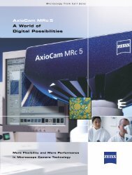
Red: YFP-labeled cell body Cyan: CFP-labeled peroxisomes Multi-channel image: red and cyan channels superimposed<br />
Primary neurons (rat) in culture.<br />
Objective: EC Plan-NEOFLUAR 40x/0.75.<br />
Y. Okada, Dept. Cell Biol. & Anatomy, Grad.Sch.Med,<br />
Univ. Tokyo Hongo, Tokyo, Japan<br />
possible degree. Seven new filter sets and multi-color<br />
combinations with double and triple filter sets were developed<br />
especially for combination with new fluorescing<br />
proteins. The trend toward red dyes such as mRFP,<br />
mCherry, mPlum, mTomato was considered in optimizing<br />
the range of LED options integrated into Colibri. As a<br />
result, the energy of the available LEDs can be completely<br />
exploited.<br />
Light sources for every requirement<br />
For <strong>Axio</strong> <strong>Imager</strong> you can select exactly the light source<br />
which ideally meets the specific demands of your fluorescence<br />
application.<br />
• The self-adjusting HBO lamp has been the illumination<br />
source of choice for all standard fluorescence<br />
applications since 2004. After each lamp change and<br />
each time the device is switched on, it centers itself<br />
automatically such that uniform illumination is guaranteed.<br />
• Metal halide lamps such as HXP 120 exhibit an emission<br />
spectrum similar to HBO lamps. Remote coupling via<br />
liquid light guide, minimizes heat transfer to the stand<br />
making it ideal for live cell imaging.<br />
• Exact intensity control and thus ideal specimen<br />
protection, Specific wavelength selection, and flexible<br />
mixing of different wavelengths, long lifetime, and –<br />
above all – switching time in the microsecond range<br />
characterize the Colibri LED light source. It is ideal for<br />
complex applications at extremely high speeds.<br />
• HXP 120 and Colibri can also be used in combination.<br />
In this manner dyes for which there is no LED today<br />
can be excited.<br />
There are 11 different LEDs available for Colibri: from UV to dark red<br />
Each LED is continuously adjustable and can<br />
be switched in the microsecond range<br />
7
Applications<br />
Infinite Diversity.<br />
The more diverse the applications, the more flexible the imaging platform – that<br />
is what <strong>Axio</strong> <strong>Imager</strong> stands for. The modular architecture of <strong>Axio</strong> <strong>Imager</strong> 2 allows<br />
you to use a technology that optimally supports your application. And which grows<br />
with your performance requirements.<br />
Pathology<br />
<strong>Axio</strong> <strong>Imager</strong>.A2 with LED illumination, the coded stand<br />
with Fixed Köhler illumination is ideal for pathology. In<br />
conjunction with EC Plan-NEOFLUAR or Plan-APOCHROMAT<br />
objectives it is the standard equipment for histological<br />
evaluation. The economical LED illumination has a long<br />
service life, consumes little energy, and requires no maintenance<br />
or adjustment. It provides incredible images, for<br />
instance with the typical H.-E-, DAB- or Azan staining<br />
techniques. Its constant color temperature ensures uniform<br />
light quality and brilliant image presentation over<br />
the entire intensity range.<br />
Human genetics<br />
For the diagnosis of diseases which are due to a mutation<br />
in genetic material, genome analysis is a standard tool<br />
in human genetics. Karyograms are acquired and analyzed<br />
in transmitted light brightfield. The Fluorescence<br />
In Situ Hybridization (FISH) method identifies the gene<br />
loci on the chromosomes based on the DNA probes used<br />
and helps detect deviations from the healthy condition.<br />
In this context the <strong>Axio</strong> <strong>Imager</strong> provides complete support:<br />
the apochromatically corrected IC2S beam path<br />
illuminates the object field uniformly for all colors. The<br />
integrated light traps eliminate stray light in the illumination<br />
and imaging beam path. The 6-position reflector<br />
turret for <strong>Axio</strong> imager.A2 and <strong>Axio</strong> <strong>Imager</strong>.M2 as well as<br />
the 10-position reflector turret for <strong>Axio</strong> <strong>Imager</strong>.D2 and<br />
<strong>Axio</strong> <strong>Imager</strong>.Z2 allow rapid multi-channel image acquisition,<br />
the basis for FISH analyses. Control with <strong>Axio</strong>Vision<br />
or MetaCyte from MetaSystems make the use of such<br />
complex applications as simple and reliable as possible.<br />
Salivary gland: azan staining; Orange: cytoplasm,<br />
Red: nuclei, Blue: collagen.<br />
Objective: Plan-APOCHROMAT 20x/0.8<br />
Multi-color FISH preparation.<br />
Objective: Plan-APOCHROMAT 63x/1.4 Oil<br />
Histological section – brightfield. Red: Anti-CD. Blue: nuclear<br />
counterstaining.<br />
Objective: Plan-APOCHROMAT 63x/1.4 Oil<br />
8
Histological section – Red: CD61. Blue: nuclear counterstaining.<br />
Objective: EC Plan-NEOFLUAR 20x/0.5<br />
Histological section – Red: MPOX2. Blue: nuclear counterstaining.<br />
Objective: EC Epiplan-NEOFLUAR 10x/0.3.<br />
A. Schmitt-Gräff, Pathology, Univ. Freiburg, Germany<br />
Arabidopsis root thread – DIC superimposed fluorescence<br />
Green: GFP. Objective: EC Plan-NEOFLUAR 40x/0.75<br />
Histology<br />
The requirements in histology and anatomy are optimum<br />
resolution in the image, perfect color presentation in the<br />
documentation of details and overviews and rapid, precise<br />
relocalization of diagnostically conclusive locations<br />
in the specimen. Ideally tailored to this: the EC Plan-<br />
NEOFLUAR and Plan-APOCHROMAT objectives in conjunction<br />
with motorized stages.<br />
Cell biology<br />
The investigation of subcellular compartments such as<br />
the cell nucleus, mitochondria, vesicles or dynamic processes<br />
such as motility, mobility and cell division make<br />
special demands on the respective microscope systems.<br />
<strong>Axio</strong> <strong>Imager</strong> allows brilliant DIC, phase contrast, darkfield<br />
applications, and optical sections with ApoTome as well<br />
as fluorescence at the highest resolution. DIC and fluorescence<br />
can be combined most conveniently with the<br />
motorized <strong>Axio</strong> <strong>Imager</strong>.Z2 stand.<br />
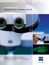
Neurobiology<br />
The samples are as different as the diverse range of<br />
topics in neurobiology: meaningful results must be obtained<br />
from individual cells and thin sections to thicker<br />
brain sections up to entire brains. <strong>Axio</strong> <strong>Imager</strong> is the ideal<br />
platform for this: excellent image quality in brightfield<br />
and fluorescence, high-resolution DIC for thick preparations<br />
and high-contrast DIC images for very thin sections.<br />
MosaiX provides high-resolution overview images<br />
of large specimens. The motorization of all important<br />
components and the use of the motorized DIC turret<br />
on <strong>Axio</strong> <strong>Imager</strong>.Z2 allow the storage of all important<br />
HeLa cells – multichannel image. Green: GFP Red: alpha-tubulin Blue: cellular nuclei (DAPI)<br />
Objective: Plan-APOCHROMAT 63x/1.4 Oil.<br />
L. Pelletier and T. Hyman,<br />
MPI for Molecular Cell Biology and Genetics, Dresden,<br />
Germany<br />
9
Brain section (rat) – multichannel image with ApoTome.<br />
Green: GFP-labeled astrocytes. Blue: cell nuclei (DAPI).<br />
Objective: Plan-APOCHROMAT 20x/0.6.<br />
E. Fuchs, S. Bauch, DPC, Göttingen, Germany<br />
Drosophila larval stage. Red: fibrillarin. Green:<br />
Venus-CG 8571-Transgene. Blue: cell nucleus (DAPI).<br />
Objective: EC Plan-NEOFLUAR 40x/0.75. M. Buszcak,<br />
A. Spradling, CIW-Dept. Embryology, MD, USA<br />
CHO cell culture. Green: GFP-histone. Red: dsRed,<br />
Objective: EC Plan-NEOFLUAR 40x/0.75.<br />
S. Haxelmans, R. Nitschke, Inst. Biologie I. Univ. Freiburg,<br />
Germany<br />
settings for reproducible imaging and subsequent image<br />
analysis tasks.<br />
Developmental biology<br />
The documentation and analysis of the processes which<br />
result in differentiation, regeneration or growth of cells,<br />
tissues and organisms make particularly high demands<br />
on a microscope system. Regardless of the animal model<br />
used, the highest performance of color fidelity, resolution<br />
and contrast is critical. <strong>Axio</strong> <strong>Imager</strong> provides you with the<br />
ideal uniform illumination in the common transmitted<br />
light contrast techniques, the best optical resolution, the<br />
extremely sample protecting fluorescence illumination<br />
with optimum signal-noise ratio to ensure brilliant image<br />
quality. With <strong>Axio</strong> <strong>Imager</strong> as the basis for an imaging<br />
system the processes to be investigated can be imaged<br />
at high spatial and temporal resolution and analyzed<br />
with different <strong>Axio</strong>Vision Modules. The motorization of<br />
<strong>Axio</strong> <strong>Imager</strong> M- and Z-stands allows efficient and reproducible<br />
imaging. Beyond this the manipulation of the<br />
sample is clearly facilitated and the sample turnover increased<br />
with the help of the docking station and scanning<br />
stage.<br />
Interphase Metaphase Telophase<br />
HeLa cells – mitosis stages. Red: Alexa Fluor 594-DM1-alpha. Green: Alexa Fluor 488-Mad2. Blue: DNA (DAPI). Objective: EC Plan-NEOFLUAR 100x/1.3 Oil<br />
H.Y. Li, Y. Xheng, HHMI & CIW, Dept. Embryology, MD, USA<br />
10
Imaging Systems.<br />
From Simple Observation to Analysis.<br />
The type of task determines the system solution. <strong>Axio</strong> <strong>Imager</strong> 2 provides the<br />
appropriate system for every requirement of life science research. Sophisticated<br />
modularity and a wide spectrum of perfectly coordinated components guarantee<br />
perfect results. Quickly. At any time.<br />
Preconfigured and individual: the systems<br />
The demands on the relevant systems are as different<br />
as the nature of the tasks in life science research. The<br />
modular architecture of <strong>Axio</strong> <strong>Imager</strong> 2 allows you to<br />
make an individual configuration which is exactly tuned<br />
to your requirements. For the digital image documentation<br />
of 3 (x, y, z) to 6 dimensions (additionally<br />
t, λ and x,y location) <strong>Axio</strong> <strong>Imager</strong> can be expanded<br />
with highly sensitive cameras from the <strong>Axio</strong>Cam family.<br />
<strong>Axio</strong>Vision offers a large number of specific modules for<br />
subsequent image analysis.<br />
Digital intelligence: <strong>Axio</strong>Vision<br />
<strong>Axio</strong>Vision is the high-performance software for useroriented<br />
solutions in digital imaging. From image acquisition<br />
and processing to image analysis and archiving.<br />
<strong>Axio</strong>Vision is practice-oriented, can be operated intuitively,<br />
and can be easily adapted to individual requirements.<br />
The modular design of the Carl Zeiss imaging software<br />
can be expanded in many ways. For example, for Z-<br />
stack, multi-channel fluorescence or time-lapse images.<br />
<strong>Axio</strong>Vision is the solution for growing demands.<br />
Stand Standard Equipment Options Fields of Application Materials Applications<br />
A2 LED • LED – Fixed Köhler Illumination<br />
transmitted light<br />
• Lightmanager<br />
• Encoded<br />
• Transmitted light beampath<br />
with manual filter wheel<br />
• Reflected light beampath<br />
• ApoTome<br />
• Encoded stage<br />
• Pathology<br />
• Histology<br />
• Cytology<br />
A2<br />
D2<br />
M2p<br />
• Universal stand transmitted light<br />
• Lightmanager<br />
• Encoded<br />
• Neutral density filter wheel<br />
• Universal stand transmitted light<br />
• Encoded<br />
• Partly motorizable:<br />
Reflector turret<br />
• LED – Fixed Köhler illumination<br />
transmitted light<br />
• Convenience-Motorization:<br />
Parfocality, Condenser<br />
• Encoded nosepiece<br />
• Motorized z-drive with 25 nm<br />
step size<br />
• Reflected light beampath<br />
• ApoTome<br />
• Encoded and 2-plate scanning<br />
stages<br />
• Reflected light beampath<br />
• Reflector turret 6x or 10x<br />
• ApoTome<br />
• Encoded and 2-plate scanning<br />
stages<br />
• Transmitted light beampath with<br />
motorized luminous field stop<br />
• Reflected light beampath<br />
• TFT<br />
• ApoTome<br />
• LSM (entry level)<br />
• 2- and 3-Plate scanning stages<br />
• Biosciences research<br />
• Medical sciences<br />
research<br />
• Industrial research<br />
• Bio-material-research<br />
• Human Genetics<br />
• Animal Genetics<br />
• FISH-applications<br />
• Pathology<br />
• Histology<br />
• Cytology<br />
• Histological staining<br />
• Antibody staining<br />
• Fluorescence In situ<br />
Hybridisation (FISH)<br />
• Live cell staining on<br />
samples of<br />
- Living cells<br />
- Fixed cells<br />
- Tissue sections<br />
- Whole-mount-samples<br />
• Evaluation<br />
• Fast routine work<br />
• Observation<br />
• Image acquisition and reporting<br />
• Interactive measurements<br />
• Evaluation<br />
• Image acquisition and reporting<br />
• Semiautomatic measurements<br />
• Evaluation<br />
• Image acquisition and reporting<br />
• Fast routine work<br />
• Confocal Imaging (entry level)<br />
M2<br />
• Universal stand transmitted light<br />
• Motorized: Luminous field stop<br />
• Lightmanager<br />
• Contrastmanager<br />
• Motorized z-drive with 25 nm<br />
step size<br />
• Reflected light beampath<br />
• ACR for objectives<br />
• ApoTome<br />
• 2- und 3-plate scanning stages<br />
• 2 TV Tube motorized<br />
• Biosciences research<br />
• Medical sciences<br />
research<br />
• Industrial research<br />
• Bio-material-research<br />
• Automatic image acquisition and<br />
analysis<br />
• 3D Imaging<br />
• Medium sample throughput<br />
• Multi-User environment<br />
Z2<br />
• High performance stand transmitted<br />
light<br />
• Motorized: Luminous field stop<br />
• Lightmanager<br />
• Contrastmanager<br />
• Motorized focus drive:<br />
- 10 nm step size<br />
- designed for loads up to max.<br />
9 kg<br />
- designed for continuous<br />
operation<br />
• Reflected light beampath<br />
• ACR for objectives and filter<br />
cubes<br />
• ApoTome<br />
• 2- and 3-plate scanning stages<br />
• LSM<br />
• Biosciences research<br />
• Medical sciences<br />
research<br />
• Industrial research<br />
• Bio-material-research<br />
• Automatic image acquisiton and<br />
analysis<br />
• Certified image acquisition and<br />
archiving (CFR 21 part 11)<br />
• 3D imaging<br />
• DIC-Fluorescence Imaging<br />
• Confocal Imaging<br />
• High sample throughput<br />
• Multi-User environment
Observation, reporting and interactive measurement<br />
3D Imaging<br />
ApoTome<br />
Camera<br />
Camera<br />
Imaging<br />
PC<br />
<strong>Axio</strong>Vision<br />
software<br />
Scanning stage<br />
Imaging<br />
PC<br />
<strong>Axio</strong>Vision<br />
software<br />
<strong>Axio</strong> <strong>Imager</strong><br />
<strong>Axio</strong> <strong>Imager</strong><br />
Proven and appreciated: the <strong>Axio</strong>Cam family<br />
Carl Zeiss offers a broad spectrum of digital cameras in<br />
different performance classes. The monochrome cameras<br />
are characterized by optimum resolution and highest<br />
sensitivity (12 or 14 bit dynamics) particularly in cases of<br />
faint fluorescent samples. The color cameras stand for<br />
the best color reproduction and highest resolution up to<br />
12 megapixels per color channel. All the cameras have<br />
thermoelectric cooling and provide the option of rapid<br />
shutter synchronization. All <strong>Axio</strong>Cam cameras are characterized<br />
by rapid live image and complete integration in<br />
the Carl Zeiss system world.<br />
Highly stress resistant: motorized focus and<br />
high-performance focus<br />
The <strong>Axio</strong> <strong>Imager</strong> offers you two different versions of the<br />
z-motor. The standard design with a step size of 25 nm<br />
at a reproducibility of ± 75 nm is always part of the<br />
M-stand configuration. And for the highest requirements<br />
such as LSM or Z-stack imaging with small intervals, a highperformance<br />
focus is available for the <strong>Axio</strong> <strong>Imager</strong>.Z2.<br />
It has a step size of only 10 nm with a reproducibility of<br />
± 10 nm – and that at a 3-fold higher traverse rate. It<br />
was specifically developed for continuous use (24 hours /<br />
7 days) and even with large stages guarantees absolutely<br />
precise focus movements over long periods.<br />
Optical sections with ApoTome<br />
ApoTome has firmly established itself as the standard<br />
method in high-end research in the life sciences. For<br />
the first time it can be used with all the stands in the<br />
<strong>Axio</strong> <strong>Imager</strong> 2 family: The ApoTome slider is simply inserted<br />
in the luminous field diaphragm plane of the reflected<br />
light beam path. Via the principle of fringe projection,<br />
precise optical sections are created online. With elevated<br />
contrast and clearly increased axial resolution. The ideal<br />
solution for tissue sections and thicker, fixed samples.<br />
No stray light ever again:<br />
<strong>Axio</strong>Vision 3D deconvolution<br />
Deconvolution from Carl Zeiss calculates mathematically<br />
the stray light from outside the focal plane back to its<br />
origin. In this manner the object recorded in the 3D image<br />
stack is “unfolded”. The result is a first-class image<br />
quality particularly in samples with extremely weak fluorescence<br />
where a high light yield is essential.<br />
Fast image acquisition / Live Cell Imaging<br />
High-end research system<br />
with <strong>Axio</strong> Vision<br />
Camera<br />
Incubation<br />
Camera<br />
Piezo focusing unit<br />
<strong>Axio</strong> <strong>Imager</strong><br />
Imaging<br />
PC<br />
<strong>Axio</strong>Vision<br />
software
Reporting and automatic measurement<br />
Confocal imaging<br />
LSM<br />
ZEN<br />
software<br />
Incubation<br />
Camera<br />
Camera<br />
Scanning stage<br />
Imaging<br />
PC<br />
<strong>Axio</strong>Vision<br />
software<br />
Scanning stage<br />
Controller<br />
Imaging<br />
PC<br />
<strong>Axio</strong>Vision<br />
software<br />
<strong>Axio</strong> <strong>Imager</strong><br />
<strong>Axio</strong> <strong>Imager</strong><br />
Precisely on the spot: motorized stages<br />
and z-piezo insert<br />
They allow a precisely accurate approach to positions and<br />
the highest degree of reproducibility. Via highly sensitive<br />
piezo or step motor every desired position can be exactly<br />
set and relocated.<br />
• Piezo stage: step size 0.2 μm, reproducibility:<br />
+/- 0.6 μm<br />
• Mechanical stage: step size 0.1 μm, reproducibility:<br />
+/- 0.3 μm<br />
• New stage control for stages with DC motors for<br />
direct coupling with the motorized stages (magnification-dependent<br />
traverse rate): highest reproducibility<br />
and precision in high-end applications<br />
• z-Piezo focusing insert with 100 µm focusing range<br />
for rapid imaging with Colibri and z stack images;<br />
resolution 5 nm, reproducibility: +/- 1 nm, max.<br />
additional load 2 kg, for frame size 222 x 139 mm,<br />
available mounting frames for all common preparation<br />
shapes<br />
The scanning stages are the prerequisite for all automated<br />
imaging techniques such as MosaiX or Mark&Find.<br />
New stimuli for your research: the LSM family<br />
Confocal microscopy at the highest level: LSM 700, LSM<br />
710 and LSM 7 MP belong to the seventh device generation<br />
of the Laser Scanning Microscope from Carl Zeiss.<br />
The use of the same first-class system components and<br />
the same software in the entire device class ensures outstanding<br />
performance and image quality without any<br />
compromises. The result: an excellent price-performance<br />
ratio. A novel beam path ensures excellent laser suppression<br />
and maximum registration of emission and results in<br />
breathtaking images. Demanding tasks such as spectral<br />
imaging, FRET, FRAP or colocalization analysis are easily<br />
managed with unprecedented image quality and high<br />
scanning speed.<br />
LSM 710 NLO and LSM 7 MP are ideally suitable for highly<br />
sensitive deep examination of living preparations or organisms.<br />
Both systems are characterized by unrivalled sensitivity.<br />
Highly effective non-descanned detection ensures<br />
efficient depiction in deep tissue layers. These are the<br />
systems of choice for long-term developmental studies,<br />
patch-clamp- and uncaging-experiments.<br />
Precision in z: the closed loop system<br />
<strong>Axio</strong> <strong>Imager</strong>.Z2 with the focus linear sensor offers anyone<br />
who has to fulfill extremely high requirements precision<br />
of ±1nm in the z-direction. On the one hand, the application-independent<br />
movements of the microscope stage<br />
are detected and readjusted automatically. And, on the<br />
other hand, highly precise and reproducible Z-stacks are<br />
ensured with z-steps of equal size, which gives maximum<br />
control and reliability.<br />
LSM 710 with<br />
<strong>Axio</strong> <strong>Imager</strong>.Z2<br />
13
Ergonomy and Ease of Operation.<br />
Efficient and Relaxed Working.<br />
<strong>Axio</strong> <strong>Imager</strong> is intelligent technology with a trendsetting control concept. Even the<br />
most demanding experiments and long working sessions at the microscope become<br />
simple and efficient. Automated procedures allow rapid, intuitive control with either<br />
manual or motorized components, depending on the individual requirements.<br />
Efficient, rapid, comfortable: the Touch Screen<br />
A good thing has been made even better: The control<br />
software of <strong>Axio</strong> <strong>Imager</strong> 2 collects all of the critical functions<br />
on one touch-sensitive TFT display. All motorized<br />
components are controlled with a touch of your finger,<br />
and their status is also displayed. The integrated light and<br />
contrast managers constantly adjust the light and contrast<br />
settings optimally.<br />
• The Contrast Manager’s control and user guidance<br />
adhere to the logic and workflow of all applications.<br />
• Motorized components can be optionally switched<br />
to automated or manual control.<br />
• The Favorites Page allows access to frequently used<br />
functions when switching ON the microscope.<br />
• Individual settings can be defined for up to 10 different<br />
users.<br />
Ergonomically well-conceived: Control buttons<br />
and exchangeable fine drive<br />
Ease of operation redefined: the control buttons which<br />
have been ergonomically arranged around the focus<br />
drive can be easily distinguished by their tactile surfaces.<br />
The two different fine drive buttons of the focus drive<br />
are exchangeable and can be optionally used for right or<br />
left. The motorized stand has ten freely assignable control<br />
buttons. The manual stand allows the simple setting<br />
of light intensity as well as switching of the motorized<br />
shutter in reflected and transmitted light via five preconfigured<br />
buttons.<br />
Ergophototube for perfect convenience<br />
Ergonomically distributed control buttons<br />
Ideal arrangement of the diaphragm slider and filter wheel<br />
in reflected light<br />
14
ACR reflector module<br />
ACR objective. ACR detects objectives and reflector<br />
modules automatically<br />
Communication connections<br />
Provides mobility: the control panel<br />
<strong>Axio</strong> <strong>Imager</strong> can also be remote controlled via a free positionable<br />
control panel. Among other things, this panel<br />
has a focus drive and a brightness control. Additional<br />
arbitrary functions can be programmed. The panel provides<br />
an interface for the TFT and for the x, y-control of<br />
the motorized mechanical stage.<br />
Error-free control with ACR<br />
ACR (Automatic Component Recognition) stands for the<br />
innovative concept of automatic recognition of objectives<br />
and reflector modules on the <strong>Axio</strong> <strong>Imager</strong>.Z2.<br />
When changed, the replaced components are immediately<br />
registered in the system. An important advantage<br />
for ease of operation and safety: operating errors and<br />
time-consuming programming are avoided.<br />
Absolutely stable: the Imaging Cell<br />
The key elements of <strong>Axio</strong> <strong>Imager</strong> such as the nosepiece,<br />
z-guidance and the stage are decoupled from the remainder<br />
of the stand as a stable cell. The entire unit has been<br />
designed to be practically vibration-free and insensitive<br />
to thermal influences. Even in the long-term it provides<br />
the highest possible stability and absolute freedom from<br />
vibration. Ideal preconditions for imaging, particularly in<br />
long-term experiments and in time lapse imaging.<br />
Switching of illumination on the TFT<br />
The TFT display on the stand or in the docking station<br />
provides a transparent menu guide for control and<br />
configuration<br />
Control of the motorized stages via the Docking<br />
Station<br />
15
Stand Design.<br />
Flexibility Times 6.<br />
Advanced technology assures that the user will select the appropriate system. The sophisticated<br />
stand design of <strong>Axio</strong> <strong>Imager</strong> 2 and well-conceived, preconfigured packages guarantee<br />
you an appropriate, configuration that meets the most demanding applications.<br />
Convincing technology: the stand<br />
Progressive down to the smallest detail – even in the<br />
basic configuration, all stands have an interface to the<br />
control computer. The parameters of encoded or motorized<br />
components can be read out or controlled directly<br />
by <strong>Axio</strong>Vision.<br />
• <strong>Axio</strong> <strong>Imager</strong>.A2 LED<br />
Ideally appropriate for brightfield applications in transmitted<br />
light: an LED light source ensures a constant<br />
color temperature across the entire intensity range.<br />
• <strong>Axio</strong> <strong>Imager</strong>.A2 and M2<br />
More flexibility: interfaces for sliders in the reflected<br />
light beam path allow convenient working with either<br />
aperture or field stop diaphragms or an attenuator<br />
in fluorescence. Optional on the <strong>Axio</strong> <strong>Imager</strong>.M2:<br />
motorized filter wheels and diaphragm sliders in<br />
reflected light (M2m) or in transmitted light (M2).<br />
<strong>Axio</strong> <strong>Imager</strong>.A2 LED <strong>Axio</strong> <strong>Imager</strong>.A2 <strong>Axio</strong> <strong>Imager</strong>.D2<br />
16
• <strong>Axio</strong> <strong>Imager</strong>.M2p<br />
Automatic parfocality compensation, a light manager,<br />
the motorized condenser and manual objective<br />
changing make routine work, e. g., in pathology<br />
comfortable and efficient.<br />
• <strong>Axio</strong> <strong>Imager</strong>.D2<br />
The manual high-end stand can be equipped with a<br />
6x- or 10x-motorized reflector turret, which, above<br />
all, make fluorescence applications comfortable and<br />
fast.<br />
• <strong>Axio</strong> <strong>Imager</strong>.Z2<br />
The stand has been developed to meet the most stringent<br />
requirements. A high-performance focus allows<br />
constant operation with a high sample throughput.<br />
It ensures precise focusing movements over long<br />
periods and also when using large and heavy sample<br />
stages up to 9 kg.<br />
<strong>Axio</strong> <strong>Imager</strong>.M2 <strong>Axio</strong> <strong>Imager</strong>.M2p <strong>Axio</strong> <strong>Imager</strong>.Z2<br />
17
<strong>Axio</strong> <strong>Imager</strong> 2 – Flexibility for all application areas<br />
Component Option A2 LED A2 M2p M2 D2 Z2 A2m M2m D2m Z2m<br />
Stand manual + + - - + - + - + -<br />
motorized - - + + O* + - + O* +<br />
Encoding readout by computer + + + + + + + + + +<br />
Tube lens turret encoded O O O O O O O O O O<br />
motorized - - O O - O - O - O<br />
Reflector turret 6x encoded O O O O O O O O O O<br />
6x motorized - - O O O O - + O O<br />
6x motorized ACR - - - - - O - - - O<br />
10x motorized ACR** - - - - O O - - O O<br />
Nosepiece 6x encoded POL O O - O O O O O O O<br />
6x encoded HD DIC O O - O O O O O O O<br />
6x motorized HD DIC - - - O - O - O - O<br />
6x motorized HD DIC ACR - - - O - O - O - O<br />
7x encoded HD O O + O O O O O O O<br />
7x motorized HD - - - O - O - O - O<br />
Modulator turret for C-DIC/TIC manual O O O O O O O O O O<br />
motorized***** - - - O - O - O - O<br />
Modulator turret for DL- DIC motorized***** - - - - - O - - - O<br />
Stage carrier with condenser carrier, detachable 0 mm - 25 mm Sample height + + + + + O O O O O<br />
Stage carrier detachable, for attachable condenser carrier 0 mm - 45 mm Sample height O O O O O O O O O O<br />
Stage carrier reflected light, detachable 0 mm - 63 mm Sample height O O O O O O O O O O<br />
Transmitted light beam path manual - + - - + - O O O O<br />
motorized - - - + - + - - - O<br />
LED transmitted light - + O + O O O O O O O<br />
Double filter wheel transmitted light manual - + - O O O O O O O<br />
motorized - - - O - O - - - O<br />
Reflected light beam path manual*** O O O O O O + - + -<br />
motorized*** - - - - - O - + - +<br />
Luminous field stop slider reflected light manual O O O O O O + O + O<br />
motorized - - - - - O - O - O<br />
Aperture stop slider reflected light manual O O O O O O O O O O<br />
motorized - - - - - O - O - O<br />
Double filter wheel reflected light manual O O O O O O O O O O<br />
motorized - - - O - O - O - O<br />
Fluorescence attenuator manual O O O O O O O O O O<br />
motorized - - - - - O - O - O<br />
Lamp switch reflected light/transmitted light manual + + - - + - + - + -<br />
software - - + + - + - + - +<br />
Mixed light with additional external power supply manual + + - - + - + - + -<br />
software - - + + - + - + - +<br />
Focus (z-Axis) manual + + - - + - + - + -<br />
motorized 25 nm Step size - - + + - - - + - -<br />
High Performance Focus<br />
motorized 10 nm Step size<br />
- - - - - + - - - +<br />
TFT-Display - - - O + - + - + - +<br />
ApoTome - O O O O O O O O O O<br />
Power supply external - - + + - + - + - +<br />
internal + + - - + - + - + -<br />
Mechanical stage CAN motorized**** O O O O O O O O O O<br />
Scanning stages Piezo O O O O O O O O O O<br />
DC / Stepper motors O O O O O O O O O O<br />
Fast z-piezo insert with manual stage O O O O O O O O O O<br />
with scanning stage O O O O O O O O O O<br />
2 TV tube head motorized - - - O O - O - O - O<br />
Condenser manual O O O O O O O O O O<br />
motorized - - O O - O - O - O<br />
+ = Included in stand<br />
O = Optionally available<br />
- = Not available<br />
* = Motorized (6x und 10x) reflector revolver<br />
can be used<br />
** = ACR function not possible with<br />
„<strong>Axio</strong> <strong>Imager</strong>“ D1 and D1m<br />
*** = A motorized shutter is included in every<br />
reflected light illumination.<br />
For fluorescence applications this can<br />
optionally be replaced by a High speed<br />
shutter<br />
**** = For the use at the „<strong>Axio</strong> <strong>Imager</strong>“ A2 LED,<br />
A2, A2m, D2 and D2m USB/CAN Converter<br />
432909 is required<br />
***** = Only in combination with motorized<br />
objective nosepiece<br />
m = Optimized for materials applications<br />
18
<strong>Axio</strong> <strong>Imager</strong> 2.<br />
Even More Highlights.<br />
The optics<br />
• IC²S beam path for high contrast<br />
• Highest possible resolution through high-performance objectives<br />
The fluorescence<br />
• Combination of DIC and fluorescence with the motorized DIC turret<br />
• Excellent image quality due to the optimized beam path<br />
• Triggerable LED light source<br />
• Several light sources for uniform illumination<br />
The stands<br />
• Preconfigured packages for the most common applications<br />
• Coded and motorized components<br />
• Modular and individually upgradable<br />
The imaging<br />
• Motorized DIC turret: Combination of fluorescence and DIC for absolute artifact-free images<br />
• Rapid image acquisition in up to 6 dimensions<br />
• Motorized scanning stages, motorized z-focus and high-performance focus (<strong>Axio</strong> <strong>Imager</strong>.Z2) for the<br />
highest precision and positioning accuracy<br />
Carl Zeiss MicroImaging GmbH<br />
07740 Jena, Germany<br />
BioSciences | Göttingen location<br />
Phone: +49 551 5060 660<br />
Telefax: +49 551 5060 464<br />
E-Mail: micro@zeiss.de<br />
www.zeiss.de/axioimager<br />
Information subject to change.<br />
Printed on environmentally friendly<br />
paper bleached without cholorine.<br />
60-2-0037/e – printed 05.09