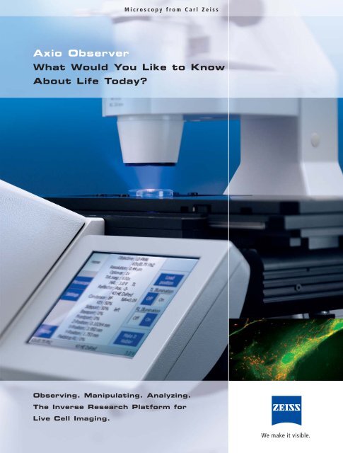
AXIO-OBSERVER BROCHURE.pdf - Focuspi.com
AXIO-OBSERVER BROCHURE.pdf - Focuspi.com
AXIO-OBSERVER BROCHURE.pdf - Focuspi.com

You also want an ePaper? Increase the reach of your titles
YUMPU automatically turns print PDFs into web optimized ePapers that Google loves.
How Do You Increase Access tothe Most Demanding Life ScienceApplications?The human genome was decoded in 2001, which wasa scientific revolution. This prompted a flurry of questionsin the scientific <strong>com</strong>munity. Since that time, internationalresearch teams looking into processes insideand between living cells have been concerned lesswith the “What” and more with the “How”. How domolecules or proteins, lipids, enzymes, DNA and RNAfunction? How do they interact? And why?In their search for answers to these questions,researchers focus their efforts on the most sophisticatedmicroscopic techniques for observation, manipulationand analysis. None more so than those involvingfluorescence. The development of such techniques hasbe<strong>com</strong>e a mission for Carl Zeiss, and one that the <strong>com</strong>panyhas assigned a name: FluoresScience. For manyyears, this initiative has been providing the leadingmicroscope systems for applied and basic research,and helping researchers to open new pathways of discoveryin science.Now a technical innovation is moving science a stepfurther: Axio Observer, the inverted research microscopefrom Carl Zeiss. It has been developed formaximum flexibility in the Live Cell techniques oftoday and tomorrow and realized as a fully integratedresearch platform for cell observation, cellmanipulation and cell analysis. It can be expandedcost-effectively from a basic stand for tissue cultureand fluorescence, to one for high speed, laser scanningmicroscopy or microdissection. Axio Observer: whatbetter way is there to meet the high demands of LifeScience projects today?2
Axio Observer3
The ApoTome and PlasDIC were awardedthe R&D 100 Award.Have Optics Reached Their LimitsWith Living Cells?Carl Zeiss has been continuously stretching the possibilitiesof its research optics to their very limits.With Axio Observer we are further strengtheningour technological leadership position – with newdevelopments in contrasting methods, objectivesand numerous innovative details. At the limits ofvisibility, these developments provide greaterinformation content, better adjustment to versatileLive Cell applications, and even more convenientprocesses.Each series in a class of its own:the objectivesThe “eyes” of the inverted Axio Observer researchplatform – developed to perform the differenttasks in Live Cell Imaging brilliantly. Each objectiveseries is in a class of its own:• LCI Objectives LCI Plan-Neofluar 25x, 63xand LD LCI Plan-Apochromat 25x. Superbmulti-immersion objectives with optimalcorrection options for spherical aberration,tailored to the special temperature spectrumin Life Science experiments. The LD variantmakes it possible to focus even more deeplyinto the sample.• Insulated objectives i LCI Plan-Neofluar 25xand 63x and i Plan-Apochromat 63x. Thermalinsulation is the only way to ensure the perfecttemperature at the specimen.• LD Plan-Neofluar Ph1 Ph2- Corr, the innovation:positive and negative phase contrast <strong>com</strong>binedin one objective.• C-Apochromat und LD C-Apochromat,perfect for un<strong>com</strong>promising high resolution.Ideal for LSM, ApoTome and Deconvolution.• Plan-Apochromat for extremely high demandson image leveling and color correction.• LD Plan-Neofluar, sophistication and versatility,useable for all cover glass specimens (0.17 mm)and plastic culture plates with a bottom thicknessof up to 2 mm.• EC Plan-Neofluar Neofluar for outstandingcontrast and multiple image techniques.• LD A-Plan, the favorable, flexible standardobjectives from Carl Zeiss for inverted microscopy.Optimal for the entire field of view:the new DICThe new generation of Differential InterferenceContrast provides brilliance and homogeneous illuminationacross the entire field of view. Imagedetails are resolved well with highest resolution and1.DICDIC2. 3.PlasDIC41. Forebrain neurons (rat) cultured on poly-D-lysine/⁄laminin coated glass coverslips.J. Perron, Columbia University, Columbia, USA.2. MDCK cells (dog) after short incubation period.R. Nitschke, Life Imaging Center, University of Freiburg, Germany.
outstanding contrast across a 25 mm field of view.A further innovation is the <strong>com</strong>bination of polarizerand Nomarski prism in a sandwich design. Theadvantage to you: there is no need to engage thepolarizer manually.Practical, economic, impressive:PlasDICGlass or plastic? The economic relief contrast fromCarl Zeiss is insensitive to birefringent materialsand is therefore just as suitable for plastic dishesas it is for glass bottom vessels. In terms of application,PlasDIC is re<strong>com</strong>mended for use withthicker adherent cells or oocytes and is ideallysuited to Intracytoplasmic Sperm Injection (ICSI)due to the excellent relief display. PlasDIC is easyto operate and already functions with the costeffectiveLD A-Plan objectives. If your requirementsgrow, we re<strong>com</strong>mend the LD Plan-Neofluarobjectives.Phase + Phase -4. 5.Innovation in plus and minus:the phase contrastFamiliar, yet surprisingly different: the new phasecontrast in an economic 2-in-1 solution and a newarea of application. The negative phase contrastworks well with thick cell areas or stages of division.The <strong>com</strong>bination of positive and negative phasecontrast now makes it possible to contrast all thestructures of your object perfectly using oneobjective. Switching is easy, involving a simplediaphragm change. You will appreciate the newphase contrast for normal monitoring, as well asfor special morphological problems.Automatically quicker:the new transmitted light shutterA new standard since the introduction ofAxio Observer: faster switching, silent operationand low vibration. Time is saved and operationmade easier thanks to a new integrated shutterconcept. Automated in transmitted light for thefirst time, with a short switching time making itperfect for the configuration of time lapse experiments,e.g. for the rapid switch between transmittedlight and fluorescence.+ Ph - Ph + Ph3. Human embryos (four-cell stage).S. Mittmann, IVF-Labor, Göttingen, Germany.4. und 5.: MDCK cells (dog) – thick cell areas are displayed better usingnegative phase contrast.R. Nitschke, Life Imaging Center, University of Freiburg, Germany.5
Why Does Everything Revolve AroundOptimum Fluorescence?Highly differentiated fluorescence techniques – inresearch on living cells these are be<strong>com</strong>ing thestandard. Carl Zeiss is focusing its knowledge andinnovation on further developing these techniques.So that they may be made accessable to newapplications all the time. At the forefront of thedevelopment of inverted microscopes stands is theAxio Observer – with the most efficient, flexible andgentlest fluorescence, protecting your cells andleading to brilliant results.Perfected for all wavelengths:the new fluorescence beam pathAxio Observer represents a new level of quality influorescence. Corrected apochromatically, it offersconsistently good contrasting and homogeneousillumination right into the periphery at almost anyexcitation wavelength. The advantage to you:From center to edge, in multichannel fluorescenceall wavelengths are equally balanced in intensityand signal to noise. There is also the alternativeoption of the beam path with an extended transmissionspectrum optimized for 340 nm transmission.Faster and more flexible:the reflector turretAdjustment of the reflector modules to the experimentin question is quicker, saving time, which is animportant consideration with the growing diversityof fluorescent proteins. The Axio Observer reflectorturret has been optimized in triplicate for this purpose:six filter positions offer more flexibility.Exchanging filter sets without removing the turretbypasses an entire work step. And finally, theAxio Observer turret with position change in under200 ms is simply faster. With ACR* the filtermodules are read and identified automaticallyby the system – <strong>com</strong>pletely dispensing with theneed for configuration on the microscope or inthe software. Advantages: faster process andreliable documentation. A perfect solution for themultiuser environment.* Automatic Component Recognition. Available for Axio Observer.Z1Fluorescence – the operation6Reflector turret: in a matter of seconds, filter sets are now replaced withoutremoving the reflector turret.Easy to access and to operate: the diaphragm sliderfor Axio Observer.
14Beam path1 Intermediate imageplane/phototube2 Eyepiece3 Intermediate imageplane/front port4 Intermediate imageplane/base port5 Switching beam pathbetween base port/frontport/vis. observation6 Side port prisms7 Tube lens8 Analyzer9 Reflector cube10 Field diaphragm11 Aperture stop12 Filter slider13 HBO lamp14 HAL lamp15 Field diaphragm16 Polarizer17 Aperture stop18 Condenser19 Objective23115161718199876510 1112134Diversity for cell observation:the light sourcesFor Axio Observer, there is the option of a wide spectrumof high-performance light sources. Top on thelist is the self-adjusting HBO lamp. This innovativedevelopment from Carl Zeiss saves the user the timeconsumingtask of re-adjustment: it automaticallyaligns itself and provides a consistently homogeneousillumination of the field of view. In addition,you can also opt for the traditional HBO, or for variousdurable metal halide lamps, which are coupled viafibers. If you require extremely fast excitation changesin High Speed Imaging (Cell Observer ® HS), thexenon-lamp-based light sources, Sutter Lambda DG-4or Till Polychrome V, can be coupled.Standard or high speed:the fluorescence shuttersTwo shutters for fluorescence – more options foryour experiment. Besides the standard shutter,Axio Observer also features an external, extremelydurable high speed variant (5 million switchingcycles). Controlled by the camera using triggerpulses, this shutter only opens immediately priorto image acquisition. The exposure of the cells isreduced to an absolute minimum. A key <strong>com</strong>ponentparticularly for live cell experiments.Self-adjusting HBO1. 2.1. Cytoskeleton and nucleus labelled with quantum dots, HeLa cells.2. Forebrain neurons (rat) cultured on poly-D-lysine/laminin coated glass coverslips.Labelled with DAPI (blue), TUJ1 anti-beta-tubulin (green) and anti-ActRII(H65) (red). J. Perron, Columbia University, Columbia, USA.7
Variability and reproducibility:the diaphragm slidersRectangular diaphragm, FL attenuator* and iris diaphragm*:the three diaphragm sliders demonstratethe versatility of Axio Observer in Live Cell Imaging.When used in <strong>com</strong>bination with a motorized irisdiaphragm and FL attenuator, Axio Observer reachesa high degree of motorization in the fluorescencebeam path. The motorized FL attenuator automaticallysets the desired fluorescence intensity,depending on the filter set and objective.Brilliant development:the high-efficiency filter setsUp to 50% shorter exposure times – the high efficiency(HE) fluorescence filters offer a significantlyimproved signal to noise ratio and provide gentlerfluorescence imaging of living cells. The increasedtransmission during excitation and emission,together with extremely steep edges, leads to aclear signal separation and optimum yield.* Available as a mechanical or motorized optionDiaphragm slidersNumerous options for your individual workstation:the manual or motorized diaphragm sliders.Push&Click00:00:00 00:12:00 00:25:008HeLa cells expressing Histone-2B-DsRed (red), transfected with HIV-1-REV-YEP (green). Background:phase contrast. After treatment with Leptomycine B the Rev-protein accumulates in the nucleus.H. Wolff, GSF Institute for Molecular Virology, Neuherberg, Germany.
WorkflowWho Re-thinks the WorkflowWhen Applications are Be<strong>com</strong>ingIncreasingly Complex?The days when microscopes only differed in terms ofoptical performance are long gone. The <strong>com</strong>plexity ofcurrent applications in all areas of Life Sciencesmakes ease of use a factor for the success of researchwork. The challenge for Carl Zeiss is to revise theworkflow from planning to monitoring and analysisand to <strong>com</strong>bine it with an intelligent operating concept.Axio Observer now defines the benchmark forease of operation and efficiency. A must if theimmense performance potential of this high-endplatform is to be exploited quickly and economically.Recognizable increase inconvenience: TFT and LCDInput and monitoring station at the same time –the touch screen TFT display for the motorizedAxio Observer.Z1 stand is opening up a newdimension in automated operation. Control andmonitoring have been merged radically. Theresult is unique: the entire microscope can beoperated using an extremely short and clearmenu guide, which is also valid for the incubation<strong>com</strong>ponents that control the cultivation conditions.You are sure to be impressed by the ability totrigger activation of the contrast manager or ofthe full range of personal user settings on the PCwith the tip of your finger. For the D1 stand,a new LCD display has been developed with<strong>com</strong>prehensive status presentation. Chosen objective,shutter position, etc.: all the settings can beviewed with one quick upward glance. The LCD isalso a considerable help with system configuration.Freedom to operate:the docking stationA space-saving and immensely practical solution ifyou operate the system directly from the PC. TheAxio Observer docking station gives you access tothe <strong>com</strong>plete menu guide of the TFT as well as allthe control elements for sample positioning*. Thecontrol elements are identical to the elementslocated on the stand – no need to switch to differenttypes of buttons. The merging of all the controlelements into one <strong>com</strong>pact unit represents asignificant advantage, particularly with <strong>com</strong>plexsystem accessories.* In conjunction with the CAN scanning stageTFT displayDocking stationThe TFT display on the stand or in the docking station provides atransparent menu guide for control and configuration.9
Zooplankton (Brachionus plicatilis) feed on phytoplankton (Heterocapsa triquetra).A. Hagiwara, T. Oda, Faculty of Fisheries, Nagasaki University, Nagasaki, Japan.Small details with a big impact:the manual control elementsYou notice the difference as soon as you touchthem: the control elements of Axio Observer arewell thought-out, more intelligent and simplerthan anything you have ever known. The buttonsin the “keyring” can be reached without takingyour hand off the z-drive. They can also be freelyconfigured* and operated without looking. Youcan control the entire microscope with ease –without taking your eyes off the eyepiece. Thewheels for controlling the intensity of the illuminationand opening the field diaphragm in transmittedlight enable very sensitive operation andtherefore perfect adjustment to the specimen inquestion. Graphics on the stand and in the TFTdisplay help you to reach your goal more easilyand with greater speed.* For D1 with PC onlyA chip makes the crucial difference. All the filter sets andobjectives are available in a version for Automatic ComponentRecognition ACR.You make the change,who configures it? ACRAutomatic Component Recognition ACR standsfor the innovative concept of automatic recognitionof objectives and reflector modules fromCarl Zeiss. This optional additional <strong>com</strong>ponent forAxio Observer.Z1 monitors newly inserted filter setsand objectives and adopts them into the systemconfiguration automatically. This saves you time,reduces the risk of error and provides greater convenience– both in everyday microscopy whenchanging filter sets and during the joint use offilter sets on several stands. A perfect solution forthe multiuser environment.Simply more efficient: the contrast<strong>com</strong>binations in transmitted lightOne objective, three techniques, more informationin less time: the contrast <strong>com</strong>binations in transmittedlight have been expanded further forAxio Observer. PlasDIC & Phase & DIC or negativePhase & positive Phase & DIC with one objectiveare examples of how you can obtain more informationand varied information about your sample,or simply gain flexibility for different applicationswith great efficiency.Automatic Component Recognition ACR10
WorkflowAutomatic in the chosen contrast:the contrast managerA perceptible plus in your workflow. You choosethe contrast or contrast <strong>com</strong>bination, and the standensures the correct setting for the necessary <strong>com</strong>ponents,even if you change the magnification. Thisproves to be a pleasant and extremely time-savingfeature, particularly for direct observation, since iteliminates several previously required adjustmentsteps.Staying relaxed for longer:the ergotubeThe solution for long hours using the microscope.With 50 mm height adjustment and a fixed viewingangle of 25° (which is ergonomically ideal), theergotube for Axio Observer satisfies the higheststandards in terms of <strong>com</strong>fort; allowing you toadopt a relaxed posture, even during direct observationfor several hours.Transmitted and reflected lightregulated well: the light managerThe Axio Observer generation of light manageroffers more than just effective protection againstexcessively high light intensities in transmitted light.It correctly adjusts the light intensity during a changein magnification both in transmitted light and inreflected light. The fluorescence channels are alsotaken into account, and the intensity is adjustedaccordingly. For cells, this means additional protectionagainst high excitation intensity. This alsomakes direct observation more pleasant and lesstiring for your eyes.With the docking station, the familiar control elements forcontrolling the microscope are located right next to your PC.LCDErgotubez-drive with keyringLCD display, ergotube or keyring: the entire Axio Observer operating concept has beendesigned for quick and convenient workflow.11
What Does the Quantum Leapin Cell Incubation Bring?Axio Observer signals the dawn of a new era in cellincubation. One in which temperature, CO 2 , O 2 andair humidity are firmly under control. A significantleap forward in performance with trend-setting innovations.The most notable of these being the fullintegration into AxioVision. This means that theentire progression of environmental data is documenteddirectly with the image acquisition, significantlyimproving the reliability of the temperatureconditions during analysis of the experiment.Dynamic experiments (temperature levels) are nowfreely programmable. For example, you can characterizeprotein mutants that are sensitive to temperatureand perform heat-shock experiments.Better in every regard:cell incubation from Carl Zeiss• The new stack concept for the controlmodulesSaving an impressive amount of space andproviding maximum effectiveness during thepreparation of environment parameters.• The new PM S1 incubatorFor improved incubation conditions with extremelysmall volume for reduced airflow andsignificant space saving. Compatible with Petridishes and multiwell plates. There is also avariant available for micromanipulation in aCO 2 atmosphere.• The seamless integration into theTFT operating concept and AxioVisionIdeal for brand new experiment guides. Clearmonitoring of the environment parametersenables a quick overview and increased reliability.A particularly important feature is theability to store the experiment guide andretrieve it again at any time.• The <strong>com</strong>ponent upgrade and retrofitconceptGreatly simplified and even more flexible: oneof many examples is the easily upgradeable O 2module for reducing the O 2 concentration.PM S1 incubatorWith thermally-insulated objectives, aninsulation ring blocks the temperature flow.12Optimum incubation conditions, more space: the PM S1 incubator forPetri dishes and multiwell plates.
Incubation• The control sensor for measuring temperaturedirectly inside the cultivation dishFor the first time you can measure the temperatureat the observation location precisely,thereby validating your experiment. A majorplus, e.g. in dynamic temperature experiments.• The thermally-insulated objectivesFor optimized temperature at the sample locationand dynamic temperature experiments.The innovative Carl Zeiss principle: reductionin the quantity of material for heating orcooling – the temperature flow to the rear ofthe objective and to the objective nosepiece iseffectively interrupted. A further advantage isthat with experiments involving cooling thereis no need for any measures to prevent condensationon the rearmost objective lens. Thecold does not reach that far in the first place.• The flexibility of two conceptsElectric heating or flow-induced heating/cooling.The flow principle is also re<strong>com</strong>mended in electrophysiologyas it eliminates disturbing influencesbased on electronics.Big in every respect: the incubatorsDeveloped specifically for the wide-range ofCell Biology and Pharmacology applications, theCarl Zeiss incubation generation spans a widerange of different models. From the <strong>com</strong>pactPM S1 incubator for maximum freedom to operateto the large incubators of the XL S1 category andtheir special DARK, TIRF and LSM versions. Theseare joined by various models for micromanipulationin a CO 2 atmosphere, offering optimum flexibility.The same applies to the operating concept: withAxio Observer.Z1 the entire incubation can be controlledvia the TFT display, or optionally via the PC.Keeping everything dry:Aqua Stop IIEffective microscope protection with absolute freedomto operate: Aqua Stop II for Axio Observerhas been perfectly adapted to the microscope<strong>com</strong>ponents, and is now even more reliablethanks to <strong>com</strong>prehensive protection and optimizedassembly. If you change Petri dishes on aregular basis or conduct perfusion experiments,Aqua Stop II is really a must, to protect sensitive<strong>com</strong>ponents, such as filter sets, analyzers and thebeam path, against dirt and fluid spills.XL S1 incubatorAqua Stop IIThe XL S1 incubator provides peak stability.The Aqua Stop II – perfectsafety concept for Axio Observer.13
How Does a Cell Research Station GrowWith Your Requirements?Never before has the integration of microscope,software and external <strong>com</strong>ponents reached such ahigh level of performance. And never before havesystems for observing, manipulating and analyzingbeen so flexible. The advanced architecture ofAxio Observer opens the system up to the unlimitedintegration of external <strong>com</strong>ponents, turning theinnovative cell research stations from Carl Zeiss intosolutions that you can also use to realize any demandingapplications that lie just around the corner.Modular intelligence:documentation with AxioVisionDeveloped in order to make even <strong>com</strong>plex applicationswith living cells economical, the microscopesoftware from Carl Zeiss is setting the standards inuser guidance, individualization and range of performance.From the basic to the most sophisticatedpackage, AxioVision literally grows as your tasksdictate. Module by module. For multidimensionalimage acquisition, e.g. with Multichannel, Time Lapse,Z-stack or Mark&Find. Even the basic package canhandle dynamic temperature experiments.Targeting the result more purposefully:analysis with AxioVisionThe most demanding tasks simplified intelligently –the analysis modules of AxioVision offer the mosttechnically advanced solution for almost any application.They can also be used in ongoing experiments.For example, the Physiology module undertakesthe intensity <strong>com</strong>parison within the channelsin defined regions (ROI) including the graphicalrepresentation. There are also numerous othermodules available to you with AxioVision – forinstance, standard intensity measurements or positionanalysis of different fluorophores (colocalization).Even if the aim is to allocate emissions tofluorophores (Unmixing), or for intelligent timelapse processing, including the display of movingobject regions with details of speed and acceleration.AxioCam14
Integrated SystemSeeing and documenting:the Carl Zeiss bluesCarl Zeiss cameras are re<strong>com</strong>mended in all performanceclasses – particularly in monochrome forfluorescence imaging. There is the AxioCam HRmwith its extremely high resolution in 14 bit dynamics,the AxioCam MRm with its high sensitivityand diverse range of uses, and the AxioCam HSm,which transfers moving images to the hard drivein real time with up to 360 images per second*.One thing that all models have in <strong>com</strong>mon is providingyou with perfectly integrated camera technologythat optimally supports your system’sperformance.Everything you want for Live CellImaging, right through to highspeed: Cell Observer ®Cell Observer ® has long been the established <strong>com</strong>pletesolution in Live Cell Imaging. The newCell Observer ®HS (High Speed) option has beenperfected for the documentation of quick processesand long-period observation in Live CellImaging. It is particularly useful for calcium studies,cilia movements, vesicle transport and the emergenceof microtubuli – i.e. highly dynamic processesthat place extreme demands on the entiresystem. All the elements of Cell Observer ® HS arecontrolled directly using hardware triggers, sothere is no delay. With perfect coordination, everysingle <strong>com</strong>ponent – microscope, camera, lightsource, shutter and focusing system – is optimizedfor maximum speed. Often a logical additionto the high-speed system is the AxioVisionPhysiology module for calculating emission intensities(ratio experiments).Superb optical sections:ApoTomeThe solution for the glare-free 3D Imaging of thicksamples and tissue sections. ApoTome impresseswith outstanding image quality and a conceivablysimple operating concept. Available to theAxio Observer D1 and Z1 stands, the microscopeinsert is simply installed in the plane of the field diaphragmof the reflected light beam path. The principleof grid projection is used to create preciseoptical sections online, with increased contrast andgreatly improved axial resolution.LSM DuoSatisfying even the highest demands ofconfocal microscopy: the <strong>com</strong>bination ofLSM Meta and LSM Live leaves no wishesunfulfilled.* With 5x5 binning, full camera field of viewApoTome15
0:00 2:45 3:00Hormone-induced gene expression in HeLa cells. The mRFP-marked protein (yellow) is hormonallyinduced, and in turn induces the expression of the YFP-marked reporter protein (blue).H. Wolff, GSF Institute of Molecular Virology, Neuherberg, Germany.At the high-end of 3D:the LSM systemsUnsurpassed in spectral resolution, temporal resolutionand sensitivity: the laser scanning systems fromCarl Zeiss embody high-end technology enabling youto look into the depth of your cells. LSM 510 METAeven records the spectral signature of every singlepixel – for perfect unmixing. LSM 5 LIVE, with> 100 frames/sec., adds a new dimension to thescan speed – and with even greater sensitivity. Idealfor observing transport processes in cells andorganisms, e.g. the movement of blood cells in thevascular system or the change in dendritic spines onneurons. LSM 510 NLO, with its multiphoton excitation,makes it possible to penetrate into thespecimen to unprecedented depths – with several100 µm as an ideal prerequisite.Highly pure samples for exactresults: PALM MicroBeamEnormously versatile and 100% contact-free forthe isolation of extremely small tissue samples,such as chromosomes, organelles, cells or smallorganisms: PALM MicroBeam is the solution forLaser Microdissection and Pressure Catapulting(LMPC), <strong>com</strong>bining laser microdissection withtransport by laser light for the first time ever. Thisunique, patented technique enables you to cut andremove analysis material without ever touching thesample. Because the technique is contact-free, andtherefore also contamination-free, you obtain thepurest, clearly defined sample material for all typesof downstream processes from genetic analysis tothe cultivation of insulated cells. In DNA and RNAappraisal, protein analysis and research with livingcells, this system offers science new possibilitiesand perspectives.Maximum resolution in theevanescent field: Laser TIRFLaser TIRF from Carl Zeiss represents new findingsrelating to transport processes near the membraneor coupled to the membrane. The focus is also oncell-free systems, for studying protein-proteininteractions, for example. This <strong>com</strong>pact <strong>com</strong>pletesolution for Total Internal Reflection Fluorescenceoffers top image quality in all wavelengths – withoutre-adjustment of the TIRF angle. Space-savingapproach: the TIRF slider is simply inserted into thefield diaphragm. Perfectly conceived, right downto all typical peripherals like the special TIRF incubatorswith integrated laser safety.PALM MicroBeamLiving cell after lasertransport pulseTIRF1.161. B16/F1 melanoma cells (mouse) – TIRF illumination. Blue: CFP-actin. Excitation 458 nm, green: DsRed-clathrinLight Chain A. Excitation 514 nm. Alpha Plan-Fluar 100x/1.45 oil.Oberbanscheidt, van den Boom, Bähler, Institute for General Zoology and Genetics, University of Münster, Germany.
CDA. TIRFB. Laser portC. Laser (Catapulting, Tweezer)D. EpifluorescenceABNew flexibility in fluorescenceapplications: the laser portDesigned specifically for demanding laser applicationssuch as FRAP, Uncaging, or for the targetedablation of cellular structures: the laser port forAxio Observer. A receptacle for your own couplingsolution. It enables you to work simultaneouslywith reflected light fluorescence, to change toTIRF applications quickly. It provides new flexibilityin the application – without the need to extendthe infinity beam path, and without <strong>com</strong>promisingoptical quality in any way.Micromanipulation – EppendorfBasis for higher success rates:Axio Observer in micromanipulationAxio Observer is the perfect platform for stem cellsor In-vitro-Fertilization (IVF). The extremely high stabilityand wide-ranging mounting options for manipulatorsis PlasDIC, the innovative relief contrastideally designed for the implementation ofIntracytoplasmic Sperm Injection (ICSI). Withimpressive quality and ease of operation, it is alreadyin use in a number of IVF laboratories. If your applicationsrequire it, the optimized traditional DIC,with the highest resolution of detail, is ideal forimproved success rates, e.g. in sperm assessmentand embryo viability. A further important detail isthe Thermo Plate glass mounting frame, whichensures homogeneous distribution of temperatureon the culture plate. The entire surface of the stageis perfectly flat, making handling of the culturedishes simple and safe.Micromanipulation – Narishige17
Why Is The Basis for All Cell ResearchSystems More Than a Microscope Stand?The demands on a research microscope in Life Sciencesare as varied as the applications. The Axio Observerexpansion concept is designed to meet that. There arethree stands designed for different application focalpoints. This concept is essential if you want to realizeany system solution economically with Axio Observer.Three options offer more freedomto make decisions: the stand typesFrom the economic starter in the research categoryto the high-end dimensions of Live Cell Imaging:the Axio Observer stand concept gives you the freedomto choose a stand that suits your requirementsand budget. Whatever your decision, you will begetting a high-performance microscope for a reasonableprice.• Axio Observer.A1: for demanding routinetasks in Live Cell Imaging and particularly idealfor micromanipulation. The manual standoffers the same high optical quality as theother types.• Axio Observer.D1: greater ease of operation,more flexibility. The D1 stand enables motorizedselection of reflector turret, condenser andfluorescence beam path.• Axio Observer.Z1: this stand, the height ofperfection in inverse research microscopy, currentlyoffers the very best in terms of ease ofoperation and flexibility for automated onlineexperiments.Axio Observer: three stands, three operating conceptsAxio Observer.A1The basic solution for manual operationAxio Observer.D1Above standard: semi-motorized with a freely configurable“keyring”*, LCD display and light manager* With PC onlyAxio Observer.Z1Optimum convenience: fully motorized with two freelyconfigurable keyrings, TFT display on the stand or in thedocking station, light manager and contrast manager18
1:122
423646423646423646System Overview23
Stative187646425297468,5613150294386Axio ObserverComponent Option A1 D1 Z1Stand manual + + -motorized - o* +Coding readoutable from the stand - + +Tube lense mount 1x + o oOptovar nosepiece 3x cod - o -3x mot - - oObjective nosepiece 3x H/3x H DIC man + - -6x H DIC cod - • -6x H DIC mot - - o6x H DIC ACR mot - - oReflector turret 6x man o o -6x cod - o o6x mot - o o6x mot ACR - o oCondenser N.A. 0.35 man o o oN.A. 0.55 man o o oN.A. 0.55 mot - o oN.A. 0.8 man o o oN.A. 1.4 man o o oReflected-light beam path apochromatic man o o oapochromatic mot - o oUV optimized man o o oShutter fast Uniblitz shutter TL - o ostandard shutter RL o o ofast Uniblitz shutter RL - o oDiaphragm slider or FL attenuator man o o omot - o oDocumentation side port (left) o o oside port (right) - o ophototube o o obase port/front port - o oz-focus man + + -mot - - +Display LCD display - + -TFT display - - +docking station for TFT - - oLaser port - + +Switching mirror for 2 illuminators man o o omot - o oExcitation filter wheel - o oAqua Stop II o o oLaser safety facility TIRF/LSM - o oImaging AxioCam/AxioVision o o oApoTome - o oCell Observer ® - o oTIRF - o oConfocal LSM 510 - - oLSM Exciter - - oConfoCor 2 - - o- = not possible+ = included with stando = optional• = necessary* = optional: reflector turret, condenser andfluorescence beam path19
Win-Win SituationThe microscope• New flexibility in the inverse research category• Developed for the observation, manipulationand analysis of living cellsThe optics• High-performance objectives for the differenttasks involved in Live Cell Imaging, with specialLCI and thermally-insulated objectives leadingthe way• Optimized Differential Interference Contrastfor homogeneous illumination across theentire field of view• Innovative and open to new areas of applications:the <strong>com</strong>bination of positive and negativephase contrast in one objective• Perceptibly more intelligent in manual operation• Automatic Component Recognition ACR forobjectives and reflector modules• Simply more efficient due to greater contrast<strong>com</strong>binations in transmitted light• Optimally regulated by means of contrast andlight management• Working more relaxed for longer periods oftime with the ergotube• Automated to save time, and greater ease ofoperation provided by the new shutter conceptThe safety• Unshakable: the reliable pyramid design• Effective microscope protection with absolutefreedom to operate provided by Aqua Stop IIThe fluorescence• Absolute brilliance right into the peripheryprovided by the newly designed fluorescencebeam path• Apochromatic correction for optimum imagingof all wavelengths• Up to 70% higher excitation intensity due tohigh-performance filter sets• Up to 50% shorter exposure times due to HEfilter sets• 6x reflector turret, faster at changing position(< 200 ms) with a new quick change conceptfor the filter sets• Versatile illumination spectrum from the selfadjustingHBO to the high-speed light sourceThe workflow• Ease of operation with new dimensions forcontrol and monitoring via TFT display• Flexible system control via stand, TFT, dockingstation or PCThe cell research station• Unique flexibility for any level of application,from routine to high-end• Superb integration at all system levels• A new level of performance in cell incubation• Open system architecture for easy integrationof external <strong>com</strong>ponentsThe system• Options: Cell Observer ® or Cell Observer ® HS• AxioVision imaging software• TIRF, LSM, Microdissection, laser port for FRAPand Uncaging• Unique solutions for incubationThe expansion concept• Optimum performance economically feasible:the variable expansion concept in three stands• Designed for different requirements and applicationfocal points• Growth platform for now and for the futureCarl Zeiss MicroImaging GmbHP.O.B. 4041, 37030 Göttingen, GermanyPhone: +49 551 5060 660Fax: +49 551 5060 464E-mail: micro@zeiss.dewww.zeiss.de/axioobserverPrinted on environmentally-friendlypaper, bleached without the use ofchlorine. Subject to change in designand standard equipment, as well astechnical improvement.46-0111 e 09.2006