Annual Report 2010
Annual Report 2010
Annual Report 2010

You also want an ePaper? Increase the reach of your titles
YUMPU automatically turns print PDFs into web optimized ePapers that Google loves.
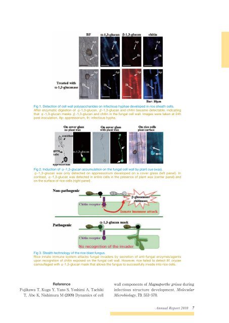
Fig 1. Detection of cell wall polysaccharides on infectious hyphae developed in rice sheath cells.<br />
After enzymatic digestion of α-1,3-glucan, β-1,3-glucan and chitin became detectable, indicating<br />
that α-1,3-glucan masks β-1,3-glucan and chitin in the fungal cell wall. Images were taken at 24h<br />
post inoculation. Ap: appressorium, Ih: infectious hypha.<br />
Fig 2. Induction of α-1,3-glucan accumulation on the fungal cell wall by plant cue (wax).<br />
α-1,3-glucan was only detected on appressorium developed on a cover glass (left panel). In<br />
contrast, α-1,3-glucan was detected in entire cells in the presence of plant wax (center panel) and<br />
on the surface of rice cells (right panel) .<br />
Fig 3. Stealth technology of the rice blast fungus.<br />
Rice innate immune system attacks fungal invaders by secretion of anti-fungal enzymes/agents<br />
upon recognition of chitin exposed on the fungal cell wall. However, rice failed to detect M. oryzae<br />
camouflaged with α-1,3-glucan mask that allows the fungus to successfully invade into rice cells.<br />
Reference<br />
Fujikawa T, Kuga Y, Yano S, Yoshimi A, Tachiki<br />
T, Abe K, Nishimura M (2009) Dynamics of cell<br />
wall components of Magnaporthe grisea during<br />
infectious structure development. Molecular<br />
Microbiology, 73: 553-570.<br />
<strong>Annual</strong> <strong>Report</strong> <strong>2010</strong> 7