Role of the endothelium in viral hemorrhagic fevers - Benh Vien ...
Role of the endothelium in viral hemorrhagic fevers - Benh Vien ...
Role of the endothelium in viral hemorrhagic fevers - Benh Vien ...

Create successful ePaper yourself
Turn your PDF publications into a flip-book with our unique Google optimized e-Paper software.
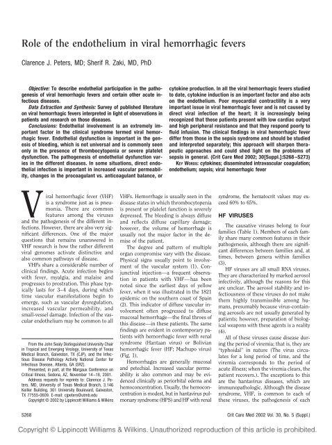
<strong>Role</strong> <strong>of</strong> <strong>the</strong> endo<strong>the</strong>lium <strong>in</strong> <strong>viral</strong> <strong>hemorrhagic</strong> <strong>fevers</strong><br />
Clarence J. Peters, MD; Sherif R. Zaki, MD, PhD<br />
Objective: To describe endo<strong>the</strong>lial participation <strong>in</strong> <strong>the</strong> pathogenesis<br />
<strong>of</strong> <strong>viral</strong> <strong>hemorrhagic</strong> <strong>fevers</strong> and certa<strong>in</strong> o<strong>the</strong>r acute <strong>in</strong>fectious<br />
diseases.<br />
Data Extraction and Syn<strong>the</strong>sis: Survey <strong>of</strong> published literature<br />
on <strong>viral</strong> <strong>hemorrhagic</strong> <strong>fevers</strong> <strong>in</strong>terpreted <strong>in</strong> light <strong>of</strong> observations <strong>in</strong><br />
patients and research on those diseases.<br />
Conclusions: Endo<strong>the</strong>lial <strong>in</strong>volvement is an extremely important<br />
factor <strong>in</strong> <strong>the</strong> cl<strong>in</strong>ical syndrome termed <strong>viral</strong> <strong>hemorrhagic</strong><br />
fever. Endo<strong>the</strong>lial dysfunction is important <strong>in</strong> <strong>the</strong> genesis<br />
<strong>of</strong> bleed<strong>in</strong>g, which is not universal and is commonly seen<br />
only <strong>in</strong> <strong>the</strong> presence <strong>of</strong> thrombocytopenia or severe platelet<br />
dysfunction. The pathogenesis <strong>of</strong> endo<strong>the</strong>lial dysfunction varies<br />
<strong>in</strong> <strong>the</strong> different diseases. In some situations, direct endo<strong>the</strong>lial<br />
<strong>in</strong>fection is important <strong>in</strong> <strong>in</strong>creased vascular permeability,<br />
changes <strong>in</strong> <strong>the</strong> procoagulant vs. anticoagulant balance, or<br />
cytok<strong>in</strong>e production. In all <strong>the</strong> <strong>viral</strong> <strong>hemorrhagic</strong> <strong>fevers</strong> studied<br />
to date, cytok<strong>in</strong>e <strong>in</strong>duction is an important factor and also acts<br />
on <strong>the</strong> endo<strong>the</strong>lium. Poor myocardial contractility is a very<br />
important issue <strong>in</strong> <strong>viral</strong> <strong>hemorrhagic</strong> fever and is not caused by<br />
direct <strong>viral</strong> <strong>in</strong>fection <strong>of</strong> <strong>the</strong> heart; it is <strong>in</strong>creas<strong>in</strong>gly be<strong>in</strong>g<br />
recognized that <strong>the</strong>se patients present with low cardiac output<br />
and high peripheral resistance and that <strong>the</strong>y respond poorly to<br />
fluid <strong>in</strong>fusion. The cl<strong>in</strong>ical f<strong>in</strong>d<strong>in</strong>gs <strong>in</strong> <strong>viral</strong> <strong>hemorrhagic</strong> fever<br />
differ from those <strong>in</strong> <strong>the</strong> sepsis syndrome and should be studied<br />
and <strong>in</strong>terpreted separately; this approach will sharpen <strong>the</strong>rapeutic<br />
approaches and could shed light on <strong>the</strong> problems <strong>of</strong><br />
sepsis <strong>in</strong> general. (Crit Care Med 2002; 30[Suppl.]:S268–S273)<br />
KEY WORDS: cytok<strong>in</strong>es; dissem<strong>in</strong>ated <strong>in</strong>travascular coagulation;<br />
endo<strong>the</strong>lium; sepsis; <strong>viral</strong> <strong>hemorrhagic</strong> fever<br />
Viral <strong>hemorrhagic</strong> fever (VHF)<br />
is a syndrome just as is pneumonia.<br />
There are common<br />
features among <strong>the</strong> viruses<br />
and <strong>the</strong> pathogenesis <strong>of</strong> <strong>the</strong> different <strong>in</strong>fections.<br />
However, <strong>the</strong>re are also very significant<br />
differences. One <strong>of</strong> <strong>the</strong> major<br />
questions that rema<strong>in</strong>s unanswered <strong>in</strong><br />
VHF research is how <strong>the</strong> ra<strong>the</strong>r different<br />
<strong>viral</strong> genomes activate dist<strong>in</strong>ctive and<br />
also common pathways <strong>of</strong> disease.<br />
VHFs share a considerable number <strong>of</strong><br />
cl<strong>in</strong>ical f<strong>in</strong>d<strong>in</strong>gs. Acute <strong>in</strong>fection beg<strong>in</strong>s<br />
with fever, myalgia, and malaise and<br />
progresses to prostration. This phase typically<br />
lasts for 3–4 days, dur<strong>in</strong>g which<br />
time vascular manifestations beg<strong>in</strong> to<br />
emerge, such as vascular dysregulation,<br />
<strong>in</strong>creased vascular permeability, and<br />
small-vessel damage. Infection <strong>of</strong> <strong>the</strong> vascular<br />
endo<strong>the</strong>lium may be common to all<br />
From <strong>the</strong> John Sealy Dist<strong>in</strong>guished University Chair<br />
<strong>in</strong> Tropical and Emerg<strong>in</strong>g Virology, University <strong>of</strong> Texas<br />
Medical Branch, Galveston, TX (CJP), and <strong>the</strong> Infectious<br />
Disease Pathology Activity National Center for<br />
Infectious Disease, Atlanta, GA (SRZ).<br />
Presented, <strong>in</strong> part, at <strong>the</strong> Margaux Conference on<br />
Critical Illness, Sedona, AZ, November 14–18, 2001.<br />
Address requests for repr<strong>in</strong>ts to: Clarence J. Peters,<br />
MD, University <strong>of</strong> Texas Medical Branch, 3.146<br />
Keiller Build<strong>in</strong>g, 301 University Boulevard, Galveston,<br />
TX 77555-0609. E-mail: cjpeters@utmb.edu<br />
Copyright © 2002 by Lipp<strong>in</strong>cott Williams & Wilk<strong>in</strong>s<br />
VHFs. Hemorrhage is usually seen <strong>in</strong> <strong>the</strong><br />
disease states <strong>in</strong> which thrombocytopenia<br />
is present or platelet function is severely<br />
depressed. The bleed<strong>in</strong>g is always diffuse<br />
and reflects diffuse capillary damage;<br />
however, <strong>the</strong> volume <strong>of</strong> hemorrhage is<br />
usually not <strong>the</strong> major factor <strong>in</strong> <strong>the</strong> demise<br />
<strong>of</strong> <strong>the</strong> patient.<br />
The degree and pattern <strong>of</strong> multiple<br />
organ compromise vary with <strong>the</strong> disease.<br />
Physical signs usually po<strong>in</strong>t to <strong>in</strong>volvement<br />
<strong>of</strong> <strong>the</strong> vascular system (1). Conjunctival<br />
<strong>in</strong>jection—a frequent observation<br />
<strong>in</strong> patients with VHF—has been<br />
noted s<strong>in</strong>ce <strong>the</strong> earliest days <strong>of</strong> yellow<br />
fever, when it was illustrated <strong>in</strong> <strong>the</strong> 1821<br />
epidemic on <strong>the</strong> sou<strong>the</strong>rn coast <strong>of</strong> Spa<strong>in</strong><br />
(2). This <strong>in</strong>dicator <strong>of</strong> diffuse vascular <strong>in</strong>volvement<br />
<strong>of</strong>ten progressed to diffuse<br />
mucosal hemorrhage—<strong>the</strong> f<strong>in</strong>al throes <strong>of</strong><br />
this disease—<strong>in</strong> <strong>the</strong>se patients. The same<br />
f<strong>in</strong>d<strong>in</strong>gs are evident <strong>in</strong> contemporary patients<br />
with <strong>hemorrhagic</strong> fever with renal<br />
syndrome (Hantaan virus) or Bolivian<br />
<strong>hemorrhagic</strong> fever (HF; Machupo virus)<br />
(Fig. 1).<br />
Hemorrhages are generally mucosal<br />
and petechial. Increased vascular permeability<br />
is also common and may be evidenced<br />
cl<strong>in</strong>ically as periorbital edema and<br />
hemoconcentration. Usually, <strong>the</strong> hemoconcentration<br />
is modest, but <strong>in</strong> hantavirus pulmonary<br />
syndrome (HPS) and HF with renal<br />
syndrome, <strong>the</strong> hematocrit values may exceed<br />
60% to 65%.<br />
HF VIRUSES<br />
The causative viruses belong to four<br />
families (Table 1). Members <strong>of</strong> each family<br />
share many common features <strong>in</strong> <strong>the</strong>ir<br />
pathogenesis, although <strong>the</strong>re are significant<br />
differences between families and, at<br />
times, between genera with<strong>in</strong> families<br />
(3).<br />
HF viruses are all small RNA viruses.<br />
They are characterized by marked aerosol<br />
<strong>in</strong>fectivity, although <strong>the</strong> reasons for this<br />
are unclear. The aerosol stability and <strong>in</strong>fectiousness<br />
<strong>of</strong> <strong>the</strong>se viruses do not make<br />
<strong>the</strong>m highly transmissible among humans,<br />
presumably because virus-conta<strong>in</strong><strong>in</strong>g<br />
aerosols are not usually generated by<br />
patients; however, preparation <strong>of</strong> biological<br />
weapons with <strong>the</strong>se agents is a reality<br />
(4).<br />
All <strong>of</strong> <strong>the</strong>se viruses cause disease dur<strong>in</strong>g<br />
<strong>the</strong> period <strong>of</strong> viremia; that is, <strong>the</strong>y are<br />
“typhoidal” <strong>in</strong> nature (The virus circulates<br />
for a long period <strong>of</strong> time, and <strong>the</strong><br />
viremia corresponds to <strong>the</strong> period <strong>of</strong><br />
acute illness; when <strong>the</strong> viremia clears, <strong>the</strong><br />
patient recovers.). The exceptions to this<br />
are <strong>the</strong> hantavirus diseases, which are<br />
immunopathologic. Although <strong>the</strong> disease<br />
syndrome, VHF, is common to each <strong>of</strong><br />
<strong>the</strong>se viruses, <strong>the</strong> pathogenesis <strong>of</strong> each<br />
S268<br />
Crit Care Med 2002 Vol. 30, No. 5 (Suppl.)
esponse. Most <strong>of</strong> <strong>the</strong>se diseases are worst<br />
while <strong>the</strong> virus is actively replicat<strong>in</strong>g <strong>in</strong> <strong>the</strong><br />
organs and released to circulate <strong>in</strong> <strong>the</strong><br />
blood. Only <strong>in</strong> <strong>the</strong> hantavirus diseases and<br />
<strong>in</strong> dengue HF does <strong>the</strong> classic, virusspecific<br />
immune response seem to play a<br />
role <strong>in</strong> caus<strong>in</strong>g <strong>the</strong> cl<strong>in</strong>ical manifestations,<br />
and <strong>in</strong> those diseases, immunopathology is<br />
an important part <strong>of</strong> <strong>the</strong> disease process.<br />
In <strong>the</strong> case <strong>of</strong> dengue virus, <strong>in</strong> <strong>the</strong> overwhelm<strong>in</strong>g<br />
majority <strong>of</strong> cases, HF follows sequential<br />
<strong>in</strong>fections with different serotypes;<br />
it is not <strong>in</strong>cluded <strong>in</strong> Table 1 because it is not<br />
one <strong>of</strong> <strong>the</strong> primary VHFs. There is now<br />
considerable <strong>in</strong>formation that suggests that<br />
immune enhancement and immunopathology<br />
are <strong>the</strong> major factors <strong>in</strong> <strong>the</strong> genesis<br />
<strong>of</strong> this syndrome, which is primarily a vascular<br />
permeability disease.<br />
ARENAVIRUSES<br />
Figure 1. Top, patient with <strong>hemorrhagic</strong> fever with renal syndrome show<strong>in</strong>g subconjunctival hemorrhage<br />
<strong>of</strong> <strong>the</strong> bulbar conjunctivae and vascular congestion <strong>of</strong> <strong>the</strong> palpebral conjunctivae. Bottom,<br />
patient with Bolivian <strong>hemorrhagic</strong> fever, a South American arenavirus <strong>hemorrhagic</strong> fever, with diffuse<br />
mucosal bleed<strong>in</strong>g. Reproduced with permission from Peters et al (1).<br />
Table 1. Viral <strong>hemorrhagic</strong> <strong>fevers</strong><br />
Arenaviridae Lassa fever and South American<br />
HF (Argent<strong>in</strong>e, Bolivian, etc.)<br />
Bunyaviridae<br />
Phlebovirus Rift Valley fever<br />
Nairovirus Crimean Congo HF<br />
Hantavirus HF with renal syndrome and<br />
Hantavirus pulmonary<br />
syndrome<br />
Filovirus Marburg HF and Ebola HF<br />
Flavivirus Yellow fever, KFD, and Omsk<br />
HF<br />
HF, <strong>hemorrhagic</strong> fever; KFD, Kyasanur Forest<br />
disease.<br />
differs, and <strong>the</strong> ways <strong>in</strong> which <strong>the</strong> viruses<br />
<strong>in</strong>teract with cells are similarly different<br />
(Tables 2 and 3).<br />
PATHOGENESIS OF HF<br />
Some <strong>of</strong> <strong>the</strong>se viruses cause almost no<br />
cytopathic effects, whereas o<strong>the</strong>rs are<br />
highly destructive to <strong>the</strong> cells <strong>the</strong>y <strong>in</strong>fect.<br />
Pro-<strong>in</strong>flammatory cytok<strong>in</strong>e <strong>in</strong>duction (particularly<br />
tumor necrosis factor [TNF]-), <strong>in</strong><br />
cases <strong>in</strong> which it has been measured, is a<br />
general f<strong>in</strong>d<strong>in</strong>g. Dissem<strong>in</strong>ated <strong>in</strong>travascular<br />
coagulation (DIC) occurs <strong>in</strong> some but<br />
Crit Care Med 2002 Vol. 30, No. 5 (Suppl.)<br />
Table 2. Similarities among <strong>hemorrhagic</strong> fever<br />
viruses<br />
Similarities<br />
Vascular syndrome<br />
Dysregulation<br />
Increased permeability<br />
Diffuse damage<br />
Small RNA viruses, 1–2 10 6 d<br />
Lipid envelope<br />
Aerosol <strong>in</strong>fectivity<br />
Persist <strong>in</strong> nature <strong>in</strong>dependently <strong>of</strong> humans<br />
Table 3. Differences among <strong>hemorrhagic</strong> fever<br />
viruses<br />
Differences<br />
Replication strategy<br />
Virion structure and morphogenesis<br />
Cytopathic effects <strong>in</strong> mammalian cells<br />
Sensitivity to anti<strong>viral</strong> effects <strong>of</strong> <strong>in</strong>terferon<br />
Pathogenesis <strong>of</strong> <strong>in</strong>fection<br />
Human immune response<br />
Survival strategy <strong>in</strong> nature<br />
not <strong>in</strong> o<strong>the</strong>rs. Involvement <strong>of</strong> <strong>the</strong> liver also<br />
varies. HF is a s<strong>in</strong>gle syndrome but not a<br />
stereotypical disease (3, 5).<br />
One <strong>of</strong> <strong>the</strong> important pathogenetic differences<br />
<strong>in</strong>volves <strong>the</strong> role <strong>of</strong> <strong>the</strong> immune<br />
The arenaviruses, which cause Lassa<br />
fever <strong>in</strong> Africa and Argent<strong>in</strong>e and Bolivian<br />
HF <strong>in</strong> South America, are particularly<br />
<strong>in</strong>terest<strong>in</strong>g; fur<strong>the</strong>rmore, arenavirus <strong>in</strong>fections<br />
occur reasonably frequently <strong>in</strong><br />
predictable sites, provid<strong>in</strong>g a pool <strong>of</strong> patients<br />
for study. Throat swabs from patients<br />
demonstrate a small amount <strong>of</strong> virus,<br />
but titers are low and <strong>in</strong>constant.<br />
Person-to-person transmission is not<br />
very high, except possibly through sexual<br />
<strong>in</strong>tercourse <strong>in</strong> convalescence, but patients<br />
develop effusions quite regularly.<br />
High-titered virus is present <strong>in</strong> <strong>the</strong><br />
Lassa fever effusion. Arenavirus <strong>in</strong>fection<br />
<strong>of</strong> <strong>the</strong> meso<strong>the</strong>lial cells on <strong>the</strong> surface <strong>of</strong><br />
<strong>the</strong> organs ba<strong>the</strong>d <strong>in</strong> <strong>the</strong> effusion suggests<br />
a direct effect <strong>of</strong> <strong>the</strong> <strong>in</strong>fection on <strong>the</strong><br />
exudation <strong>of</strong> fluid (5). Figure 2 shows <strong>the</strong><br />
pleural surface <strong>of</strong> a patient with Lassa<br />
fever. The meso<strong>the</strong>lium is very <strong>in</strong>tensely<br />
sta<strong>in</strong>ed for <strong>viral</strong> antigen. Arenaviruses<br />
also <strong>in</strong>fect <strong>the</strong> capillary endo<strong>the</strong>lium <strong>of</strong><br />
many organs <strong>in</strong> <strong>the</strong> body.<br />
The arenaviruses are not highly cytopathic,<br />
and thus, it is not clear what<br />
mechanism is <strong>in</strong>volved <strong>in</strong> <strong>the</strong> alteration<br />
<strong>of</strong> vascular endo<strong>the</strong>lial function or how<br />
much <strong>of</strong> <strong>the</strong> functional change is caused<br />
by direct <strong>in</strong>fection and how much is from<br />
<strong>the</strong> cytok<strong>in</strong>e activation that is evident <strong>in</strong><br />
<strong>the</strong> arena<strong>viral</strong> HF. Pro-<strong>in</strong>flammatory cytok<strong>in</strong>es<br />
are elevated, and <strong>in</strong> Argent<strong>in</strong>e<br />
HF, <strong>the</strong>re is a correlation between mortality<br />
and <strong>the</strong> levels <strong>of</strong> TNF- and <strong>in</strong>terferon<br />
(IFN) (6–8). Arenavirus <strong>in</strong>fection<br />
has also been shown to cause a loss <strong>of</strong><br />
cellular function—such as growth hormone<br />
secretion <strong>in</strong> <strong>the</strong> mur<strong>in</strong>e pituitary<br />
or neurohumoral transmitter secretion<br />
S269
The cl<strong>in</strong>ical f<strong>in</strong>d<strong>in</strong>gs<br />
<strong>in</strong> <strong>viral</strong> <strong>hemorrhagic</strong><br />
fever differ<br />
from those <strong>in</strong> <strong>the</strong> sepsis syndrome<br />
and should be studied<br />
and <strong>in</strong>terpreted separately;<br />
this approach will sharpen<br />
<strong>the</strong>rapeutic approaches and<br />
could shed light on <strong>the</strong> problems<br />
<strong>of</strong> sepsis <strong>in</strong> general.<br />
Figure 2. Pleural surface <strong>of</strong> patient with Lassa fever.<br />
<strong>in</strong> cultured neurotransmitter cells—<br />
without any detectable sign <strong>of</strong> cellular<br />
damage or <strong>in</strong>terruption <strong>of</strong> <strong>the</strong> basic life<br />
functions <strong>of</strong> <strong>the</strong> cell (9). Hence, <strong>the</strong>se<br />
viruses may exert reasonably selective<br />
and specific actions on function without<br />
any overt signs <strong>of</strong> cellular damage.<br />
The role <strong>of</strong> soluble mediators <strong>in</strong> VHF<br />
was first <strong>in</strong>vestigated <strong>in</strong> gu<strong>in</strong>ea pigs <strong>in</strong>fected<br />
with Pich<strong>in</strong>de virus (10). (Pich<strong>in</strong>de<br />
virus is an arenavirus that does not cause<br />
disease <strong>in</strong> humans and thus provides a<br />
useful model for <strong>the</strong> laboratory study <strong>of</strong><br />
arenavirus HF.) The lesions visible on<br />
detailed histopathologic study were not<br />
sufficient to suggest <strong>the</strong> cause <strong>of</strong> death,<br />
and <strong>the</strong> marked extension <strong>of</strong> <strong>in</strong>fection<br />
beyond any zone <strong>of</strong> morphologic damage<br />
suggested that disease is associated with<br />
an alteration <strong>of</strong> function ra<strong>the</strong>r than<br />
overt cytopathology—and direct <strong>viral</strong> <strong>in</strong>terference<br />
with endo<strong>the</strong>lial cell function<br />
is a possibility (9). A role for soluble mediators<br />
presumed to be <strong>in</strong>volved <strong>in</strong> <strong>the</strong><br />
pathogenesis <strong>of</strong> septic or late <strong>hemorrhagic</strong><br />
shock has also been considered,<br />
and several have been found to be markedly<br />
altered <strong>in</strong> Pich<strong>in</strong>de virus <strong>in</strong>fections,<br />
<strong>in</strong>clud<strong>in</strong>g leukotrienes (11), plateletactivat<strong>in</strong>g<br />
factor (12), and endorph<strong>in</strong>s<br />
(13). TNF- is also activated <strong>in</strong> <strong>the</strong><br />
Pich<strong>in</strong>de-<strong>in</strong>fected gu<strong>in</strong>ea pig (14). Supportive<br />
evidence for <strong>the</strong> role <strong>of</strong> soluble<br />
factors comes from <strong>the</strong> observations that<br />
cardiovascular depression is an important<br />
component <strong>of</strong> <strong>the</strong> disease <strong>in</strong> <strong>the</strong> absence<br />
<strong>of</strong> direct cardiac <strong>in</strong>fection virus (15).<br />
Intensive studies <strong>of</strong> coagulation have<br />
failed to identify major isolated defects <strong>in</strong><br />
coagulation pathways, fibr<strong>in</strong>olysis, or <strong>the</strong><br />
presence <strong>of</strong> DIC to expla<strong>in</strong> bleed<strong>in</strong>g <strong>in</strong><br />
arenavirus disease (16). In any case,<br />
bleed<strong>in</strong>g is marked <strong>in</strong> <strong>the</strong> South American<br />
diseases, presumably related to vascular<br />
damage and <strong>the</strong> associated severe<br />
thrombocytopenia. Bleed<strong>in</strong>g occurs less<br />
frequently <strong>in</strong> Lassa fever, but when it<br />
does, it is <strong>in</strong> a sett<strong>in</strong>g <strong>of</strong> mild thrombocytopenia<br />
but marked loss <strong>of</strong> platelet<br />
function measured <strong>in</strong> vitro (17, 18).<br />
RIFT VALLEY FEVER<br />
Rift Valley fever is perhaps unique <strong>in</strong><br />
hav<strong>in</strong>g a highly variable cl<strong>in</strong>ical presentation<br />
that <strong>in</strong>cludes VHF, encephalitis,<br />
and ret<strong>in</strong>al disease (19). Development <strong>of</strong><br />
Rift Valley fever is <strong>of</strong>ten associated with<br />
marked hepatitis. The pathogenesis <strong>in</strong><br />
humans is not known, but extensive studies<br />
<strong>in</strong> monkeys have shown that an important<br />
determ<strong>in</strong>ant is late secretion <strong>of</strong><br />
IFN- (20). If IFN- is detectable with<strong>in</strong><br />
6–12 hrs <strong>of</strong> <strong>in</strong>fection, <strong>the</strong> course will be<br />
mild. Later onset <strong>of</strong> <strong>the</strong> IFN response is<br />
associated with more severe disease, DIC,<br />
microangiopathic hemolytic anemia, and<br />
<strong>in</strong>travascular deposition <strong>of</strong> fibr<strong>in</strong> thrombi<br />
(21). The virus itself causes rapid cell<br />
death <strong>in</strong> all mammalian cells tested, <strong>in</strong>clud<strong>in</strong>g<br />
human endo<strong>the</strong>lial cell cultures<br />
(19). It seems likely that <strong>the</strong> delay <strong>in</strong><br />
IFN- response is sufficient to allow<br />
more extensive <strong>in</strong>fection <strong>of</strong> vascular endo<strong>the</strong>lium<br />
and that direct <strong>viral</strong> damage<br />
destroys <strong>the</strong> antithrombotic l<strong>in</strong><strong>in</strong>g <strong>of</strong> capillaries,<br />
<strong>in</strong>clud<strong>in</strong>g <strong>the</strong> glomeruli.<br />
CRIMEAN CONGO HF<br />
Crimean Congo HF virus causes a severe<br />
HF but is not highly cytopathic. The<br />
mechanism for hemorrhage is not<br />
known, but <strong>the</strong>re is cl<strong>in</strong>ical evidence <strong>of</strong><br />
DIC (22). Of all <strong>the</strong> VHFs, this <strong>in</strong>fection is<br />
associated with <strong>the</strong> most florid hemorrhage<br />
and also seems to be associated<br />
with <strong>the</strong> highest frequency <strong>of</strong> large ecchymoses.<br />
More detailed clott<strong>in</strong>g and cytok<strong>in</strong>e<br />
studies would be <strong>of</strong> considerable<br />
<strong>in</strong>terest. Immunohistochemical and <strong>in</strong><br />
situ hybridization studies have primarily<br />
focused on liver tissues and have shown<br />
<strong>in</strong>volvement <strong>of</strong> hepatocytes and endo<strong>the</strong>lial<br />
cells (23). The o<strong>the</strong>r tissues studied<br />
showed a few endo<strong>the</strong>lial cells and occasional<br />
mononuclear phagocytes to be<br />
positive. There is modest cellular necrosis<br />
generally associated with <strong>the</strong> presence<br />
<strong>of</strong> antigen <strong>in</strong> hepatocytes. As with all <strong>the</strong><br />
nonhantavirus diseases discussed here,<br />
<strong>the</strong>re is marked cl<strong>in</strong>ical improvement at<br />
<strong>the</strong> onset <strong>of</strong> <strong>the</strong> immune response and<br />
disappearance <strong>of</strong> viremia.<br />
HANTAVIRUSES<br />
Hantaviruses from American rodents<br />
cause HPS, which is associated with a<br />
marked and abrupt <strong>in</strong>crease <strong>in</strong> pulmonary<br />
capillary permeability (24). HPS is<br />
conf<strong>in</strong>ed to <strong>the</strong> Americas. Unlike most<br />
VHFs, <strong>the</strong> disease process <strong>in</strong> HPS is<br />
ma<strong>in</strong>ly conf<strong>in</strong>ed to <strong>the</strong> thoracic cavity.<br />
There is an <strong>in</strong>itial febrile prodrome last<strong>in</strong>g<br />
4–5 days. Patients subsequently develop<br />
bilateral alveolar pulmonary<br />
edema, which progresses rapidly. Patients<br />
exhibit thrombocytopenia—and this is<br />
S270<br />
Crit Care Med 2002 Vol. 30, No. 5 (Suppl.)
useful diagnostically—although hemorrhage<br />
is not common. The development<br />
<strong>of</strong> shock and severe arterial desaturation<br />
ushers <strong>in</strong> <strong>the</strong> phase dur<strong>in</strong>g which about<br />
40% <strong>of</strong> patients will die. Five to 7 days<br />
after <strong>the</strong> onset <strong>of</strong> shock and arterial desaturation,<br />
patients who survive <strong>the</strong> <strong>in</strong>itial<br />
24–48 hrs will usually be extubated,<br />
breath<strong>in</strong>g well, and shortly <strong>the</strong>reafter,<br />
able to leave <strong>the</strong> hospital. Clearly, this<br />
syndrome cannot reflect endo<strong>the</strong>lial cell<br />
death, but ra<strong>the</strong>r functional changes <strong>in</strong><br />
permeability. This is re<strong>in</strong>forced by <strong>the</strong> <strong>in</strong><br />
vitro f<strong>in</strong>d<strong>in</strong>gs that <strong>the</strong> virus itself has no<br />
effect on permeability <strong>of</strong> microvascular<br />
pulmonary endo<strong>the</strong>lial cells (25).<br />
Patients present with anti<strong>viral</strong> antibody<br />
and large numbers <strong>of</strong> circulat<strong>in</strong>g<br />
activated CD8 lymphocytes, some <strong>of</strong><br />
which can be cloned and are virus specific;<br />
when stimulated with <strong>viral</strong> antigen,<br />
<strong>the</strong>y produce <strong>in</strong>terleuk<strong>in</strong>-2 and IFN-. In<br />
tissues from patients dy<strong>in</strong>g from HPS,<br />
virtually all <strong>of</strong> <strong>the</strong> pulmonary endo<strong>the</strong>lium<br />
is <strong>in</strong>fected with <strong>the</strong> virus. Many <strong>of</strong><br />
<strong>the</strong> lymphoid cells <strong>in</strong>filtrat<strong>in</strong>g <strong>the</strong> lung<br />
parenchyma are CD8 T lymphocytes<br />
(26), and many are produc<strong>in</strong>g <strong>in</strong>terleuk<strong>in</strong>-2,<br />
TNF-, and IFN- (27). These lung<br />
lymphocytes are responsible for <strong>in</strong>duc<strong>in</strong>g<br />
a reversible <strong>in</strong>crease <strong>in</strong> permeability, mediated<br />
at least <strong>in</strong> part by TNF-. Table 4<br />
shows <strong>the</strong> markedly elevated cytok<strong>in</strong>e<br />
measurements <strong>in</strong> patients <strong>in</strong> New Mexico<br />
with HPS (28). Increases <strong>in</strong> TNF- have<br />
also been observed, particularly <strong>in</strong> those<br />
patients who die (FT Koster, unpublished<br />
observations) (28).<br />
Although <strong>the</strong>re are clear parallels <strong>in</strong><br />
<strong>the</strong> <strong>in</strong>flammatory cytok<strong>in</strong>e pr<strong>of</strong>ile <strong>in</strong> sera<br />
from patients with acute respiratory distress<br />
syndrome and sepsis, patients who<br />
die from HPS usually do not have overt<br />
multiple organ failure. Indeed, one <strong>of</strong> <strong>the</strong><br />
<strong>in</strong>terest<strong>in</strong>g developments after <strong>the</strong> identification<br />
<strong>of</strong> <strong>the</strong> hanta<strong>viral</strong> etiology <strong>of</strong> <strong>the</strong><br />
1993 epidemic was f<strong>in</strong>d<strong>in</strong>g a clearly demarcated<br />
syndrome with<strong>in</strong> <strong>the</strong> acute respiratory<br />
distress syndrome category.<br />
This previously unrecognized syndrome,<br />
HPS, differed from acute respiratory distress<br />
syndrome <strong>in</strong> etiology, pathogenesis,<br />
cl<strong>in</strong>ical f<strong>in</strong>d<strong>in</strong>gs, and pathology.<br />
FILOVIRUSES<br />
The filovirus family comprises Ebola<br />
and Marburg viruses and exercises a considerable<br />
grip on <strong>the</strong> public’s imag<strong>in</strong>ation.<br />
New <strong>in</strong>formation has emerged on<br />
<strong>the</strong> molecular pathogenesis <strong>of</strong> filovirus<br />
<strong>in</strong>fections (Fig. 3) (29). The Ebola Zaire<br />
subtype is <strong>the</strong> most pathogenic <strong>of</strong> <strong>the</strong><br />
filoviruses, with a case fatality <strong>of</strong> around<br />
90%. It is a very <strong>in</strong>vasive, cytopathic virus<br />
with a primary target <strong>of</strong> macrophages,<br />
although it rapidly spreads to <strong>in</strong>volve <strong>the</strong><br />
endo<strong>the</strong>lium and parenchymal cells <strong>of</strong><br />
many organs (30); cytok<strong>in</strong>es are also activated<br />
(31).<br />
Molecular studies <strong>of</strong> <strong>the</strong> pathogenesis<br />
<strong>of</strong> <strong>the</strong> related Marburg filovirus have been<br />
reported (29, 32, 33). Dur<strong>in</strong>g <strong>the</strong> early<br />
stages <strong>of</strong> pathogenesis, Marburg virus–<br />
<strong>in</strong>fected macrophages release cytok<strong>in</strong>es<br />
that lead to endo<strong>the</strong>lial cell disorganization<br />
and <strong>in</strong>creased permeability. TNF- is<br />
clearly a major player <strong>in</strong> <strong>the</strong> <strong>in</strong>creased<br />
endo<strong>the</strong>lial permeability as judged by <strong>the</strong><br />
ability <strong>of</strong> specific antibodies to block <strong>the</strong><br />
pathogenic effects <strong>of</strong> <strong>in</strong>fected macrophage<br />
supernatants. The permeability defects<br />
are accompanied by disruption <strong>of</strong><br />
some <strong>of</strong> <strong>the</strong> molecules important for <strong>in</strong>tercellular<br />
adhesion, <strong>in</strong>clud<strong>in</strong>g caten<strong>in</strong>s,<br />
cadher<strong>in</strong>s, and plakoglob<strong>in</strong>. Cytok<strong>in</strong>e secretion<br />
has also been implicated <strong>in</strong> <strong>the</strong><br />
severe apoptosis <strong>in</strong> lymphoid organs <strong>of</strong><br />
Ebola virus model systems and may play a<br />
role <strong>in</strong> patients (30, 31, 34). However,<br />
cytok<strong>in</strong>e elaboration is but one <strong>of</strong> <strong>the</strong><br />
possible causes <strong>of</strong> death <strong>in</strong> fatal filovirus<br />
<strong>in</strong>fections. Macrophages certa<strong>in</strong>ly secrete<br />
pro-<strong>in</strong>flammatory cytok<strong>in</strong>es, but <strong>the</strong> virus<br />
also directly <strong>in</strong>fects endo<strong>the</strong>lium<br />
(with consequent virus-<strong>in</strong>duced damage<br />
and DIC), and <strong>the</strong>re is, <strong>in</strong> addition, extensive<br />
cytopathic <strong>in</strong>volvement <strong>of</strong> <strong>the</strong> parenchymal<br />
cells <strong>of</strong> multiple organs.<br />
HF VIRUS SUMMARY<br />
There are both common <strong>the</strong>mes and<br />
differences <strong>in</strong> <strong>the</strong> pathogenesis <strong>of</strong> VHF<br />
(Table 5). Endo<strong>the</strong>lial <strong>in</strong>fection is common<br />
and can be limited or widespread. In<br />
Table 4. Immune markers <strong>in</strong> sera <strong>of</strong> patients with<br />
hantavirus pulmonary syndrome a<br />
Immune Marker Survive Fatal<br />
Soluble CD4 NS 1<br />
Soluble CD8 1 1<br />
IL-2 NS NS<br />
IFN- NS 1<br />
IL-4 1 1<br />
IL-6 NS 1<br />
Soluble IL-2 receptor NS 1<br />
NS, not significant; 1, markedly <strong>in</strong>creased;<br />
IFN-, <strong>in</strong>terferon-; IL, <strong>in</strong>terleuk<strong>in</strong>; NS, not significantly<br />
different from acute respiratory disease<br />
syndrome or sepsis controls.<br />
a Data reproduced with permission from<br />
Simpson et al (28).<br />
Figure 3. Pathogenesis <strong>of</strong> Marburg virus (MBGV) <strong>in</strong>fection. BM, basement membrane; CAM, cell<br />
adhesion molecule; E, erythrocyte; EC, endo<strong>the</strong>lial cell; N, nucleus; V, vacuole; Vir, virus. Reproduced<br />
with permission from Feldmann and Klenk (29).<br />
Crit Care Med 2002 Vol. 30, No. 5 (Suppl.)<br />
S271
<strong>the</strong> case <strong>of</strong> Rift Valley fever virus, <strong>the</strong><br />
virus has a highly destructive <strong>in</strong>teraction<br />
with any cell type <strong>in</strong>fected, and although<br />
<strong>the</strong>re are no good human data, it seems<br />
to destroy endo<strong>the</strong>lium and lead to DIC<br />
on a background <strong>of</strong> extensive liver damage.<br />
The cytopathic and <strong>in</strong>vasive nature<br />
<strong>of</strong> filovirus <strong>in</strong>fections contrasts sharply<br />
with <strong>the</strong> f<strong>in</strong>d<strong>in</strong>gs from arenaviruses.<br />
Ebola (Zaire subtype) is highly damag<strong>in</strong>g<br />
to <strong>the</strong> cells it <strong>in</strong>fects, and this is important<br />
<strong>in</strong> <strong>in</strong>duc<strong>in</strong>g vascular leakage, which<br />
can be multiplied by DIC. Arenaviruses,<br />
<strong>in</strong> contrast, <strong>in</strong>fect <strong>the</strong> endo<strong>the</strong>lium but<br />
cause little direct cell damage; <strong>the</strong>y may<br />
<strong>in</strong>duce cytok<strong>in</strong>es or perturb endo<strong>the</strong>lial<br />
cell function, but this is <strong>in</strong> <strong>the</strong> absence <strong>of</strong><br />
obvious morphologic signs. O<strong>the</strong>r viruses,<br />
such as Hantaviruses, are virtually<br />
noncytopathic, can <strong>in</strong>fect cells <strong>in</strong> vitro<br />
without <strong>in</strong>duc<strong>in</strong>g any permeability or<br />
o<strong>the</strong>r major change, and depend on <strong>the</strong><br />
host immune response to <strong>in</strong>duce changes<br />
<strong>in</strong> vascular permeability. The flaviviruses,<br />
such as yellow fever, are important<br />
agents that require additional study<br />
(Dengue HF differs from <strong>the</strong> rest <strong>of</strong> <strong>the</strong><br />
flaviviruses <strong>in</strong> hav<strong>in</strong>g an immunopathologic<br />
basis.).<br />
High concentrations <strong>of</strong> pro-<strong>in</strong>flammatory<br />
cytok<strong>in</strong>es clearly play an important<br />
role <strong>in</strong> <strong>the</strong> different HF <strong>in</strong>fections, but<br />
<strong>the</strong> mechanisms for cytok<strong>in</strong>e <strong>in</strong>duction<br />
and <strong>the</strong>ir exact role are different and<br />
poorly understood among <strong>the</strong> virus taxons.<br />
The clott<strong>in</strong>g defects are also quite<br />
variable <strong>in</strong> both <strong>the</strong>ir obvious manifestations<br />
and <strong>the</strong>ir pathogeneses.<br />
VIRUSES AND BACTERIAL<br />
SEPSIS<br />
Table 5. Pathogenesis <strong>of</strong> <strong>viral</strong> <strong>hemorrhagic</strong> <strong>fevers</strong> (VHF)<br />
VHF<br />
Cytopathic<br />
Effect<br />
Critical care and pulmonary specialists<br />
<strong>in</strong>itially regard VHF as simply ano<strong>the</strong>r<br />
subset <strong>of</strong> <strong>the</strong> diseases that <strong>the</strong>y deal<br />
with daily. This is overly simplified and<br />
overly reductionist. There are some strik<strong>in</strong>g<br />
similarities, such as <strong>in</strong>flammatory cytok<strong>in</strong>e<br />
secretion and coagulation activation;<br />
compar<strong>in</strong>g and contrast<strong>in</strong>g <strong>the</strong><br />
different conditions is likely to provide<br />
important <strong>in</strong>sights <strong>in</strong>to both pathogenesis<br />
and appropriate <strong>the</strong>rapeutic <strong>in</strong>tervention.<br />
Often, no obviously lethal morphologic<br />
lesions are present <strong>in</strong> VHF or <strong>in</strong><br />
sepsis, and patients with VHF (with <strong>the</strong><br />
exception <strong>of</strong> immunopathologic hantavirus<br />
diseases and dengue HF) and sepsis<br />
die with lymphoid depletion that is an<br />
anatomic correlate <strong>of</strong> immunosuppression.<br />
However, <strong>the</strong> hemodynamic patterns<br />
that are classic <strong>in</strong> sepsis are not<br />
typical <strong>of</strong> VHFs studied so far. The pathogenesis<br />
<strong>of</strong> <strong>the</strong> low-output, high-resistance<br />
state seen <strong>in</strong> patients with hantavirus<br />
and dengue HF (35, 36), and <strong>in</strong><br />
arenavirus model <strong>in</strong>fections (15), is unknown,<br />
and additional studies <strong>in</strong> o<strong>the</strong>r<br />
VHFs would be valuable (19). Multiple<br />
organ dysfunction syndrome is not <strong>the</strong><br />
rule <strong>in</strong> <strong>the</strong> VHF, fur<strong>the</strong>r <strong>in</strong>dicat<strong>in</strong>g a pattern<strong>in</strong>g<br />
<strong>in</strong> <strong>the</strong> different diseases. It seems<br />
that <strong>the</strong> key similarity may be endo<strong>the</strong>lial<br />
<strong>in</strong>volvement, ei<strong>the</strong>r direct or through <strong>the</strong><br />
mediation <strong>of</strong> cytok<strong>in</strong>es. The most important<br />
questions that rema<strong>in</strong> are how to<br />
measure <strong>the</strong> dysfunction <strong>of</strong> <strong>the</strong> vascular<br />
bed, why it occurs, and how it manifests<br />
differentially <strong>in</strong> dist<strong>in</strong>ct organs.<br />
OTHER INFECTIONS WITH<br />
ENDOTHELIAL INVOLVEMENT<br />
Endo<strong>the</strong>lial<br />
Infection<br />
Cytok<strong>in</strong>e<br />
Activation DIC Liver<br />
South American HF 2 4 No <br />
Lassa 2 3 No 1–<br />
2<br />
Rift Valley fever 4 ? ? Yes 2–<br />
3<br />
Crimean Congo HF 1 1 ? Yes 2–<br />
3<br />
Hantavirus pulmonary syndrome 0 4 4 No <br />
Filovirus HF 3 3 3 Yes 3<br />
Yellow fever 2 ? ? No 4<br />
DIC, dissem<strong>in</strong>ated <strong>in</strong>travascular coagulation; HF, <strong>hemorrhagic</strong> fever; ?, unknown; , variable, not<br />
marked; 1, occurs regularly; 2, occurs regularly and is marked; 3, occurs regularly and is<br />
moderately severe; 4, occurs regularly and is severe.<br />
O<strong>the</strong>r acute <strong>in</strong>fectious diseases that<br />
specifically <strong>in</strong>volve <strong>the</strong> endo<strong>the</strong>lium <strong>in</strong><br />
<strong>the</strong>ir pathogenesis <strong>in</strong>clude rickettsiae,<br />
paramyxoviruses, and anthrax. Rickettsiae<br />
are an important group <strong>of</strong> pathogenic<br />
agents that specifically <strong>in</strong>fect endo<strong>the</strong>lium<br />
and resemble VHF <strong>in</strong> <strong>the</strong>ir<br />
cl<strong>in</strong>ical manifestations (37). The newly<br />
discovered Nipah virus is a paramyxovirus<br />
with epidemic potential <strong>in</strong> pigs and<br />
that crosses species to kill humans. It<br />
<strong>in</strong>fects endo<strong>the</strong>lium <strong>in</strong> <strong>the</strong> lung to cause<br />
pulmonary disease and also ga<strong>in</strong>s entry to<br />
<strong>the</strong> central nervous system by <strong>in</strong>fect<strong>in</strong>g<br />
capillary endo<strong>the</strong>lium with formation <strong>of</strong><br />
giant cells, resultant clott<strong>in</strong>g, and micro<strong>in</strong>farcts,<br />
followed by direct spread to<br />
bra<strong>in</strong> neurons (38).<br />
Anthrax is <strong>of</strong> particular concern <strong>in</strong> today’s<br />
climate <strong>of</strong> potential bioterrorism.<br />
The typical patient with <strong>the</strong> <strong>in</strong>halational<br />
form <strong>of</strong> anthrax dies with massive endo<strong>the</strong>lial<br />
swell<strong>in</strong>g, tissue edema, hemoconcentration,<br />
and coagulation defects (39).<br />
SUMMARY<br />
The VHFs and several o<strong>the</strong>r <strong>in</strong>fectious<br />
diseases provide <strong>in</strong>terest<strong>in</strong>g models <strong>of</strong> <strong>the</strong><br />
participation <strong>of</strong> endo<strong>the</strong>lium <strong>in</strong> disease<br />
pathogenesis. Comparison to <strong>the</strong> sepsis<br />
model can be enlighten<strong>in</strong>g to students <strong>of</strong><br />
both.<br />
REFERENCES<br />
1. Peters CJ, Zaki SR, Roll<strong>in</strong> PE: Viral <strong>hemorrhagic</strong><br />
<strong>fevers</strong>. In: Atlas <strong>of</strong> Infectious Diseases.<br />
Vol. 8: External Manifestations <strong>of</strong> Systemic<br />
Infections. Mandell GL (Ed). Philadelphia,<br />
Current Medic<strong>in</strong>e, 1997, pp 10.1–10.26<br />
2. Bally, Francoise, Pariset: Histoire Medicale<br />
de la Fievre Jaune, observee en espagne et<br />
particulierment en Catalogne, dans l“anee<br />
1821. First Edition. Paris, L’imprimerie Royale,<br />
1823, pp 664<br />
3. Peters CJ, Zaki SR: Viral <strong>hemorrhagic</strong> fever:<br />
An overview. In: Tropical Infectious Diseases:<br />
Pr<strong>in</strong>ciples, Pathogens, and Practice. Guerrant<br />
RL, Walker DH, Weller PF (Eds). New<br />
York, WB Saunders, 1999, pp 1180–1188<br />
4. Peters CJ: Are <strong>hemorrhagic</strong> fever viruses<br />
practical agents for biological terrorism? In:<br />
Emerg<strong>in</strong>g Infections. Vol 4. Scheld WM,<br />
Craig WA, Hughes JM (Eds). Wash<strong>in</strong>gton DC,<br />
ASM Press, 2000, pp 203–211<br />
5. Zaki SR, Peters CJ: Viral <strong>hemorrhagic</strong> <strong>fevers</strong>.<br />
In: The Pathology <strong>of</strong> Infectious Diseases.<br />
Connor DH, Chandler FW, Schwartz DA, et<br />
al (Eds). Stamford, CT, Appleton and Lange,<br />
1997, pp 347–364<br />
6. Marta RF, Montero VF, Hack CE, et al: Pro<strong>in</strong>flammatory<br />
cytok<strong>in</strong>es and elastase-alpha-<br />
1antitryps<strong>in</strong> <strong>in</strong> Argent<strong>in</strong>e <strong>hemorrhagic</strong> fever.<br />
Am J Trop Med Hyg 1999; 60:85–89<br />
7. Mahanty S, Bausch D, Thomas RL, et al: Low<br />
levels <strong>of</strong> IL-8 and IP-10 <strong>in</strong> serum are associated<br />
with fatal <strong>in</strong>fections <strong>in</strong> acute Lassa fever.<br />
J Infect Dis 2001; 183:1713–1721<br />
8. Heller MV, Saavedra MC, Falc<strong>of</strong>f R, et al:<br />
S272<br />
Crit Care Med 2002 Vol. 30, No. 5 (Suppl.)
Increased tumor necrosis factor-alpha levels<br />
<strong>in</strong> Argent<strong>in</strong>e <strong>hemorrhagic</strong> fever. J Infect Dis<br />
1992; 166:1203–1204<br />
9. de la Torre JC, Borrow P, Oldstone MB: Viral<br />
persistence and disease: Cytopathology <strong>in</strong> <strong>the</strong><br />
absence <strong>of</strong> cytolysis. Br Med Bull 1991; 47:<br />
838–851<br />
10. Connolly BM, Jensen AB, Peters CJ, et al:<br />
Pathogenesis <strong>of</strong> Pich<strong>in</strong>de virus <strong>in</strong>fection <strong>in</strong><br />
stra<strong>in</strong> 13 gu<strong>in</strong>ea pigs: An immunocytochemical,<br />
virologic, and cl<strong>in</strong>ical chemistry study.<br />
Am J Trop Med Hyg 1993; 49:10–24<br />
11. Liu CT, Jahrl<strong>in</strong>g PB, Peters CJ: Evidence for<br />
<strong>the</strong> <strong>in</strong>volvement <strong>of</strong> sulfidopeptide leukotrienes<br />
<strong>in</strong> <strong>the</strong> pathogenesis <strong>of</strong> Pich<strong>in</strong>de virus<br />
<strong>in</strong>fection <strong>in</strong> stra<strong>in</strong> 13 gu<strong>in</strong>ea pigs. Prostagland<strong>in</strong>s<br />
Leukot Med 1986; 24:129–138<br />
12. Qian C, Ch<strong>in</strong>g-Tong L, Peters CJ: Increased<br />
platelet-activat<strong>in</strong>g factor (PAF) concentrations<br />
<strong>in</strong> hearts and lungs <strong>of</strong> Pich<strong>in</strong>de virus<strong>in</strong>fected<br />
gu<strong>in</strong>ea pigs. J Lipid Mediat 1992;<br />
5:261–270<br />
13. Guo ZM, Liu CT, Peters CJ: Possible <strong>in</strong>volvement<br />
<strong>of</strong> endogenous beta-endorph<strong>in</strong> <strong>in</strong> <strong>the</strong><br />
pathophysiological mechanisms <strong>of</strong> Pich<strong>in</strong>de<br />
virus-<strong>in</strong>fected gu<strong>in</strong>ea pigs. Proc Soc Exp Biol<br />
Med 1992; 200:343–348<br />
14. Aronson JF, Herzog NK, Jerrells TR: Tumor<br />
necrosis factor and <strong>the</strong> pathogenesis <strong>of</strong><br />
Pich<strong>in</strong>de virus <strong>in</strong>fection <strong>in</strong> gu<strong>in</strong>ea pigs. Am J<br />
Trop Med Hyg 1995; 52:262–269<br />
15. Qian C, Jahrl<strong>in</strong>g PB, Peters CJ, et al: Cardiovascular<br />
and pulmonary responses to<br />
Pich<strong>in</strong>de virus <strong>in</strong>fection <strong>in</strong> stra<strong>in</strong> 13 gu<strong>in</strong>ea<br />
pigs, Lab Anim Sci 1994; 44:600–607<br />
16. Heller MV, Marta RF, Sturk A, et al: Early<br />
markers <strong>of</strong> blood coagulation and fibr<strong>in</strong>olysis<br />
activation <strong>in</strong> Argent<strong>in</strong>e <strong>hemorrhagic</strong> fever.<br />
Thromb Haemost 1995; 73:368–373<br />
17. Fisher-Hoch S, McCormick JB, Sasso D, et al:<br />
Hematologic dysfunction <strong>in</strong> Lassa fever.<br />
J Med Virol 1988; 26:127–135<br />
18. Cumm<strong>in</strong>s D, Fisher-Hoch SP, Walshe KJ, et<br />
al: A plasma <strong>in</strong>hibitor <strong>of</strong> platelet aggregation<br />
<strong>in</strong> patients with Lassa fever. Br J Haematol<br />
1989; 72:543–548<br />
19. Peters CJ: Pathogenesis <strong>of</strong> <strong>viral</strong> <strong>hemorrhagic</strong><br />
<strong>fevers</strong>. In: Viral Pathogenesis. Nathanson N,<br />
Ahmed R, Gonzalez-Scarano F, et al (Eds).<br />
Philadelphia, Lipp<strong>in</strong>cott-Raven, 1997, pp<br />
779–799<br />
20. Morrill JC, Jenn<strong>in</strong>gs GB, Johnson AJ, et al:<br />
Pathogenesis <strong>of</strong> Rift Valley fever <strong>in</strong> rhesus<br />
monkeys: <strong>Role</strong> <strong>of</strong> <strong>in</strong>terferon response. Arch<br />
Virol 1990; 110:195–212<br />
21. Cosgriff TM, Morrill JC, Jenn<strong>in</strong>gs GB, et al:<br />
The hemostatic derangement produced by<br />
Rift Valley fever virus <strong>in</strong> rhesus monkeys.<br />
Rev Infect Dis 1989; 11(Suppl 4):S807–S814<br />
22. Swanepoel R, Gill DE, Shepherd AJ, et al: The<br />
cl<strong>in</strong>ical pathology <strong>of</strong> Crimean-Congo <strong>hemorrhagic</strong><br />
fever. Rev Infect Dis 1989; 11(Suppl<br />
4):S794–S800<br />
23. Burt FJ, Swanepoel R, Shieh WJ, et al: Immunohistochemical<br />
and <strong>in</strong> situ localization<br />
<strong>of</strong> Crimean-Congo <strong>hemorrhagic</strong> fever virus<br />
<strong>in</strong> human tissues and pathogenic implications.<br />
Arch Pathol Lab Med 1997; 121:<br />
839–846<br />
24. Duch<strong>in</strong> JS, Koster FT, Peters CJ, et al: Hantavirus<br />
pulmonary syndrome: A cl<strong>in</strong>ical description<br />
<strong>of</strong> 17 patients with a newly recognized<br />
disease. N Engl J Med 1994; 330:<br />
949–955<br />
25. Sundstrom JB, McMullan LK, Spiropoulou<br />
CF, et al: Hantavirus <strong>in</strong>fection <strong>in</strong>duces <strong>the</strong><br />
expression <strong>of</strong> RANTES and IP-10 without<br />
caus<strong>in</strong>g <strong>in</strong>creased permeability <strong>in</strong> human<br />
lung microvascular endo<strong>the</strong>lial cells. J Virol<br />
2001; 75:6070–6085<br />
26. Zaki SR, Greer PW, C<strong>of</strong>field LM, et al: Hantavirus<br />
pulmonary syndrome: Pathogenesis<br />
<strong>of</strong> an emerg<strong>in</strong>g <strong>in</strong>fectious disease. Am J<br />
Pathol 1995; 146:552–579<br />
27. Mori M, Rothman AL, Kurane I, et al: High<br />
levels <strong>of</strong> cytok<strong>in</strong>e-produc<strong>in</strong>g cells <strong>in</strong> <strong>the</strong> lung<br />
tissues <strong>of</strong> patients with fatal hantavirus pulmonary<br />
syndrome. J Infect Dis 1999; 179:<br />
295–302<br />
28. Simpson SQ, Mapel V, Koster FT, et al: Evidence<br />
for lymphocyte activation <strong>in</strong> <strong>the</strong> hantavirus<br />
pulmonary syndrome. Chest 1995;<br />
108:97S<br />
29. Feldmann H, Klenk HD: Filoviruses: Marburg<br />
and Ebola. In: Advances <strong>in</strong> Virus Research.<br />
Vol. 47. Maramorosch K, Murphy FA,<br />
Shatk<strong>in</strong> AJ (Eds). New York, Academic Press,<br />
1996, pp 1–52<br />
30. Zaki SR, Goldsmith CS: Pathologic features<br />
<strong>of</strong> filovirus <strong>in</strong>fections <strong>in</strong> humans. Curr Top<br />
Microbiol Immunol 1999; 235:97–116<br />
31. Vill<strong>in</strong>ger F, Roll<strong>in</strong> PE, Brar SS, et al: Markedly<br />
elevated levels <strong>of</strong> <strong>in</strong>terferon (IFN)-<br />
gamma, IFN-alpha, <strong>in</strong>terleuk<strong>in</strong> (IL)-2, IL-10,<br />
and tumor necrosis factor-alpha associated<br />
with fatal Ebola virus <strong>in</strong>fection. J Infect Dis<br />
1999; 179:S188–S191<br />
32. Feldmann H, Bugany H, Mahner F, et al:<br />
Filovirus-<strong>in</strong>duced endo<strong>the</strong>lial leakage triggered<br />
by <strong>in</strong>fected monocytes/macrophages.<br />
J Virol 1996; 70:2208–2214<br />
33. Schnittler HJ, Feldmann H: Molecular<br />
pathogenesis <strong>of</strong> filovirus <strong>in</strong>fections: <strong>Role</strong> <strong>of</strong><br />
macrophages and endo<strong>the</strong>lial cells. Curr Top<br />
Microbiol Immunol 1999; 235:175–204<br />
34. Geisbert TW, Hensley LE, Gibb TR, et al:<br />
Apoptosis <strong>in</strong>duced <strong>in</strong> vitro and <strong>in</strong> vivo dur<strong>in</strong>g<br />
<strong>in</strong>fection by Ebola and Marburg viruses. Lab<br />
Invest 2000; 80:171–196<br />
35. Wali JP, Biswas A, Chandra S, et al: Cardiac<br />
<strong>in</strong>volvement <strong>in</strong> dengue haemorrhagic fever.<br />
Int J Cardiol 1998; 64:31–36<br />
36. Peters CJ, Simpson GL, Levy H: Spectrum <strong>of</strong><br />
hantavirus <strong>in</strong>fection: Hemorrhagic fever<br />
with renal syndrome and hantavirus pulmonary<br />
syndrome. Annu Rev Med 1999; 50:<br />
531–545<br />
37. Wolbach SB: The etiology and pathology <strong>of</strong><br />
Rocky Mounta<strong>in</strong> spotted fever. (Third Prelim<strong>in</strong>ary<br />
Report.) The occurrence <strong>of</strong> <strong>the</strong> parasite<br />
and <strong>the</strong> pathology <strong>of</strong> <strong>the</strong> disease <strong>in</strong> man.<br />
Additional notes on <strong>the</strong> parasite. J Med Res<br />
1917; 37:499–508<br />
38. Chua KB, Bell<strong>in</strong>i WJ, Rota PA, et al: Nipah<br />
virus: A recently emergent deadly paramyxovirus.<br />
Science 2000; 288:1432–1435<br />
39. Gr<strong>in</strong>berg LM, Abramova FA, Yampolskaya<br />
OV, et al: Quantitative pathology <strong>of</strong> <strong>in</strong>halation<br />
anthrax I: Quantitative microscopic<br />
f<strong>in</strong>d<strong>in</strong>gs. Mod Pathol 2001; 14:482–495<br />
Crit Care Med 2002 Vol. 30, No. 5 (Suppl.)<br />
S273