Orthodontic and Nonorthodontic Root Resorption - Journal of Dental ...
Orthodontic and Nonorthodontic Root Resorption - Journal of Dental ...
Orthodontic and Nonorthodontic Root Resorption - Journal of Dental ...

You also want an ePaper? Increase the reach of your titles
YUMPU automatically turns print PDFs into web optimized ePapers that Google loves.
for implants. So the maxillary lateral incisors were<br />
not extracted (Figure 2D). No further root resorption<br />
had occurred over the two years <strong>of</strong> orthodontics<br />
(Figure 2E). An Essix retainer was worn in the<br />
maxillary arch to help stabilize the teeth. After two<br />
years, when the patient had completed her growth,<br />
the maxillary lateral incisor roots had not shortened<br />
further, <strong>and</strong> the teeth were relatively stable (Figure<br />
2F). So they were not extracted. Thirteen years after<br />
completion <strong>of</strong> the orthodontic treatment, the patient<br />
was now twenty-eight years <strong>of</strong> age, <strong>and</strong> the lateral<br />
incisors were still stable with no further root resorption<br />
(Figure 2G, H, <strong>and</strong> I).<br />
Case 3<br />
This fourteen-year-old female had been under<br />
the supervision <strong>of</strong> a general dentist since she was<br />
eight years <strong>of</strong> age (Figure 3A). However, for the past<br />
four years she had not been seen by the dentist due to<br />
the divorce <strong>of</strong> her parents <strong>and</strong> custody issues regarding<br />
their children. Current periapical radiographs<br />
show that this patient has had severe resorption <strong>of</strong> the<br />
maxillary right <strong>and</strong> left lateral incisors (Figure 3B).<br />
A review <strong>of</strong> the periapical radiographs taken at ten<br />
years <strong>of</strong> age showed that the root resorption occurred<br />
gradually as the maxillary canines erupted (Figure<br />
3C). This girl was unhappy with the hypoplastic appearance<br />
<strong>of</strong> her maxillary anterior teeth <strong>and</strong> wanted<br />
to have her smile improved. The general dentist<br />
planned to temporarily bond the facial surfaces <strong>of</strong><br />
the central incisors with composite <strong>and</strong> to restore<br />
these teeth with porcelain veneers when she was<br />
older. But what should be done with the maxillary<br />
lateral incisors? This girl was still growing <strong>and</strong> too<br />
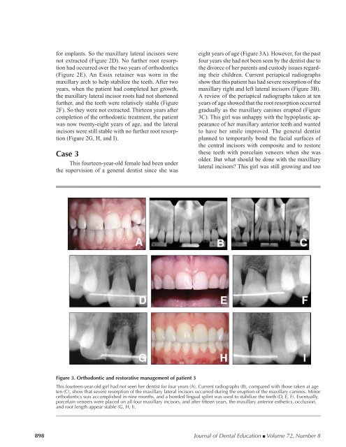
Figure 3. <strong>Orthodontic</strong> <strong>and</strong> restorative management <strong>of</strong> patient 3<br />
This fourteen-year-old girl had not seen her dentist for four years (A). Current radiographs (B), compared with those taken at age<br />
ten (C), show that severe resorption <strong>of</strong> the maxillary lateral incisors occurred during the eruption <strong>of</strong> the maxillary canines. Minor<br />
orthodontics was accomplished in nine months, <strong>and</strong> a bonded lingual splint was used to stabilize the teeth (D, E, F). Eventually,<br />
porcelain veneers were placed on all four maxillary incisors, <strong>and</strong> after fifteen years, the maxillary anterior esthetics, occlusion,<br />
<strong>and</strong> root length appear stable (G, H, I).<br />
898 <strong>Journal</strong> <strong>of</strong> <strong>Dental</strong> Education ■ Volume 72, Number 8