Orthodontic and Nonorthodontic Root Resorption - Journal of Dental ...
Orthodontic and Nonorthodontic Root Resorption - Journal of Dental ...
Orthodontic and Nonorthodontic Root Resorption - Journal of Dental ...
You also want an ePaper? Increase the reach of your titles
YUMPU automatically turns print PDFs into web optimized ePapers that Google loves.
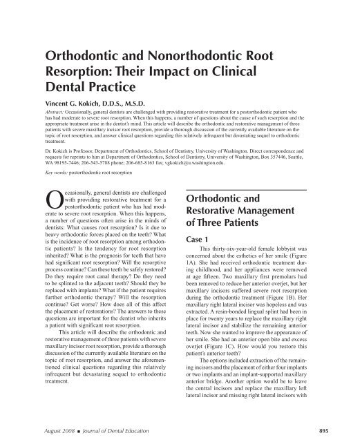
<strong>Orthodontic</strong> <strong>and</strong> <strong>Nonorthodontic</strong> <strong>Root</strong><br />
<strong>Resorption</strong>: Their Impact on Clinical<br />
<strong>Dental</strong> Practice<br />
Vincent G. Kokich, D.D.S., M.S.D.<br />
Abstract: Occasionally, general dentists are challenged with providing restorative treatment for a postorthodontic patient who<br />
has had moderate to severe root resorption. When this happens, a number <strong>of</strong> questions about the cause <strong>of</strong> such resorption <strong>and</strong> the<br />
appropriate treatment arise in the dentist’s mind. This article will describe the orthodontic <strong>and</strong> restorative management <strong>of</strong> three<br />
patients with severe maxillary incisor root resorption, provide a thorough discussion <strong>of</strong> the currently available literature on the<br />
topic <strong>of</strong> root resorption, <strong>and</strong> answer clinical questions regarding this relatively infrequent but devastating sequel to orthodontic<br />
treatment.<br />
Dr. Kokich is Pr<strong>of</strong>essor, Department <strong>of</strong> <strong>Orthodontic</strong>s, School <strong>of</strong> Dentistry, University <strong>of</strong> Washington. Direct correspondence <strong>and</strong><br />
requests for reprints to him at Department <strong>of</strong> <strong>Orthodontic</strong>s, School <strong>of</strong> Dentistry, University <strong>of</strong> Washington, Box 357446, Seattle,<br />
WA 98195-7446; 206-543-5788 phone; 206-685-8163 fax; vgkokich@u.washington.edu.<br />
Key words: postorthodontic root resorption<br />
Occasionally, general dentists are challenged<br />
with providing restorative treatment for a<br />
postorthodontic patient who has had moderate<br />
to severe root resorption. When this happens,<br />
a number <strong>of</strong> questions <strong>of</strong>ten arise in the minds <strong>of</strong><br />
dentists: What causes root resorption? Is it due to<br />
heavy orthodontic forces placed on the teeth? What<br />
is the incidence <strong>of</strong> root resorption among orthodontic<br />
patients? Is the tendency for root resorption<br />
inherited? What is the prognosis for teeth that have<br />
had significant root resorption? Will the resorptive<br />
process continue? Can these teeth be safely restored?<br />
Do they require root canal therapy? Do they need<br />
to be splinted to the adjacent teeth? Should they be<br />
replaced with implants? What if the patient requires<br />
further orthodontic therapy? Will the resorption<br />
continue? Get worse? How does all <strong>of</strong> this affect<br />
the placement <strong>of</strong> restorations? The answers to these<br />
questions are important for the dentist who inherits<br />
a patient with significant root resorption.<br />
This article will describe the orthodontic <strong>and</strong><br />
restorative management <strong>of</strong> three patients with severe<br />
maxillary incisor root resorption, provide a thorough<br />
discussion <strong>of</strong> the currently available literature on the<br />
topic <strong>of</strong> root resorption, <strong>and</strong> answer the aforementioned<br />
clinical questions regarding this relatively<br />
infrequent but devastating sequel to orthodontic<br />
treatment.<br />
<strong>Orthodontic</strong> <strong>and</strong><br />
Restorative Management<br />
<strong>of</strong> Three Patients<br />
Case 1<br />
This thirty-six-year-old female lobbyist was<br />
concerned about the esthetics <strong>of</strong> her smile (Figure<br />
1A). She had received orthodontic treatment during<br />
childhood, <strong>and</strong> her appliances were removed<br />
at age fifteen. Two maxillary first premolars had<br />
been removed to reduce her anterior overjet, but her<br />
maxillary incisors suffered severe root resorption<br />
during the orthodontic treatment (Figure 1B). Her<br />
maxillary right lateral incisor was hopeless <strong>and</strong> was<br />
extracted. A resin-bonded lingual splint had been in<br />
place for twenty years to replace the maxillary right<br />
lateral incisor <strong>and</strong> stabilize the remaining anterior<br />
teeth. Now she wanted to improve the appearance <strong>of</strong><br />
her smile. She had an anterior open bite <strong>and</strong> excess<br />
overjet (Figure 1C). How would you restore this<br />
patient’s anterior teeth?<br />
The options included extraction <strong>of</strong> the remaining<br />
incisors <strong>and</strong> the placement <strong>of</strong> either four implants<br />
or two implants <strong>and</strong> an implant-supported maxillary<br />
anterior bridge. Another option would be to leave<br />
the central incisors <strong>and</strong> replace the maxillary left<br />
lateral incisor <strong>and</strong> missing right lateral incisors with<br />
August 2008 ■ <strong>Journal</strong> <strong>of</strong> <strong>Dental</strong> Education 895
Figure 1. <strong>Orthodontic</strong> <strong>and</strong> restorative management <strong>of</strong> patient 1<br />
This thirty-six-year-old patient was unhappy with the appearance <strong>of</strong> her maxillary anterior teeth (A). She had had previous orthodontic<br />
treatment, including the extraction <strong>of</strong> two maxillary first premolars. Moderate to severe root resorption had occurred,<br />
so her anterior teeth were splinted with a cast lingual splint (B). However, her root length had not diminished in twenty years.<br />
So her malocclusion was corrected (D), <strong>and</strong> the roots did not get shorter during the orthodontic retreatment (E). A provisional<br />
bridge (F) was worn for one year. A six-unit porcelain-fused-to-metal bridge, shown five years after orthodontics (G, H, I), has<br />
successfully restored esthetics <strong>and</strong> function <strong>and</strong> stabilized the teeth.<br />
implants. A third option would be to place another<br />
resin-bonded splint. A fourth option would be to<br />
remove the maxillary left lateral incisor <strong>and</strong> place a<br />
six-unit conventional bridge attached to the maxillary<br />
canines <strong>and</strong> central incisors replacing the lateral incisors.<br />
However, all plans required further orthodontic<br />
therapy in order to reduce the overjet, align the m<strong>and</strong>ibular<br />
incisors, <strong>and</strong> close the open bite.<br />
After a thorough discussion among the periodontist,<br />
orthodontist, general dentist, <strong>and</strong> patient, it<br />
was decided that extraction <strong>of</strong> the left lateral incisor<br />
<strong>and</strong> restoration <strong>of</strong> the remaining teeth with a conventional<br />
fixed bridge would not only improve the<br />
esthetics, but would also stabilize the central incisors<br />
<strong>and</strong> canines with the short roots. Since the patient<br />
had not suffered further root resorption <strong>of</strong> the central<br />
incisors during the twenty years after the original<br />
orthodontics, it was believed that further resorption<br />
was not likely. <strong>Orthodontic</strong> appliances were placed<br />
on all teeth, the maxillary right central incisor was<br />
intruded to level the gingival margins between the<br />
central incisors, the overjet was corrected, <strong>and</strong> the<br />
open bite was closed (Figure 1D). No further root<br />
resorption occurred during the orthodontic therapy<br />
(Figure 1E). A maxillary anterior provisional bridge<br />
was worn for one year after the orthodontic treatment<br />
(Figure 1F), <strong>and</strong> then a porcelain-fused-to-metal<br />
bridge was placed on the maxillary canines <strong>and</strong><br />
central incisors <strong>and</strong> has been stable for five years<br />
(Figure 1G, H, <strong>and</strong> I).<br />
896 <strong>Journal</strong> <strong>of</strong> <strong>Dental</strong> Education ■ Volume 72, Number 8
Case 2<br />
This thirteen-year-old female had been under<br />
the care <strong>of</strong> a pediatric dentist since age six. Her<br />
mother decided to transfer her daughter to their<br />
family general dentist. She was in the transitional<br />
dentition <strong>and</strong> was erupting her maxillary canines<br />
(Figure 2A).<br />
The patient was congenitally missing her<br />
maxillary right <strong>and</strong> left second premolars. Current<br />
periapical radiographs showed that the maxillary<br />
right <strong>and</strong> left lateral incisors had severely resorbed<br />
roots (Figure 2B). This girl had never had previous<br />
orthodontic treatment. Periapical radiographs taken<br />
by the pediatric dentist <strong>of</strong> the girl at age eight years<br />
showed that no root resorption had occurred up until<br />
that time (Figure 2C). So, over the past four years,<br />
the pressure <strong>of</strong> the erupting maxillary canines had<br />
caused the resorption <strong>of</strong> the maxillary lateral incisor<br />
roots. 1 How would you eventually restore this<br />
patient? She obviously needed orthodontic therapy.<br />
Would implants be necessary to replace the lateral<br />
incisors? If so, when?<br />
Since the patient was already missing her<br />
maxillary right <strong>and</strong> left second premolars, <strong>and</strong> since<br />
she was too young for implants, it was decided that<br />
the maxillary lateral incisors should not be extracted<br />
until after the orthodontic treatment, so they could<br />
provide space for eventual implants. The orthodontic<br />
treatment lasted over two years. However, at the end<br />
<strong>of</strong> orthodontic therapy the patient was only fifteen<br />
years <strong>of</strong> age, still growing, <strong>and</strong> therefore too young<br />
Figure 2. <strong>Orthodontic</strong> <strong>and</strong> restorative management <strong>of</strong> patient 2<br />
This thirteen-year-old female had been under the care <strong>of</strong> a pediatric dentist since age six years. She was now transferring to<br />
the family dentist. Current periapical radiographs (B) reveal extensive resorption <strong>of</strong> the maxillary lateral incisor roots. Comparison<br />
with radiographs taken four years earlier (C) show that the root resorption was caused by the erupting maxillary canines.<br />
Because the patient was too young for implants, the lateral incisors were maintained during the orthodontics (D), <strong>and</strong> she had<br />
no further root resorption after bracket removal (E). The teeth were not mobile nor did they need restoration, so they were left for<br />
the next thirteen years. At twenty-eight years <strong>of</strong> age, the patient’s alignment, esthetics, occlusion, <strong>and</strong> root length appear stable<br />
(G, H, I).<br />
August 2008 ■ <strong>Journal</strong> <strong>of</strong> <strong>Dental</strong> Education 897
for implants. So the maxillary lateral incisors were<br />
not extracted (Figure 2D). No further root resorption<br />
had occurred over the two years <strong>of</strong> orthodontics<br />
(Figure 2E). An Essix retainer was worn in the<br />
maxillary arch to help stabilize the teeth. After two<br />
years, when the patient had completed her growth,<br />
the maxillary lateral incisor roots had not shortened<br />
further, <strong>and</strong> the teeth were relatively stable (Figure<br />
2F). So they were not extracted. Thirteen years after<br />
completion <strong>of</strong> the orthodontic treatment, the patient<br />
was now twenty-eight years <strong>of</strong> age, <strong>and</strong> the lateral<br />
incisors were still stable with no further root resorption<br />
(Figure 2G, H, <strong>and</strong> I).<br />
Case 3<br />
This fourteen-year-old female had been under<br />
the supervision <strong>of</strong> a general dentist since she was<br />
eight years <strong>of</strong> age (Figure 3A). However, for the past<br />
four years she had not been seen by the dentist due to<br />
the divorce <strong>of</strong> her parents <strong>and</strong> custody issues regarding<br />
their children. Current periapical radiographs<br />
show that this patient has had severe resorption <strong>of</strong> the<br />
maxillary right <strong>and</strong> left lateral incisors (Figure 3B).<br />
A review <strong>of</strong> the periapical radiographs taken at ten<br />
years <strong>of</strong> age showed that the root resorption occurred<br />
gradually as the maxillary canines erupted (Figure<br />
3C). This girl was unhappy with the hypoplastic appearance<br />
<strong>of</strong> her maxillary anterior teeth <strong>and</strong> wanted<br />
to have her smile improved. The general dentist<br />
planned to temporarily bond the facial surfaces <strong>of</strong><br />
the central incisors with composite <strong>and</strong> to restore<br />
these teeth with porcelain veneers when she was<br />
older. But what should be done with the maxillary<br />
lateral incisors? This girl was still growing <strong>and</strong> too<br />
Figure 3. <strong>Orthodontic</strong> <strong>and</strong> restorative management <strong>of</strong> patient 3<br />
This fourteen-year-old girl had not seen her dentist for four years (A). Current radiographs (B), compared with those taken at age<br />
ten (C), show that severe resorption <strong>of</strong> the maxillary lateral incisors occurred during the eruption <strong>of</strong> the maxillary canines. Minor<br />
orthodontics was accomplished in nine months, <strong>and</strong> a bonded lingual splint was used to stabilize the teeth (D, E, F). Eventually,<br />
porcelain veneers were placed on all four maxillary incisors, <strong>and</strong> after fifteen years, the maxillary anterior esthetics, occlusion,<br />
<strong>and</strong> root length appear stable (G, H, I).<br />
898 <strong>Journal</strong> <strong>of</strong> <strong>Dental</strong> Education ■ Volume 72, Number 8
young for implants. She needed some minor corrective<br />
orthodontic treatment.<br />
After consultation among the general dentist,<br />
the orthodontist, <strong>and</strong> the patient <strong>and</strong> her mother, it<br />
was decided that the lateral incisors not be extracted<br />
until after orthodontics. The orthodontic treatment<br />
lasted for nine months, <strong>and</strong> the roots <strong>of</strong> the lateral incisors<br />
did not get any shorter during that time (Figure<br />
3D <strong>and</strong> F). The incisors were bonded with composite<br />
(Figure 3E), <strong>and</strong> a wire splint was bonded on the<br />
lingual to stabilize the teeth until the patient was old<br />
enough to have implants. At seventeen years <strong>of</strong> age,<br />
the patient had stopped growing <strong>and</strong> was ready for<br />
implants. However, the gingival tissue <strong>and</strong> papillae<br />
around the lateral incisors were healthy, at normal<br />
levels, <strong>and</strong> the lateral incisors were immobile with<br />
the lingual splint. So it was decided that the resorbed<br />
teeth, instead <strong>of</strong> being extracted, would be restored<br />
(along with the central incisors) with porcelain<br />
veneers <strong>and</strong> stabilized with a bonded lingual wire.<br />
Fifteen years after orthodontics, the lateral incisors<br />
were still present, the gingival <strong>and</strong> restorative esthetics<br />
were good, <strong>and</strong> the roots had not resorbed any<br />
further (Figure 3G, H, <strong>and</strong> I).<br />
Discussion<br />
This article has demonstrated the long-term<br />
treatment <strong>and</strong> management <strong>of</strong> three patients with<br />
severe root resorption. In one case, the root resorption<br />
was most likely caused by the orthodontic movement.<br />
In the other two cases, eruption <strong>of</strong> the maxillary<br />
canines caused the resorption <strong>of</strong> the lateral incisor<br />
roots. 1 However, the resorbed teeth were maintained.<br />
In two cases they were restored, <strong>and</strong> in all three cases<br />
they have remained functional <strong>and</strong> esthetically natural<br />
in appearance many years after completion <strong>of</strong> the<br />
orthodontics <strong>and</strong> restorative dentistry.<br />
When a general dentist inherits a patient with<br />
moderate to severe orthodontic or nonorthodontic<br />
root resorption, the shortened roots on the periapical<br />
radiograph make the outlook for these teeth seem<br />
hopeless. However, as one can see from these three<br />
cases, the outcome is far from hopeless. It is therefore<br />
important that general dentists underst<strong>and</strong> the<br />
incidence, cause, <strong>and</strong> outcome <strong>of</strong> root resorption<br />
in order to provide the best follow-up treatment<br />
for their patients who experience this devastating<br />
problem.<br />
The first question to address is the incidence <strong>of</strong><br />
root resorption in orthodontic patients. Several clinical<br />
studies have compared pre- <strong>and</strong> post-treatment<br />
periapical radiographs to determine the incidence<br />
<strong>of</strong> root resorption after orthodontic treatment. 2-6<br />
However, radiographs only provide a crude twodimensional<br />
assessment <strong>of</strong> root resorption <strong>and</strong> will<br />
usually underestimate the true amount <strong>of</strong> root resorption.<br />
Therefore, the only accurate assessment <strong>of</strong> root<br />
resorption must come from a histologic assessment <strong>of</strong><br />
the root surface after orthodontic movement. These<br />
studies have been accomplished in both animals 7 <strong>and</strong><br />
humans, 8-10 <strong>and</strong> they clearly show that root resorption<br />
occurs in over 90 percent <strong>of</strong> the cases when a<br />
tooth root is compressed against the alveolar socket.<br />
Therefore, root resorption is a common sequel <strong>of</strong><br />
orthodontic movement.<br />
Why does root resorption occur in response<br />
to compression <strong>of</strong> the periodontal ligament? This<br />
phenomenon is not completely understood, but recent<br />
studies have found that the presence or absence<br />
<strong>of</strong> hyaline in the periodontal ligament affects the<br />
incidence <strong>of</strong> root resorption. 11 Hyalinization is a<br />
common sequel after a compressive load is placed on<br />
the periodontal ligament. Hyaline has been termed<br />
sterile necrosis 10 <strong>and</strong> forms in the interstitial space<br />
within the periodontal ligament after a compressive<br />
load is placed on a tooth root. Hyalinization <strong>of</strong> the<br />
periodontal ligament usually occurs after a few days,<br />
<strong>and</strong> the hyaline may remain within the periodontal<br />
ligament up to four to eight weeks after initiation <strong>of</strong><br />
the compressive load. 12,13 During this time, resorption<br />
<strong>of</strong> the alveolar socket is virtually prevented, <strong>and</strong><br />
undermining resorption <strong>of</strong> the alveolar bone may<br />
occur. 11 In addition, root resorption near the areas <strong>of</strong><br />
hyalinization will occur. After about eight weeks in<br />
experimental animals, the hyaline has been removed<br />
from the periodontal ligament by macrophages, <strong>and</strong><br />
at this time resorption <strong>of</strong> the alveolar socket wall<br />
occurs that permits the tooth to move. 11 However, by<br />
this time, extensive resorption lacunae are typically<br />
found along the length <strong>of</strong> the root surface.<br />
Why don’t we see more radiographic evidence<br />
<strong>of</strong> root resorption in larger numbers <strong>of</strong> orthodontic<br />
patients? Actually, these resorption lacunae will repair<br />
themselves after the hyaline has been removed<br />
from the periodontal ligament <strong>and</strong> cementoblasts<br />
begin to secrete cellular cementum. 11,14-17 Since most<br />
<strong>of</strong> these resorption lacunae repair themselves with<br />
time, there is little or no radiographic evidence <strong>of</strong> root<br />
resorption in the majority <strong>of</strong> orthodontic patients.<br />
What if the cementoblasts do not repair the resorption<br />
lacunae? Then the patient may suffer moderate<br />
to severe root resorption.<br />
August 2008 ■ <strong>Journal</strong> <strong>of</strong> <strong>Dental</strong> Education 899
What is the incidence <strong>of</strong> moderate to severe<br />
root resorption after orthodontic treatment? Several<br />
radiographic assessments <strong>of</strong> consecutively<br />
treated populations <strong>of</strong> patients have been made to<br />
determine the prevalence <strong>of</strong> moderate to severe root<br />
resorption in both adolescent <strong>and</strong> adult populations<br />
<strong>of</strong> orthodontic patients. Moderate to severe root<br />
resorption is typically described as a greater than 20<br />
percent reduction in the original root length. Using<br />
this definition, the incidence <strong>of</strong> moderate to severe<br />
root resorption in an adolescent sample 2,5 is about 3<br />
percent. In adults, 18 researchers have shown that the<br />
incidence <strong>of</strong> moderate to severe root resorption is<br />
near 4 percent.<br />
Can root resorption be predicted? Previous<br />
studies have used statistical comparisons <strong>of</strong> gender,<br />
pre-treatment root form, pre-treatment root length,<br />
length <strong>of</strong> orthodontic treatment, premolar extraction,<br />
<strong>and</strong> linear amount <strong>of</strong> root movement as independent<br />
variables to determine if there are any accurate<br />
predictors <strong>of</strong> root resorption related to orthodontic<br />
treatment. In general, three <strong>of</strong> these variables show<br />
an association: amount <strong>of</strong> linear root movement,<br />
length <strong>of</strong> orthodontic treatment, <strong>and</strong> premolar extraction.<br />
5,6,19-22 If a patient were susceptible to significant<br />
root resorption, then the farther the tooth is moved<br />
<strong>and</strong> the longer the duration <strong>of</strong> the orthodontic treatment,<br />
the more root resorption would likely occur.<br />
In addition, epidemiological studies show that premolar<br />
extraction cases tend to demonstrate more root<br />
resorption in susceptible patients, probably because<br />
<strong>of</strong> the increased distance that the teeth move in extraction<br />
cases.<br />
Does the amount <strong>of</strong> force used during orthodontics<br />
affect the amount <strong>of</strong> root resorption? It<br />
seems logical that, in a susceptible sample, greater<br />
orthodontic forces would cause more root resorption.<br />
However, this assumption is not valid. Studies in both<br />
animals <strong>and</strong> humans have shown that the amount <strong>of</strong><br />
force placed on a tooth root has neither a positive nor<br />
negative effect on the amount <strong>of</strong> root resorption. 8,23<br />
On the other h<strong>and</strong>, studies in humans have shown<br />
that quadrupling the force on a tooth root does not<br />
produce greater root resorption, but can increase the<br />
speed <strong>of</strong> root movement through the bone. 9 Does it<br />
make a difference if the orthodontic force is continuous<br />
or intermittent? Researchers have clearly shown<br />
that although considerable variation typically exists,<br />
continuous forces tend to produce more extensive<br />
root resorption than intermittent forces. 13,14,24<br />
Is the tendency or susceptibility for root resorption<br />
an inherited trait? In the past, this question has<br />
been controversial. However, recent studies have<br />
suggested that external apical root resorption can<br />
be traced to a specific locus on a specific gene. 23,25<br />
These researchers believe that external apical root<br />
resorption is a complex condition influenced by<br />
many factors, with the IL-1B gene contributing an<br />
important predisposition to this common problem.<br />
Personally, I have treated two families <strong>of</strong> parent <strong>and</strong><br />
child, where both experienced moderate to severe root<br />
resorption during orthodontic treatment. It is evident<br />
that more studies evaluating a genetic determination<br />
<strong>of</strong> root resorption susceptibility are needed.<br />
Do specific types <strong>of</strong> orthodontic movement<br />
lead to greater root resorption in susceptible patients?<br />
Several authors have pointed out the negative impact<br />
<strong>of</strong> tooth intrusion on the severity <strong>of</strong> root resorption<br />
in orthodontic patients. 26,27 Perhaps this observation<br />
is due to the method <strong>of</strong> analyzing root resorption<br />
or root shortening on two-dimensional periapical<br />
radiographs. <strong>Resorption</strong> <strong>of</strong> the root apex after tooth<br />
intrusion can be seen easily on two-dimensional<br />
radiographs, whereas the root resorption seen on<br />
periapical radiographs after lateral root movement<br />
is not as clearly visible.<br />
What is the effect <strong>of</strong> root resorption on tooth<br />
vitality? Although no studies have analyzed this<br />
relationship, from a clinical perspective I have not<br />
encountered a tooth with moderate to severe root resorption<br />
whose pulp became nonvital. Unless there is<br />
some bacterial or traumatic insult to the tooth, pulp vitality<br />
does not seem to be related to the amount <strong>of</strong> root<br />
resorption experienced during orthodontic therapy.<br />
None <strong>of</strong> the pulps <strong>of</strong> the resorbed roots <strong>of</strong> the three<br />
patients illustrated in this article were nonvital.<br />
What happens over the long term to tooth roots<br />
that have undergone moderate to severe root resorption?<br />
Researchers have reevaluated patients with<br />
moderate to severe root resorption many years after<br />
orthodontics 28-30 <strong>and</strong> have found that root resorption<br />
stops after orthodontic treatment has been discontinued.<br />
Although there may be some remodeling <strong>of</strong> the<br />
irregular resorbed edges <strong>of</strong> the root with time due to<br />
reparative deposition <strong>of</strong> cellular cementum, this type<br />
<strong>of</strong> remodeling merely produces a smoother surface<br />
long term. However the length <strong>of</strong> the root does not<br />
continue to shorten after orthodontic appliances have<br />
been removed.<br />
Do teeth with moderate to severe root resorption<br />
require splinting? There are no studies that provide<br />
us with the answer to this important clinical question.<br />
In two <strong>of</strong> the cases presented in this article, the<br />
teeth were splinted either with a conventional bridge<br />
900 <strong>Journal</strong> <strong>of</strong> <strong>Dental</strong> Education ■ Volume 72, Number 8
or a bonded lingual wire. But these teeth required<br />
restoration. In the third patient, no permanent retention<br />
other than a removal retainer was used for the<br />
patient. After thirteen years, the patient without a fixed<br />
retainer has retained her severely resorbed maxillary<br />
lateral incisors. However, this patient has a two-millimeter<br />
overjet in centric occlusion <strong>and</strong> does not have<br />
any parafunctional or destructive occlusal habits. I<br />
believe that parafunctional habits, crown mobility,<br />
<strong>and</strong> the need for restoration tend to determine the<br />
necessity for a permanent lingual splint. If a patient<br />
has a protrusive bruxing habit with mobile maxillary<br />
incisors <strong>and</strong> will require some sort <strong>of</strong> restoration <strong>of</strong><br />
these teeth, then perhaps splinting will help to avoid<br />
the negative effects <strong>of</strong> each <strong>of</strong> these parameters.<br />
If the patient requires further orthodontic<br />
treatment, will the roots continue to resorb? This<br />
research question has not been explored. However,<br />
most orthodontists have had to retreat patients who<br />
have had root resorption during an earlier phase <strong>of</strong><br />
orthodontic treatment. The three patients described<br />
in this article all had orthodontic treatment ranging<br />
in length from nine months to over two years, <strong>and</strong> the<br />
tooth movement was started after the root resorption<br />
had occurred. None <strong>of</strong> these patients exhibited any<br />
further root resorption as a result <strong>of</strong> the orthodontic<br />
retreatment. However, in these cases I tried to limit<br />
the amount <strong>of</strong> tooth movement, limit the length <strong>of</strong><br />
orthodontic treatment, <strong>and</strong> avoid intrusive tooth<br />
movements. Also, histologic follow-up after root<br />
resorption showed that reparative dentin <strong>and</strong> cellular<br />
cementum form after the tooth movement had ceased.<br />
Perhaps the presence <strong>of</strong> a cellular cemental layer<br />
plays some role in protecting the tooth root during<br />
orthodontic retreatment.<br />
Summary <strong>and</strong> Conclusions<br />
This article has described the treatment <strong>of</strong> three<br />
orthodontic patients who had experienced severe<br />
root resorption but required further orthodontics. All<br />
three patients had successful long-term outcomes by<br />
maintaining the severely resorbed roots. In addition<br />
to illustrating the logic <strong>of</strong> the treatment plans as well<br />
as the long-term effect, I have tried to answer the<br />
key questions that arise in the mind <strong>of</strong> the general<br />
dentist when he or she inherits a patient with severe<br />
postorthodontic or nonorthodontic root resorption.<br />
Hopefully, this information will be clinically useful<br />
for the general dentist <strong>and</strong> orthodontist when planning<br />
treatment for these difficult situations.<br />
REFERENCES<br />
1. Heimisdottir K, Bosshardt D, Ruf S. Can the severity<br />
<strong>of</strong> root resorption be accurately judged by means <strong>of</strong><br />
radiographs? A case report with histology. Am J Orthod<br />
Dent<strong>of</strong>acial Orthop 2005;128:106–9.<br />
2. Sameshima GT, Sinclair PM. Characteristics <strong>of</strong> patients<br />
with severe root resorption. Orthod Crani<strong>of</strong>acial Res<br />
2004;7:108–14.<br />
3. Smale I, Artun J, Behbehani F, Doppel D, van’t H<strong>of</strong> M,<br />
Kuijpers-Jagtman AM. Apical root resorption 6 months<br />
after initiation <strong>of</strong> fixed orthodontic appliance therapy. Am<br />
J Orthod Dent<strong>of</strong>acial Orthop 2005;128:57–67.<br />
4. Artun J, Smale I, Behbehani F, Doppel D, van’t H<strong>of</strong> M,<br />
Kuijpers-Jagtman AM. Apical root resorption six <strong>and</strong><br />
twelve months after initiation <strong>of</strong> fixed orthodontic appliance<br />
therapy. Angle Orthod 2005;75:919–26.<br />
5. Nigul K, Jagomagi T. Factors related to apical root resorption<br />
<strong>of</strong> maxillary incisors in orthodontic patients.<br />
Stomatologiia 2006;8:76–9.<br />
6. Moh<strong>and</strong>esan H, Ravanmehr H, Valaei N. A radiographic<br />
analysis <strong>of</strong> external apical root resorption <strong>of</strong> maxillary<br />
incisors during active orthodontic treatment. Eur J Orthod<br />
2007;29:134–9.<br />
7. Maltha JC, van Leeuwen EJ, Dijkman GE, Kuijpers-<br />
Jagtman AM. Incidence <strong>and</strong> severity <strong>of</strong> root resorption<br />
in orthodontically moved premolars in dogs. Orthod<br />
Crani<strong>of</strong>ac Res 2004;7:115–21.<br />
8. Owman-Moll P, Kurol J, Lundgren D. Effects <strong>of</strong> a doubled<br />
orthodontic force magnitude on tooth movement <strong>and</strong> root<br />
resorption: an interindividual study in adolescents. Eur J<br />
Orthod 1996;18:141–50.<br />
9. Owman-Moll P, Kurol J, Lundgren D. The effects <strong>of</strong> a<br />
four-fold increased orthodontic force magnitude on tooth<br />
movement <strong>and</strong> root resorption: an intra-individual study<br />
in adolescents. Eur J Orthod 1996;18:287–94.<br />
10. Kurol J, Owman-Moll P. Hyalinization <strong>and</strong> root resorption<br />
during early orthodontic tooth movement in adolescents.<br />
Angle Orthod 1998;68:161–5.<br />
11. Iino S, Sakoda S, Ito G, Nishimori T, Ikeda T, Miyawaki<br />
S. Acceleration <strong>of</strong> orthodontic tooth movement by alveolar<br />
corticotomy in the dog. Am J Orthod Dent<strong>of</strong>acial Orthop<br />
2007;131:448e1–448e8.<br />
12. Von Bohl M, Maltha JC, Von Den H<strong>of</strong>f JW, Kuijpers-<br />
Jagtman AM. Focal hyalinization during experimental<br />
tooth movement in beagle dogs. Am J Orthod Dent<strong>of</strong>acial<br />
Orthop 2004;125:615–23.<br />
13. Von Bohl M, Maltha J, Von Den H<strong>of</strong>f H, Kuijpers-Jagtman<br />
AM. Changes in the periodontal ligament after experimental<br />
tooth movement using high <strong>and</strong> low continuous forces<br />
in beagle dogs. Angle Orthod 2004;74:16–25.<br />
14. Owman-Moll P, Kurol J, Lundgren D. Continuous versus<br />
interrupted continuous orthodontic force related to<br />
early tooth movement <strong>and</strong> root resorption. Angle Orthod<br />
1995;65:395–401.<br />
15. Owman-Moll P. <strong>Orthodontic</strong> tooth movement <strong>and</strong> root<br />
resorption with special reference to force magnitude<br />
<strong>and</strong> duration: a clinical <strong>and</strong> histological investigation in<br />
adolescents. Swed Dent J Suppl 1995;105:1–45.<br />
16. Owman-Moll P, Kurol J. The early reparative process <strong>of</strong><br />
orthodontically induced root resorption in adolescents: location<br />
<strong>and</strong> type <strong>of</strong> tissue. Eur J Orthod 1998;20:727–32.<br />
August 2008 ■ <strong>Journal</strong> <strong>of</strong> <strong>Dental</strong> Education 901
17. Jimenez-Pellegrin C, Arana-Chavez VE. <strong>Root</strong> resorption<br />
repair in m<strong>and</strong>ibular first premolars after rotation: a<br />
transmission electron microscopy analysis combined with<br />
immunolabeling <strong>of</strong> osteopontin. Am J Orthod Dent<strong>of</strong>acial<br />
Orthop 2007;132:230–6.<br />
18. Mirabella D, Artun J. Prevalence <strong>and</strong> severity <strong>of</strong> apical root<br />
resorption <strong>of</strong> maxillary anterior teeth in adult orthodontic<br />
patients. Eur J Orthod 1995;17:93–9.<br />
19. Mirabella D, Artun J. Risk factors for apical root resorption<br />
<strong>of</strong> maxillary anterior teeth in adult orthodontic patients.<br />
Am J Orthod Dent<strong>of</strong>acial Orthop 1995;108:48–55.<br />
20. Sameshima GT, Sinclair PM. Predicting <strong>and</strong> preventing<br />
root resorption: Part II. Treatment factors. Am J Orthod<br />
Dent<strong>of</strong>acial Orthop 2001;119:511–5.<br />
21. Fox N. Longer orthodontic treatment may result in<br />
greater external apical root resorption. Evid Based Dent<br />
2005;6:21.<br />
22. Segal GR, Schiffman PH, Tuncay OC. Meta analysisrelated<br />
factors <strong>of</strong> external apical root resorption. Orthod<br />
Crani<strong>of</strong>ac Res 2004;7:71–8.<br />
23. Al-Qawasmi RA, Hartsfield JK Jr, Everett ET, Flury L,<br />
Lui L, Foroud TM, et al. Genetic predisposition to external<br />
apical root resorption. Am J Orthod Dent<strong>of</strong>acial Orthop<br />
2003;123:242–52.<br />
24. Owman-Moll P, Kurol J, Lundgren D. Repair <strong>of</strong> orthodontically<br />
induced root resorption in adolescents. Angle<br />
Orthod 1995;65:403–8.<br />
25. Al-Qawasmi RA, Hartsfield JK Jr, Everett ET, Flury L,<br />
Lui L, Foroud TM, et al. Genetic predisposition to external<br />
apical root resorption in orthodontic patients: linkage <strong>of</strong><br />
chromosome-18 marker. J Dent Res 2003;82:356–60.<br />
26. Han G, Huang S, Von den H<strong>of</strong>f JW, Zeng X, Kuijpers-<br />
Jagtman AM. <strong>Root</strong> resorption after orthodontic intrusion<br />
<strong>and</strong> extrusion: an intra-individual study. Angle Orthod<br />
2005;75:912–8.<br />
27. Harris DA, Jones AS, Darendeliler MA. Physical properties<br />
<strong>of</strong> root cementum: Part 8. Volumetric analysis <strong>of</strong> root<br />
resorption craters after application <strong>of</strong> controlled intrusive<br />
light <strong>and</strong> heavy orthodontic forces: a microcomputed<br />
tomography scan study. Am J Orthod Dent<strong>of</strong>acial Orthop<br />
2006;130:639–47.<br />
28. Remington DN, Joondeph DR, Artun J, Riedel RA,<br />
Chapko MK. Long-term evaluation <strong>of</strong> root resorption<br />
occurring during orthodontic treatment. Am J Orthod<br />
Dent<strong>of</strong>acial Orthop 1989;96:43–6.<br />
29. Lev<strong>and</strong>er E, Malmgren O. Long-term follow-up <strong>of</strong> maxillary<br />
incisors with severe apical root resorption. Eur J<br />
Orthod 2000;22:85–92.<br />
30. Becker A, Chaushu S. Long-term follow-up <strong>of</strong> severely<br />
resorbed maxillary incisors after resolution <strong>of</strong> an etiologically<br />
associated impacted canine. Am J Orthod Dent<strong>of</strong>acial<br />
Orthop 2005;127:650–4.<br />
902 <strong>Journal</strong> <strong>of</strong> <strong>Dental</strong> Education ■ Volume 72, Number 8


