Association of Merkel Cell Polyomavirus â Specific Antibodies With ...
Association of Merkel Cell Polyomavirus â Specific Antibodies With ...
Association of Merkel Cell Polyomavirus â Specific Antibodies With ...

You also want an ePaper? Increase the reach of your titles
YUMPU automatically turns print PDFs into web optimized ePapers that Google loves.
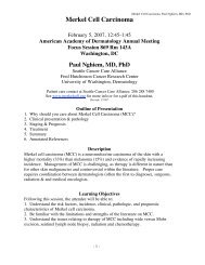
MFI<br />
35000<br />
30000<br />
25000<br />
20000<br />
15000<br />
10000<br />
5000<br />
0<br />
–5000<br />
BKV JCV WUPyV KIPyV MCPyV<br />
w162<br />
16L1<br />
Figure 6 . Distribution <strong>of</strong> seroreactivity against the major capsid proteins<br />
(VP1) <strong>of</strong> all five human polyomavirus (BK virus [BKV], JC virus [JCV],<br />
WU polyomavirus [WUPyV], KI polyomavirus [KIPyV], and <strong>Merkel</strong> cell<br />
carcinoma polyomavirus virus [MCPyV]) and human papillomavirus<br />
(HPV) 16 L1 protein among 451 women in population-based control<br />
group 2. The multiplex antibody-binding assay was used to assess<br />
seroreactivity in 451 samples previously tested for HPV-16 L1 reactivity<br />
( 18 ). The background median fluorescent intensity (MFI) values were<br />
subtracted from MFI values for each VP1 and for HPV-16 L1 and data<br />
were plotted. Dotted lines indicate the cut points <strong>of</strong> 15 000 for polyomaviruses<br />
and <strong>of</strong> 820.25 for the HPV-16 L1. The HPV-16 L1 cut point was<br />
the highest quartile <strong>of</strong> values.<br />
correlate with high levels <strong>of</strong> antibodies against VP1s from other<br />
polyomaviruses. 4) Antibody positivity to MCPyV but not the other<br />
polyomaviruses was strongly associated with <strong>Merkel</strong> cell<br />
carcinoma.<br />
The higher antibody prevalence among case patients with<br />
<strong>Merkel</strong> cell carcinoma is consistent with the hypothesis that<br />
MCPyV plays a role in the development <strong>of</strong> <strong>Merkel</strong> cell carcinoma.<br />
The lack <strong>of</strong> association between <strong>Merkel</strong> cell carcinoma<br />
and antibody positivity to the other polyomaviruses indicates that<br />
these patients are not globally susceptible to polyomavirus infection<br />
or reactivation. Although immunosuppression has been associated<br />
with <strong>Merkel</strong> cell carcinoma ( 2 ) and immunosuppression<br />
<strong>of</strong>ten leads to reactivation <strong>of</strong> polyomaviruses, higher levels <strong>of</strong><br />
antibody positivity against other polyomaviruses were not<br />
observed.<br />
Studies ( 3 , 24 , 27 , 28 ) that have tested for the MCPyV genome<br />
in <strong>Merkel</strong> cell carcinoma have found that between 15% and 30%<br />
<strong>of</strong> the tumors had no detectable viral DNA. Indeed, in this study,<br />
we detected MCPyV DNA in 24 (77%) <strong>of</strong> 31 case patients.<br />
Intriguingly, all seven case patients with <strong>Merkel</strong> cell carcinoma<br />
whose tumors were negative for MCPyV DNA had plasma that<br />
was positive for MCPyV VP1 antibodies. This result raises the<br />
possibility that MCPyV plays a role in cancer initiation but is not<br />
required for maintenance <strong>of</strong> the tumorigenic phenotype in all<br />
case patients and may be one explanation as to why some <strong>Merkel</strong><br />
cell carcinomas are negative for MCPyV DNA. Alternatively,<br />
because MCPyV infections are common, there may be MCPyVindependent<br />
pathways that lead to <strong>Merkel</strong> cell carcinoma.<br />
MCPyV has been shown to be integrated into the DNA <strong>of</strong><br />
some <strong>Merkel</strong> cell carcinoma tumors ( 3 ), analogous to the manner<br />
in which high-risk HPV DNA is integrated into the DNA <strong>of</strong><br />
cervical cancers ( 29 ). However, because some <strong>Merkel</strong> cell carcinomas<br />
do not appear to contain MCPyV DNA and other <strong>Merkel</strong><br />
cell carcinoma tumors contain much less than one viral genome<br />
per cell, the role that this virus plays in cancer development<br />
might be quite different from that <strong>of</strong> HPV in the development<br />
<strong>of</strong> cervical cancer ( 30 ). Another apparent difference in the development<br />
<strong>of</strong> these cancers is the longer time interval between<br />
initial exposure to MCPyV (before age 30 years) and development<br />
<strong>of</strong> <strong>Merkel</strong> cell carcinoma (median age = 70 years). For<br />
cervical cancer, the interval is approximately 30 years (eg, from<br />
HPV infection at age 20 years to diagnosis <strong>of</strong> cervical cancer at<br />
age 50 years) ( 31 ). The long time interval for the development<br />
<strong>of</strong> <strong>Merkel</strong> cell carcinoma suggests that rare genetic event(s)<br />
might be occurring in addition to the common event <strong>of</strong> MCPyV<br />
infection ( 32 ).<br />
We have previously shown that a higher proportion <strong>of</strong><br />
patients with <strong>Merkel</strong> cell carcinoma from North America than<br />
from Australia were positive for MCPyV DNA ( 24 ). This difference<br />
may be due to differences in sun exposure, the presence <strong>of</strong><br />
an undetectable MCPyV strain, or heterogeneous distributions<br />
<strong>of</strong> MCPyV worldwide. Future studies should examine the prevalence<br />
<strong>of</strong> MCPyV antibodies in different geographic populations.<br />
Furthermore, if the findings in our study are similar to<br />
those <strong>of</strong> other case – control studies, a prospective study with<br />
samples collected before diagnosis <strong>of</strong> <strong>Merkel</strong> cell carcinoma<br />
would be an important step to establishing causality. In this<br />
study, samples from case patients were collected after diagnosis<br />
<strong>of</strong> <strong>Merkel</strong> cell carcinoma, so the disease process may have had<br />
an impact on the antibody level. However, this study was<br />
matched on sex and age, which reduces possible confounding by<br />
these factors.<br />
In comparison to the results from MCPyV strain w162, only<br />
two <strong>of</strong> 41 case patients had detectable levels <strong>of</strong> antibodies against<br />
MCPyV strain 350. By mutating each <strong>of</strong> three discordant residues<br />
in the VP1 sequence for MCPyV strain 350 (residues 288,<br />
316, and 366) to those <strong>of</strong> strain w162, alone or in combination,<br />
we found that both residues 288 and 316 were required to confer<br />
MCPyV w162 – like binding by human serum to the strain 350<br />
VP1 sequence. It is likely that these two mutations altered the<br />
folding <strong>of</strong> VP1 from MCPyV 350 to ablate the formation <strong>of</strong><br />
conformation-dependent epitopes recognized by human serum,<br />
but it was not possible to determine whether MCPyV-specific<br />
antibodies recognize only native VP1 protein because denatured<br />
GST – VP1 fusion proteins did not bind to the polystyrene beads<br />
(results not shown). Sequences <strong>of</strong> the open reading frames <strong>of</strong><br />
MCPyV VP1 from seven additional tumors were all identical<br />
with VP1 proteins from MCPyV strains 339 and w162 at positions<br />
288, 316, and 366 (position 185 was either glutamine or<br />
glutamic acid). This finding is reminiscent <strong>of</strong> the first cloned<br />
HPV-16 L1 sequence from a cervical cancer, which had a nonconservative<br />
mutation that inhibited capsid assembly and binding<br />
by antibodies against L1 that were specific to conformationdependent<br />
epitopes ( 33 ).<br />
This is the first study, to our knowledge, to test for antibodies<br />
against all five polyomaviruses simultaneously. The prevalence <strong>of</strong><br />
antibodies against BKV and JCV, which we reported, is consistent<br />
with previous observations ( 10 , 13 , 34 ). A previous study ( 34 )<br />
reported that the prevalence <strong>of</strong> BKV decreased with increasing<br />
age and that JCV antibody prevalence peaked at approximately<br />
age 60 years. Approximately 90% <strong>of</strong> people in our control groups<br />
were positive for antibody against WUPyV and/or KIPyV, the<br />
two polyomaviruses that were recently discovered in respiratory<br />
infections ( 8 , 9 ). The prevalence <strong>of</strong> antibodies against these<br />
jnci.oxfordjournals.org JNCI | Article 11