Association of Merkel Cell Polyomavirus â Specific Antibodies With ...
Association of Merkel Cell Polyomavirus â Specific Antibodies With ...
Association of Merkel Cell Polyomavirus â Specific Antibodies With ...

You also want an ePaper? Increase the reach of your titles
YUMPU automatically turns print PDFs into web optimized ePapers that Google loves.
MFI<br />
35000<br />
30000<br />
25000<br />
20000<br />
15000<br />
10000<br />
5000<br />
0<br />
10 2 10 3 10 4 10 5 10 6<br />
Dilution <strong>of</strong> serum<br />
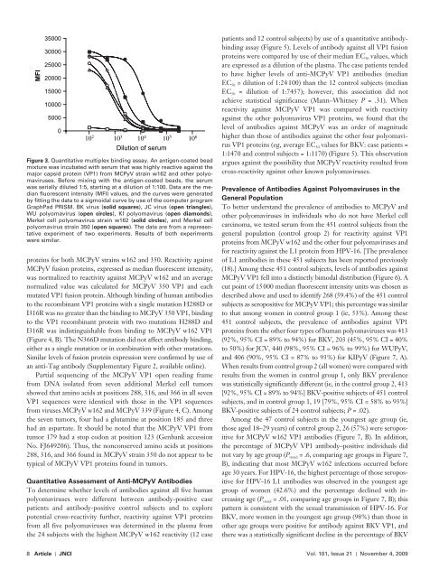
Figure 3 . Quantitative multiplex binding assay. An antigen-coated bead<br />
mixture was incubated with serum that was highly reactive against the<br />
major capsid protein (VP1) from MCPyV strain w162 and other polyomaviruses.<br />
Before mixing with the antigen-coated beads, the serum<br />
was serially diluted 1:5, starting at a dilution <strong>of</strong> 1:100. Data are the median<br />
fluorescent intensity (MFI) values, and the curves were generated<br />
by fitting the data to a sigmoidal curve by use <strong>of</strong> the computer program<br />
GraphPad PRISM. BK virus ( solid squares ), JC virus ( open triangles ),<br />
WU polyomavirus ( open circles ), KI polyomavirus ( open diamonds ),<br />
<strong>Merkel</strong> cell polyomavirus strain w162 ( solid circles ), and <strong>Merkel</strong> cell<br />
polyomavirus strain 350 ( open squares ). The data are from a representative<br />
experiment <strong>of</strong> two experiments. Results <strong>of</strong> both experiments<br />
were similar.<br />
proteins for both MCPyV strains w162 and 350. Reactivity against<br />
MCPyV fusion proteins, expressed as median fluorescent intensity,<br />
was normalized to reactivity against MCPyV w162 and an average<br />
normalized value was calculated for MCPyV 350 VP1 and each<br />
mutated VP1 fusion protein. Although binding <strong>of</strong> human antibodies<br />
to the recombinant VP1 proteins with a single mutation H288D or<br />
I316R was no greater than the binding to MCPyV 350 VP1, binding<br />
to the VP1 recombinant protein with two mutations H288D and<br />
I316R was indistinguishable from binding to MCPyV w162 VP1<br />
( Figure 4, B ). The N366D mutation did not affect antibody binding,<br />
either as a single mutation or in combination with other mutations.<br />
Similar levels <strong>of</strong> fusion protein expression were confirmed by use <strong>of</strong><br />
an anti-Tag antibody ( Supplementary Figure 2 , available online).<br />
Partial sequencing <strong>of</strong> the MCPyV VP1 open reading frame<br />
from DNA isolated from seven additional <strong>Merkel</strong> cell tumors<br />
showed that amino acids at positions 288, 316, and 366 in all seven<br />
VP1 sequences were identical with those in the VP1 sequences<br />
from viruses MCPyV w162 and MCPyV 339 ( Figure 4, C ). Among<br />
the seven tumors, four had a glutamine at position 185 and three<br />
had an aspartate. It should be noted that the MCPyV VP1 from<br />
tumor 179 had a stop codon at position 123 (Genbank accession<br />
No. FJ649206). Thus, the nonconserved amino acids at positions<br />
288, 316, and 366 found in MCPyV strain 350 do not appear to be<br />
typical <strong>of</strong> MCPyV VP1 proteins found in tumors.<br />
Quantitative Assessment <strong>of</strong> Anti-MCPyV <strong>Antibodies</strong><br />
To determine whether levels <strong>of</strong> antibodies against all five human<br />
polyomaviruses were different between antibody-positive case<br />
patients and antibody-positive control subjects and to explore<br />
potential cross-reactivity further, reactivity against VP1 proteins<br />
from all five polyomaviruses was determined in the plasma from<br />
the 24 subjects with the highest MCPyV w162 reactivity (12 case<br />
patients and 12 control subjects) by use <strong>of</strong> a quantitative antibodybinding<br />
assay ( Figure 5 ). Levels <strong>of</strong> antibody against all VP1 fusion<br />
proteins were compared by use <strong>of</strong> their median EC 50 values, which<br />
are expressed as a dilution <strong>of</strong> the plasma. The case patients tended<br />
to have higher levels <strong>of</strong> anti-MCPyV VP1 antibodies (median<br />
EC 50 = dilution <strong>of</strong> 1:24 100) than the 12 control subjects (median<br />
EC 50 = dilution <strong>of</strong> 1:7457); however, this association did not<br />
achieve statistical significance (Mann – Whitney P = .31). When<br />
reactivity against MCPyV VP1 was compared with reactivity<br />
against the other polyomavirus VP1 proteins, we found that the<br />
level <strong>of</strong> antibodies against MCPyV was an order <strong>of</strong> magnitude<br />
higher than those <strong>of</strong> antibodies against the other four polyomavirus<br />
VP1 proteins (eg, average EC 50 values for BKV: case patients =<br />
1:1470 and control subjects = 1:1170) ( Figure 5 ). This observation<br />
argues against the possibility that MCPyV reactivity resulted from<br />
cross-reactivity against other known polyomaviruses.<br />
Prevalence <strong>of</strong> <strong>Antibodies</strong> Against <strong>Polyomavirus</strong>es in the<br />
General Population<br />
To better understand the prevalence <strong>of</strong> antibodies to MCPyV and<br />
other polyomaviruses in individuals who do not have <strong>Merkel</strong> cell<br />
carcinoma, we tested serum from the 451 control subjects from the<br />
general population (control group 2) for reactivity against VP1<br />
proteins from MCPyV w162 and the other four polyomaviruses and<br />
for reactivity against the L1 protein from HPV-16. [The prevalence<br />
<strong>of</strong> L1 antibodies in these 451 subjects has been reported previously<br />
( 18 ).] Among these 451 control subjects, levels <strong>of</strong> antibodies against<br />
MCPyV VP1 fell into a distinctly bimodal distribution ( Figure 6 ). A<br />
cut point <strong>of</strong> 15 000 median fluorescent intensity units was chosen as<br />
described above and used to identify 268 (59.4%) <strong>of</strong> the 451 control<br />
subjects as seropositive for MCPyV VP1; this percentage was similar<br />
to that among women in control group 1 (ie, 53%). Among these<br />
451 control subjects, the prevalence <strong>of</strong> antibodies against VP1<br />
proteins from the other four types <strong>of</strong> human polyomaviruses was 413<br />
(92%, 95% CI = 89% to 94%) for BKV, 203 (45%, 95% CI = 40%<br />
to 50%) for JCV, 440 (98%, 95% CI = 96% to 99%) for WUPyV,<br />
and 406 (90%, 95% CI = 87% to 93%) for KIPyV ( Figure 7, A ).<br />
When results from control group 2 (all women) were compared with<br />
results from the women in control group 1, only BKV prevalence<br />
was statistically significantly different (ie, in the control group 2, 413<br />
[92%, 95% CI = 89% to 94%] BKV-positive subjects <strong>of</strong> 451 control<br />
subjects, and in control group 1, 19 [79%, 95% CI = 58% to 93%]<br />
BKV-positive subjects <strong>of</strong> 24 control subjects; P = .02).<br />
Among the 47 control subjects in the youngest age group (ie,<br />
those aged 18 – 29 years) <strong>of</strong> control group 2, 26 (57%) were seropositive<br />
for MCPyV w162 VP1 antibodies ( Figure 7, B ). In addition,<br />
the percentage <strong>of</strong> MCPyV VP1 antibody – positive individuals did<br />
not vary by age group ( P trend = .6, comparing age groups in Figure 7,<br />
B ), indicating that most MCPyV w162 infections occurred before<br />
age 30 years. For HPV-16, the highest percentage <strong>of</strong> those seropositive<br />
for HPV-16 L1 antibodies was observed in the youngest age<br />
group <strong>of</strong> women (42.6%) and the percentage declined with increasing<br />
age ( P trend = .01, comparing age groups in Figure 7, B ); this<br />
pattern is consistent with the sexual transmission <strong>of</strong> HPV-16. For<br />
BKV, more women in the youngest age group (98%) than those in<br />
other age groups were positive for antibody against BKV VP1, and<br />
there was a statistically significant decline in the percentage <strong>of</strong> BKV<br />
8 Article | JNCI Vol. 101, Issue 21 | November 4, 2009