
Create successful ePaper yourself
Turn your PDF publications into a flip-book with our unique Google optimized e-Paper software.
The role of ALLEVYN TM Ag<br />
in the management of<br />
hard-to-heal wounds<br />
Lantis J, Price P<br />
<strong>Wounds</strong> <strong>International</strong> 2011; 2(4)<br />
© <strong>Wounds</strong> <strong>International</strong> 2011
Technology and product reviews<br />
T e c h n o l o g y u p d a t e :<br />
The role of ALLEVYN TM Ag in the<br />
management of hard-to-heal wounds<br />
Author:<br />
John Lantis, Patricia Price<br />
Hard-to-heal wounds are challenging to treat and have a significant impact<br />
on a patient’s quality of life and healthcare resources [1,2,3] . For clinicians,<br />
hard-to-heal wounds pose the dual challenge of providing cost-effective<br />
management, while improving patients’ wellbeing and concordance with<br />
treatment [1,4,5] . This paper examines the impact of hard-to-heal wounds on<br />
patients and reviews the clinical efficiency and cost-effectiveness of a topical<br />
antibacterial dressing containing silver sulfadiazine (ALLEVYN TM Ag, Smith &<br />
Nephew) in the management of patients with infected hard-to-heal wounds.<br />
References<br />
1. Edwards H, Courtney M, Finlayson<br />
K, et al. Chronic venous leg ulcers:<br />
effect of a community nursing<br />
intervention on pain and healing.<br />
Nurs Stand 2005; 19(52): 47–54.<br />
2. Kotz P, Fisher J, McCluskey P, et al.<br />
Use of a new silver barrier dressing,<br />
ALLEVYN AgTM in exuding chronic<br />
wounds. Int Wound J 2009; 6(3):<br />
186–94.<br />
3. Persoon A, Heinen MM, van der<br />
Vleuten CJ, et al. Leg ulcers: a review<br />
of their impact on daily life. J Clin<br />
Nurs 2004; 13(3): 341–54.<br />
4. Jordan JL, Ellis SJ, Chambers R.<br />
Defining shared decision making<br />
and concordance: are they one and<br />
the same? Postgrad Med J 2002; 78<br />
(921): 383–4.<br />
5. Price PE. Education, psychology<br />
and ‘compliance’. Diabetes Metab Res<br />
Rev 2008; 24(Suppl 1): S101–5.<br />
Introduction<br />
The prevalence and costs of chronic wounds<br />
is increasing globally. This is reflected in<br />
the incidence of venous leg ulcers, which<br />
affect approximately 1% of the population<br />
worldwide [6] and up to 2.5 million patients per<br />
year in the US alone [7] . Pressure ulcers have an<br />
overall prevalence rate of around 10%, although<br />
this is often higher in individual facilities [8] . The<br />
number of diabetic foot ulcers is expected to<br />
reach some 380 million by 2025, representing<br />
7.1% of the adult population worldwide (www.<br />
idf.org). Venous leg ulcers alone typically<br />
consume 1–3% of healthcare budgets [9] .<br />
Defining hard-to-heal<br />
wounds<br />
In the majority of cases, wounds close<br />
following a predictable sequence of<br />
overlapping stages [6] . However, in some<br />
wounds, despite the best efforts of clinicians<br />
using standard therapies, closure is prolonged<br />
or never achieved [6] . The challenge for clinicians<br />
is to predict when a wound is likely to become<br />
hard to heal.<br />
Typically investigators have defined hard-toheal<br />
ulcers as wounds that have [10,11] :<br />
n Been present for over 12 months<br />
n A bioburden of more than 10 5 cfu/g<br />
n A wound area of more than 10cm 2 .<br />
A review by Margolis et al [12] identified that a<br />
venous leg ulcer larger than 10cm 2 and more<br />
than 12 months old has only a 22% chance<br />
of closure by 24 weeks [12] . Others have shown<br />
wound closure rates of 30–35% in ‘visually<br />
clean’ venous leg ulcers at 12 weeks using a<br />
standard care regimen [13,14,15] , while marginally<br />
improved closure rates of 55% at 12 weeks<br />
have been achieved where active therapy has<br />
been used [13] .<br />
There is a lot of data to show that older<br />
ulcers are more difficult to heal. There may be<br />
multiple reasons for this and the full picture is<br />
not clear, but as a consequence costs will be<br />
greater [16] .<br />
Such low rates of closure place a premium<br />
on:<br />
n Reducing bioburden [6,17]<br />
n Effective debridement [18]<br />
n Optimising the wound environment for<br />
closure [19] .<br />
These must all be achieved while<br />
maintaining adequate pain control [20] .<br />
White and Cutting [21] state that bioburden<br />
in a wound may be one of the most important<br />
barriers to wound closure. Bioburden refers to<br />
the bacterial load present on the surface of the<br />
wound or in the tissue. It is thought that the<br />
higher the load, the greater the risk of infection<br />
or delayed closure [22] . The bacterial diversity<br />
and density may also play a role in the delayed<br />
closure process [23] , with the presence of specific<br />
bacteria linked to closure outcomes (including<br />
Pseudomonas aeruginosa, Staphylococcus<br />
aureus and β-haemolytic Streptococcus) [23] .<br />
The presence of biofilms in the wound bed<br />
has been suggested as a major contributory<br />
2<br />
<strong>Wounds</strong> <strong>International</strong> Vol 2 | Issue 4 | ©<strong>Wounds</strong> <strong>International</strong> 2011
Technology update The role of ALLEVYN TM Ag in the management of hard-to-heal wounds<br />
factor to the failure of some acute surgical<br />
wounds to close. The presence of biofilms<br />
is also implicated in some chronic wounds<br />
becoming hard to heal [24] . Biofilms are different<br />
from normal bacterial colonies in that they are<br />
usually composed of mixed microbial species<br />
in mutually supportive complex communities<br />
attached to the surfaces of a wound and<br />
distinct from their planktonic counterparts [24] .<br />
In addition, studies have shown that<br />
elevated levels of pro-inflammatory cytokines<br />
found in some hard-to-heal wounds can lead to<br />
the degradation of newly formed extracellular<br />
matrix (ECM) and other proteins, such as<br />
growth factors and receptors [25,26] . As a result,<br />
the wound becomes stuck in the inflammatory<br />
stage, and fails to progress to the proliferative<br />
phase [27] . Some studies assume that these<br />
changes are due to a defect or disorder in the<br />
host’s ability to regulate the inflammatory<br />
processes. Other studies have shown that<br />
biofilms can ‘hijack’ the host response to<br />
infection by producing a high level of virulence<br />
factors that can either dampen or re-orient the<br />
innate and adaptive immune response that<br />
usually maintains the inflammatory process [28] .<br />
Other contributors to delayed closure<br />
include patient-related elements such as<br />
diabetes, obesity, hyperglycaemia, tissue<br />
hypoxia, old age and restrictions in mobility,<br />
all of which need to be addressed as part of a<br />
comprehensive assessment, along with other<br />
wound-related factors such as wound size<br />
and depth, anatomical location, duration and<br />
wound-bed condition [6] .<br />
Impact of wellbeing on<br />
wound closure<br />
In addition to the clinical challenges, there is<br />
increasing evidence to support a relationship<br />
between psychological and socioeconomic<br />
factors (such as a patient living alone or with<br />
poor nutritional status) and delayed wound<br />
closure [28] . Living with a wound is associated<br />
with increased anxiety and poor quality of<br />
life [3,29] .<br />
To capture patient experiences, a large-scale<br />
survey was conducted in 15 different countries<br />
with over 2,018 patients and reported in<br />
a variety of studies [30,31,32] . Data from focus<br />
group work was consistent in showing that<br />
pain is one of the symptoms that patients find<br />
particularly distressing. Pain can impact on a<br />
patient’s ability to cope, along with feelings of<br />
loss of control, ‘uncleanliness’ and a reduced<br />
sense of self-identity, which may also affect<br />
sexuality [31] . In addition, the wound may affect<br />
the patient’s ability to perform everyday<br />
activities, which can lead to social withdrawal<br />
and loss of financial independence [31] .<br />
Many patients who live with a wound over<br />
a long period of time indicate that symptom<br />
management is very important. Symptoms<br />
such as pain, odour and exudate can affect the<br />
way patients conduct their lives and they may<br />
worry that the wound will deteriorate, never<br />
heal or become infected. While patients report<br />
that their priority is for the wound to close, the<br />
ability to improve patient wellbeing appears<br />
to rely on appropriate symptom management,<br />
allowing them to get the most out of their<br />
daily living. For many patients, managing the<br />
symptom most important to them, rather<br />
than closure, can be the next step in care<br />
management [33] .<br />
The emphasis is on the need to address<br />
patient concerns through a holistic approach.<br />
Listening to patients can help clinicians<br />
gain their confidence and trust, leading to<br />
a partnership in which, for example, the<br />
patient feels able to discuss concerns about<br />
medication and clinicians can offer evidencebased<br />
advice to the patient on topics such as<br />
wound dressings and compression bandaging.<br />
A treatment plan can then be mutually<br />
agreed [4] . The quality of the relationship<br />
between the patient and the clinician can<br />
impact positively on treatment outcomes,<br />
improve quality of life and help to reduce costs<br />
by improving concordance with treatment [1,4,16] .<br />
In addition, access to care and referral to<br />
clinicians with the appropriate knowledge<br />
and skills is vital for an early diagnosis and<br />
ensuring that appropriate treatment strategies<br />
are used to either achieve closure or manage<br />
the symptoms effectively. The importance<br />
of educating staff so that they know how to<br />
develop wound-care protocols and access<br />
resources cannot be underestimated. Such<br />
factors will vary in different parts of the world<br />
according to national and local standards and<br />
priorities for healthcare delivery [6] .<br />
Clinical approaches for<br />
hard-to-heal wounds<br />
Management of wounds should focus<br />
on identifying problems early and using<br />
appropriate strategies and interventions<br />
to facilitate closure. According to several<br />
reports [34,18] hard-to-heal wounds are often<br />
treated using one strategy at a time. Due to<br />
an increase in antibiotic-resistant strains of<br />
References<br />
6. European Wound<br />
Management Association<br />
(EWMA). Position Document.<br />
Hard to heal wounds: a holistic<br />
approach. MEP Ltd, London<br />
2008.<br />
7. Brem H, Kirsner RS, Falanga<br />
V. Protocol for the successful<br />
treatment of venous ulcers. Am J<br />
Surg 2004; 188(1A Suppl): 1–8.<br />
8. <strong>International</strong> guidelines. Pressure<br />
ulcer prevention: prevalence and<br />
incidence in context. A consensus<br />
document. London: MEP Ltd, 2009.<br />
9. Rabe E, Pannier F. Societal costs<br />
of chronic venous disease in CEAP<br />
C4, C5, C6 disease. Phlebology 2010;<br />
25(Suppl 1): 64–7.<br />
10. Madsen SM, Westh H, Danielsen<br />
L, Rosdahl VT. Bacterial colonization<br />
and healing of venous leg ulcers.<br />
APMIS 1996; 104(12): 895–9.<br />
11. Vowden P, Romanelli M, Peter<br />
PR, et al. The effect of amelogenins<br />
(Xelma) on hard–to–heal venous<br />
leg ulcers. Wound Rep Regen 2006;<br />
14(3): 240–6.<br />
12. Margolis DJ, Allen–Taylor L,<br />
Hoffstad O, Berlin JA. The accuracy<br />
of venous leg ulcer prognostic<br />
models in a wound care system.<br />
Wound Repair Regen 2004; 12(2):<br />
163–8.<br />
13. Vin F, Teot L, Meaume S. The<br />
healing properties of Promogran<br />
in venous leg ulcers. J Wound Care<br />
2002; 11(9): 335–41.<br />
14. Mostow EN, Haraway GD,<br />
Dalsing M, et al. Effectiveness of an<br />
extracellular matrix graft (OASIS<br />
Wound Matrix) in the treatment of<br />
chronic leg ulcers: a randomized<br />
clinical trial. J Vasc Surg 2005; 41(5):<br />
837–43.<br />
15. Robson MC, Phillips TJ, Falanga<br />
V, et al. Randomized trial of topically<br />
applied repifermin (recombinant<br />
human keratinocyte growth<br />
factor–2) to accelerate wound<br />
healing in venous ulcers. Wound<br />
Repair Regen 2001; 9(5): 347–52.<br />
Technology and product reviews<br />
www.woundsinternational.com 3
Technology and product reviews<br />
References<br />
16. Tennvall GR, Hjelmgren J, Oien<br />
R. The costs of treating hard-toheal<br />
venous leg ulcers: results<br />
from a Swedish survey. World Wide<br />
<strong>Wounds</strong>, 2006. Available at: http://<br />
www.worldwidewounds.com/2006/<br />
november/Tennvall/Cost–of–<br />
treating–hard–to–heal–venous–<br />
leg–ulcers.html<br />
17. Bowler PG. The 10(5) bacterial<br />
growth guideline: reassessing its<br />
clinical relevance in wound healing.<br />
Ostomy Wound Manage 2003; 49(1):<br />
44–53.<br />
18. Falanga V, Brem H, Ennis WJ, et<br />
al. Maintenance debridement in<br />
the treatment of difficult–to–heal<br />
chronic wounds. Ostomy Wound<br />
Manage 2008; (Suppl) 2–13.<br />
19. EWMA. Position Document:<br />
Wound bed preparation in practice.<br />
London MEP Ltd, 2004.<br />
20. Price P, Fogh K, Glynn, C et al.<br />
Managing painful chronic wounds:<br />
the Wound Pain Management<br />
Model. Int Wound J 2007; 4(Supp<br />
1): 4–15.<br />
21. White RJ, Cutting KF. Critical<br />
colonization: the concept under<br />
scrutiny. Ostomy Wound Manage<br />
2006; 52(11): 50–6.<br />
22. Cooper RA. Understanding<br />
wound infection. In: EWMA.<br />
Position Document: Identifying<br />
criteria for wound infection.<br />
London: MEP, 2005<br />
23. Harker J. The effect of<br />
bacteria on leg ulcer healing.<br />
Br J Community Nurs 2001; 6(3):<br />
126–34.<br />
24. Phillips PL, Wolcott RD, Fletcher<br />
J, Schultz GS. Biofilms made easy.<br />
<strong>Wounds</strong> Int 2010; 1(3). Available at:<br />
http://www.woundsinternational.<br />
com/article.php?issueid=303<br />
25. Tamuzzer RW, Schultz GS.<br />
Biochemical analysis of acute and<br />
chronic wound environments.<br />
Wound Repair Regen 1996; 4(3):<br />
321–5.<br />
26. Falanga V, Grinnell F, Gilchrest<br />
B, et al. Workshop on the<br />
pathogenesis of chronic wounds.<br />
J Invest Dermatol 1994; 102(1):<br />
125–7.<br />
27. Gibson D, Cullen B, Legerstee<br />
R, et al. MMPs made easy. <strong>Wounds</strong><br />
Int 2009; 1(1). Available at: http://<br />
www.woundsinternational.com/<br />
article.php?issueid=1&contentid=<br />
123&articleid=21<br />
bacteria, wound dressings containing topical<br />
antibacterials such as silver, iodine, honey or<br />
polyhexamethylene biguanide (PHMB) are<br />
popular choices, irrespective of the quality<br />
of the in vivo efficacy data, since they have a<br />
broad theoretical spectrum of antibacterial<br />
activity [35,36,37] .<br />
However, a lack of knowledge regarding the<br />
appropriate and timely use of these products<br />
could put patients at risk of delayed closure, while<br />
untreated local infection can lead to systemic<br />
sepsis [38] . Using antibacterial dressings to stop<br />
local infection spreading may avoid unnecessary<br />
complications and costs, such as extended<br />
hospitalisation and, therefore, it is important to<br />
recognise and accurately identify the signs and<br />
symptoms of at-risk wounds [39] .<br />
The role of silver<br />
dressings<br />
When the antibacterial properties of silver<br />
are used in wound-care products, it is the<br />
silver ions rather than the atoms that exert<br />
their effect. The theory is that on contact with<br />
wound fluid, silver atoms are slowly released<br />
from the dressing as positively charged ionic<br />
silver (Ag+) [40] . These silver ions kill pathogens<br />
in a variety of ways:<br />
n Binding to the bacterial cell wall,<br />
weakening it and causing leakage from<br />
the cell and death of the bacteria [41]<br />
n Binding to bacterial cell oxidative<br />
enzymes, inhibiting their activity [42]<br />
n Binding to bacterial cell DNA to interfere<br />
with cell division and replication [43] .<br />
How far a dressing’s antibacterial effect is<br />
influenced by the amount of silver contained<br />
in a dressing and the rate of release of Ag+<br />
remains unclear [36,44] .<br />
One Cochrane review reported on three<br />
studies (n=847) using absorbent sustainedrelease<br />
silver dressings in venous leg ulcers,<br />
but failed to show faster closure rates at four<br />
weeks [36] . Similarly, the VULCAN study did<br />
not show a difference in closure rates over<br />
12 weeks for venous leg ulcers treated with<br />
a silver dressing when compared with an<br />
absorptive dressing [45] .<br />
However, the goal of using a silver dressing<br />
is not to close the wound, but rather to help<br />
reduce the bioburden and thus prepare the<br />
wound bed for closure. Therefore, large studies<br />
into the ability of a dressing that is intended to<br />
kill bacteria being used to close wounds, many<br />
of which may not have significant bacterial<br />
burden, would appear to be inappropriate.<br />
Indeed, very few studies report on bioburden,<br />
with the exception of one that examined a<br />
0.9% cadexomer iodine dressing (Iodosorb,<br />
Smith & Nephew), which was found to<br />
significantly reduce S. aureus levels over a sixweek<br />
period in venous leg ulcers [35] .<br />
Other factors such as the dressing's capacity<br />
to manage exudate, promote autolytic<br />
debridement or maintain an optimum wound<br />
environment also need to be considered when<br />
selecting a silver dressing [44] .<br />
Infected wounds are more painful and may<br />
be associated with high exudate levels [46] .<br />
This can lead to malodour and periwound<br />
maceration and leakage, requiring more<br />
frequent dressing changes. Treatment of the<br />
wound infection, by reducing bacterial load<br />
and reducing the inflammatory stimulus to<br />
the nervous system, should also result in a<br />
reduction in pain, malodour and exudate [46] .<br />
Allevyn TM Ag<br />
ALLEVYN Ag (Smith & Nephew) is described<br />
as a highly absorbent antibacterial foam<br />
dressing range that has been designed to<br />
manage exudate and provide an effective<br />
bacterial barrier [40] . It comprises a triple-layered<br />
structure of hydrocellular foam containing<br />
silver sulfadiazine, a perforated wound contact<br />
layer and an outer highly breathable top layer.<br />
Silver sulfadiazine (SSD) is a silver compound<br />
that was first developed in 1968 and is effective<br />
against a variety of pathogens [47] . It has been<br />
used by clinicians as a topical antibacterial<br />
agent for burns and other wound types,<br />
including venous leg ulcers [48,49,50] . As exudate<br />
is absorbed into the dressing and away from<br />
the wound, the SSD within the central layer<br />
is released as positively charged ions at a<br />
bactericidal concentration for up to seven<br />
days [51] .<br />
In vitro, ALLEVYN Ag has been shown to<br />
have a broad spectrum of bactericidal activity<br />
against Gram-positive and Gram-negative<br />
bacteria, antibiotic-resistant strains, anaerobes,<br />
fungi and yeast [52,53] .<br />
Clinical evidence for ALLEVYN Ag<br />
In an international study, Kotz et al [2] reported<br />
on data generated from 24 participating centres<br />
in the USA and Europe. The performance of a<br />
number of dressings, ALLEVYN Ag Adhesive,<br />
ALLEVYN Ag Non-Adhesive and ALLEVYN Ag<br />
Sacrum (Smith & Nephew), was studied for up<br />
to six dressing changes in patients with wounds<br />
of various aetiologies (median duration 8.7<br />
4<br />
<strong>Wounds</strong> <strong>International</strong> Vol 2 | Issue 4 | ©<strong>Wounds</strong> <strong>International</strong> 2011
Technology update The role of ALLEVYN TM Ag in the management of hard-to-heal wounds<br />
weeks). The primary objectives of the study<br />
were to assess dressing acceptability and<br />
dressing performance. Secondary objectives<br />
included examining changes to the wound over<br />
the course of the treatment period (median<br />
21 days). Treatment settings included wound<br />
clinics, hospitals, patients’ homes, nursing<br />
homes, medical/nurse practices and long-stay<br />
health centres.<br />
A total of 126 patients (47% males; 53%<br />
females) were recruited and data was captured<br />
using a case report form. The suitability of<br />
the dressings was assessed in 111 patients<br />
and were found to be acceptable for 88%<br />
of patients. For the majority of patients, the<br />
dressings were found to be either satisfactory<br />
or exceeded expectations in exudate<br />
management, bacterial barrier, ease of use,<br />
durability, patient comfort and convenience [2] .<br />
Over the course of the study there was a<br />
significant reduction in the percentage of<br />
patients presenting with any clinical signs<br />
of infection between the first and final<br />
assessments (p
Technology and product reviews<br />
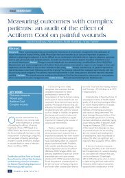
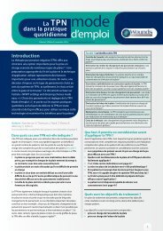
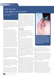
On admission : Mar 28, 2009 Week 2: April 10, 2009<br />
References<br />
38. Vowden P, Vowden K, Carville K.<br />
Antimicrobial dressings made easy.<br />
<strong>Wounds</strong> Int 2011; 2(1). Available at:<br />
http://www.woundsinternational.<br />
com/article.php?issueid=330<br />
39. Cutting K. Why use topical<br />
antiseptics? J Wound Care 2011; 4–7.<br />
40. Roberts C, Ivins N, Widgerow<br />
A. ACTICOAT and ALLEVYN Ag<br />
made easy <strong>Wounds</strong> Int 2011;<br />
2(2). Available at: http://www.<br />
woundsinternational.com/article.<br />
php?issueid=333.<br />
41. EWMA. Position Document:<br />
Identifying Criteria for Wound<br />
Infection. MEP Ltd: London, 2005.<br />
42. Lansdown AB. Silver I: its<br />
antibacterial properties and<br />
mechanisms of action. J Wound Care<br />
2002; 11(4): 125–30.<br />
43. Agranoff D, Krishna S. Metal<br />
ion homeostasis and intracellular<br />
parasitism. Mol Microbiol 1998;<br />
28(3): 403–12.<br />
44. Cutting K, White R, Hoekstra<br />
H. Topical silver–impregnated<br />
dressings and the importance of<br />
the dressing technology. Int Wound<br />
J 2009 Oct;6(5):396–402.<br />
45. Michaels JA, Campbell B, King<br />
B et al. Randomized controlled trial<br />
and cost–effectiveness analysis<br />
of silver–donating antimicrobial<br />
dressings for venous leg ulcers<br />
(VULCAN trial). Br J Surg 2009;<br />
96(10): 1147–56.<br />
46. Mudge E, Orsted H.<br />
Wound infection and pain<br />
management made easy. <strong>Wounds</strong><br />
Int 1(3). Available at www.<br />
woundsinternational.com<br />
Week 6: May 8, 2009<br />
Week 12: June 20, 2009<br />
Figure 2 – This patient had a reduction in maceration, periwound erythema and oedema with<br />
complete closure of the wound at 12 weeks.<br />
patients experienced non-progression of their<br />
ulcer [54] . The most prevalent species at initial<br />
biopsy was Enterococcus faecalis (9/24), while<br />
79% (19/24) of patients had S. aureus at some<br />
point during treatment of which 62% (15/24)<br />
had a methicillin-resistant strain.<br />
During the study, all patients had<br />
ALLEVYN Ag applied to their wounds under<br />
compression, using a multilayer bandaging<br />
system (PROFORE TM , Smith & Nephew).<br />
All wounds were assessed on a weekly<br />
basis or until wound closure [Figs 1 and 2].<br />
Debridement was performed at each weekly<br />
dressing change.<br />
Biopsies and semi-quantitative swab cultures<br />
were taken to assess bioburden. At week eight,<br />
the level of bioburden had reduced to less than<br />
10 5 cfu/g tissue in 13 patients (54%) compared<br />
with 10 patients (42%) at week two. There<br />
was also evidence of a significant reduction<br />
(p
Technology update The role of ALLEVYN TM Ag in the management of hard-to-heal wounds<br />
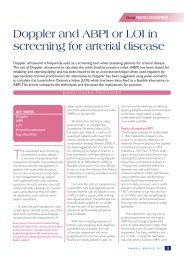
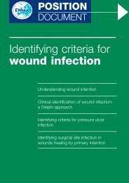
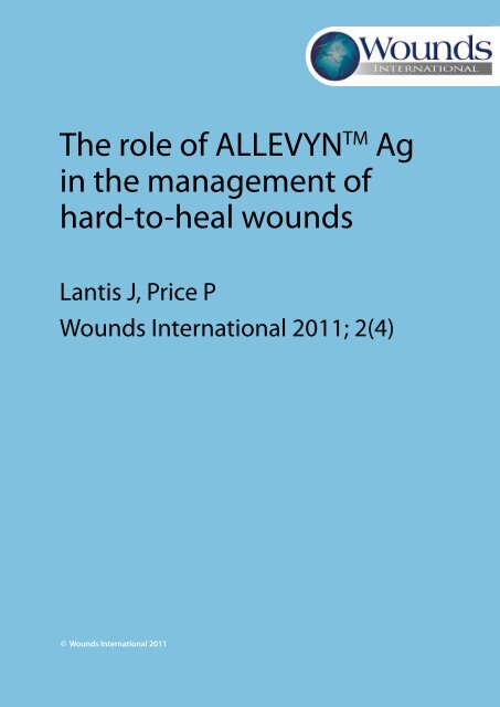
Probability of closure<br />
1.0<br />
0.9<br />
0.8<br />
0.7<br />
0.6<br />
0.5<br />
0.4<br />
0.3<br />
0.2<br />
0.1<br />
0.0<br />
0 10 20 30 40 50 60 70<br />
Time (days)<br />
Figure 3 – Kaplan-Meier plot of time-to-closure.<br />
This study provides benchmark data that<br />
may support a structured treatment protocol<br />
with frequent debridement, together with<br />
weekly dressing changes using ALLEVYN Ag in<br />
infected, hard-to-heal venous leg ulcers [54] .<br />
Cost-effectiveness of ALLEVYN Ag<br />
A cost comparison model [Table 1] comparing<br />
the wound closure rates achieved by Lantis and<br />
Gendics [54] and standard care has subsequently<br />
been extrapolated. This table makes a number<br />
of assumptions in relation to wound closure<br />
rates and frequency of dressing changes in the<br />
standard care arm. Furthermore, it assumes<br />
that, once the wound is closed, these patients<br />
do not incur any further costs and does not<br />
factor in follow-on costs associated with<br />
further clinic attendance by patients in either<br />
Parameters Compression +<br />
ALLEVYN TM Ag<br />
arm using standard care versus ALLEVYN Ag.<br />
For the purposes of this cost-comparison<br />
model, the study by Skog et al [35] has been used<br />
to provide a control baseline, as there was no<br />
standard care arm in the study by Lantis and<br />
Gendics [54] . The closure rate achieved by Skog<br />
et al [35] was 3%. To provide a more conservative<br />
measure, a 5% wound closure rate for standard<br />
care in hard-to-heal venous leg ulcers is assumed<br />
in the model. The dressing change frequency in<br />
the study by Lantis and Gendics was every 7.2<br />
days [54] . When calculating costs for the standard<br />
care arm, twice-weekly dressing changes have<br />
been assumed to reflect standard clinical practice<br />
in such wounds [16] , with an average of 32 minutes<br />
nursing time per visit.<br />
Using these assumptions, it is possible<br />
to model the costs of once-weekly versus<br />
Compression only<br />
(standard care)<br />
Source<br />
Treatment length (days 84 (12 weeks) 84 (12 weeks) Lantis and Gendics [54]<br />
Mean time to wound<br />
57.3 (eight weeks) 57.3 (eight weeks) Lantis and Gendics [54]<br />
closure (days)<br />
Wear time (days) 7.2 3.5 Lantis and Gendics [54]<br />
Tennvall et al [16]<br />
Duration of community<br />
nurse visit (minutes)<br />
30 (US$47/hour) 30 (US$47/hour) US Bureau of Labor<br />
Statistics<br />
Wound closure rate 45.8% 5% Lantis and Gendics [54]<br />
Skog et al [35]<br />
Table 1 – Cost comparison of ALLEVYN TM Ag plus compression versus standard care.<br />
References<br />
Technology and product reviews<br />
47. White RJ, Cooper<br />
RA. Silver sulphadiazine.<br />
<strong>Wounds</strong> UK 2005; 1: 51–61.<br />
48. Buckley SC, Scott S, Das<br />
K. Late review of the use of<br />
silver sulphadiazine dressings for<br />
the treatment of fingertip injuries.<br />
Injury 2000; 31(5):301–4.<br />
49. Bishop JB, Phillips LG, Mustoe<br />
TA, et al. A prospective randomized<br />
evaluator–blinded trial of two<br />
potential wound healing agents<br />
for the treatment of venous stasis<br />
ulcers. J Vasc Surg 1992; 1 6(2):<br />
251–57.<br />
50. Ulkür E, Oncül O, Karagöz H,<br />
et al. Comparison of silver–coated<br />
dressing (Acticoat), chlorehexidine<br />
acetate 0.5% (Bactigras®) and<br />
silver sulfadiazine 1% (Silverdin®)<br />
for topical antibacterial effect<br />
in Pseudomonas aeruginosa<br />
contaminated full–thickness burn<br />
wounds in rats. J Burn Care Rehabil<br />
2005; 26(5): 430–3.<br />
51. Smith & Nephew. Report<br />
reference DS/08/116/R1f. Carpenter<br />
S. Silver release of ALLEVYN<br />
dressings, June 2009.<br />
52. Smith & Nephew. Data on file<br />
report 1011017.<br />
53. Smith & Nephew. Data on file<br />
report 1011018.<br />
54. Lantis JC, Gendics C. In vivo<br />
effect of sustained–release silver<br />
sulphadiazine foam on bioburden<br />
and wound closure in infected<br />
venous leg ulcers. J Wound Care<br />
2011; 20(2): 90–6.<br />
55. Falanga V, Margolis D, Alvarez A<br />
et al. Rapid healing of venous ulcers<br />
and lack of clinical rejection with<br />
an allograft cultured human skin<br />
equivalent. Arch Dermatol 1998;<br />
134: 293–99.<br />
56. McKeown T, Hoctor B. Meeting<br />
the challenges of a nurse–led<br />
dressing clinic in a busy A&E.<br />
Enhancing Clinical Practice 2009;<br />
4–7.<br />
www.woundsinternational.com 7
Technology and product reviews<br />
Useful links and<br />
further reading<br />
ACTICOAT TM and ALLEVYN TM AG Made Easy http://<br />
www.woundsinternational.com/article.php?issuei<br />
d=333&contentid=123&articleid=9880<br />
Webcast: Improving clinical and economic<br />
outcomes in hard to heal wounds http://<br />
www.woundsinternational.com/webcasts.<br />
php?webcastid=9953<br />
Hard-to-Heal <strong>Wounds</strong>: a holistic approach<br />
http://www.woundsinternational.com/article.<br />
php?contentid=127&articleid=45<br />
twice-weekly nurse visits in the standard<br />
care arm. The resulting figures show that<br />
the once-weekly dressing changes may save<br />
US$158 per patient in material costs and a<br />
further US$314 per patient in reduced nursing<br />
time — a total saving of US$472 per patient<br />
over 12 weeks. These costs feature Medicare<br />
reimbursement and are taken from the US<br />
Bureau of Labor Statistics 2009 (www.bls.gov/<br />
home.htm). A clinical evaluation of 25 patients<br />
who were treated with ALLEVYN Ag Adhesive<br />
and Non-Adhesive in a UK accident and<br />
emergency department also illustrates the<br />
cost savings associated with the dressing [56] .<br />
This study estimated a material cost saving<br />
of 40 euros per week. Dressing changes were<br />
also reduced by 1.6 per week, resulting in 160<br />
minutes of saved nursing time for each 10<br />
patients treated with ALLEVYN Ag.<br />
It must be noted that neither of these<br />
studies were designed to validate the cost<br />
benefits of using ALLEVYN Ag in patients with<br />
chronic wounds. Therefore, the performance<br />
of larger studies with a control arm are<br />
necessary if comprehensive conclusions<br />
about any cost savings are to be drawn.<br />
Conclusion<br />
The management of hard-to-heal wounds<br />
relies on a comprehensive approach to care<br />
that involves a structured treatment protocol<br />
and allows for the practical application of<br />
available therapies. Patients with a good<br />
level of symptom management and who are<br />
concordant with therapy will often go on to<br />
achieve closure of their wounds. In wounds<br />
where healing is impaired by the presence<br />
of bioburden there is a need for clinically<br />
effective antibacterial therapies that are easy<br />
to use, effective, and that reduce the drain on<br />
scarce healthcare resources.<br />
This study featured in this article provided<br />
a ‘real-world’ clinical evaluation of a protocol<br />
for the treatment for longstanding venous<br />
leg ulcers using a dressing containing silver<br />
sulfadiazine [54] . This provides a benchmark for<br />
clinicians seeking to reduce the human and<br />
financial costs of hard-to-heal wounds.<br />
Author details<br />
John C Lantis II, is Chief of Division Vascular/<br />
Endovascular Surgery; Director of Vascular<br />
Clinical Research, St Luke’s Roosevelt Hospital<br />
and Associate Clinical Professor of Surgery,<br />
Columbia University, New York, USA<br />
Professor Patricia Price is Dean and Head<br />
of School, Healthcare Studies, Cardiff<br />
University, UK<br />
This article has been sponsored by Smith<br />
& Nephew. ALLEVYN TM and PROFORE TM are<br />
trademarks of Smith & Nephew.<br />
<strong>Wounds</strong> <strong>International</strong> invites you to<br />
wounds Understanding<br />
biofilm-based<br />
wound care: what you<br />
need to know<br />
INTERACTIVE GLOBAL<br />
WEBCAST SERIES2011<br />
Broadcasting on Wednesday 14 December 2011<br />
at 11.00 AM GMT (6.00 AM EST – USA & Canada) & 4.30 PM GMT<br />
(11.30 AM EST - USA & Canada), followed by your chance to ask the<br />
expert in a LIVE Q&A session. Watch the presentations online at:<br />
www.woundsinternational.com/webcasts.php<br />
Register at http://webcasts.woundsinternational.com<br />
Brought to you by <strong>Wounds</strong> <strong>International</strong> in conjunction with, and sponsored by Smith & Nephew<br />
international<br />
webcast series 2011<br />
<strong>Wounds</strong> <strong>International</strong> has launched a new webcast<br />
series focusing on topical issues for clinicians<br />
worldwide and providing an opportunity to ask the<br />
expert. The first webcast was broadcast on 8 June<br />
2011 and was on Improving clinical and economic<br />
outcomes in hard to heal wounds. Professor<br />
Patricia Price and Dr John Lantis discuss the impact<br />
of hard to heal wounds on patients and suggest<br />
a management algorithm for those with infected<br />
venous leg ulcers. For those of you who did not log<br />
on to view this live broadcast, you can watch the<br />
videos at: http://www.woundsinternational.com/<br />
webcasts.php<br />
To watch the second webcast in this series with<br />
Professor Gregory Schultz and Dr John Lantis<br />
speaking on Understanding biofilm-based wound<br />
management: what you need to know, register at<br />
http://www.webcasts.woundsinternational.com<br />
8<br />
<strong>Wounds</strong> <strong>International</strong> Vol 2 | Issue 4 | ©<strong>Wounds</strong> <strong>International</strong> 2011