DNA Structures - Cmgm Stanford
DNA Structures - Cmgm Stanford
DNA Structures - Cmgm Stanford

You also want an ePaper? Increase the reach of your titles
YUMPU automatically turns print PDFs into web optimized ePapers that Google loves.
<strong>DNA</strong> <strong>Structures</strong><br />
Biochemistry 201<br />
Molecular Biology<br />
January 5, 2000<br />
Doug Brutlag<br />
The Structural Conformations of <strong>DNA</strong><br />
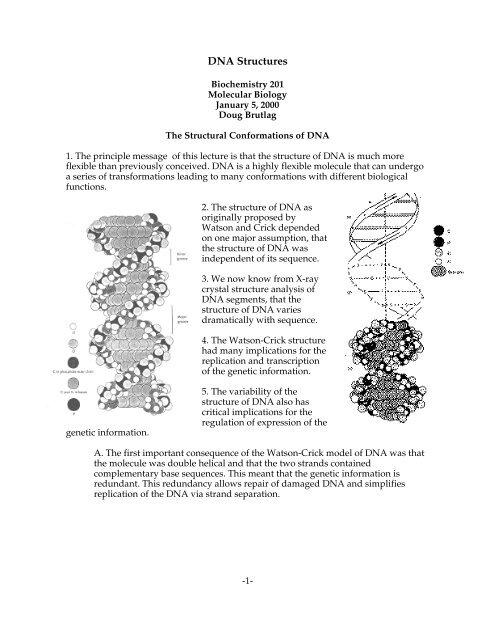
1. The principle message of this lecture is that the structure of <strong>DNA</strong> is much more<br />
flexible than previously conceived. <strong>DNA</strong> is a highly flexible molecule that can undergo<br />
a series of transformations leading to many conformations with different biological<br />
functions.<br />
2. The structure of <strong>DNA</strong> as<br />
originally proposed by<br />
Watson and Crick depended<br />
on one major assumption, that<br />
the structure of <strong>DNA</strong> was<br />
independent of its sequence.<br />
3. We now know from X-ray<br />
crystal structure analysis of<br />
<strong>DNA</strong> segments, that the<br />
structure of <strong>DNA</strong> varies<br />
dramatically with sequence.<br />
4. The Watson-Crick structure<br />
had many implications for the<br />
replication and transcription<br />
of the genetic information.<br />
genetic information.<br />
5. The variability of the<br />
structure of <strong>DNA</strong> also has<br />
critical implications for the<br />
regulation of expression of the<br />
A. The first important consequence of the Watson-Crick model of <strong>DNA</strong> was that<br />
the molecule was double helical and that the two strands contained<br />
complementary base sequences. This meant that the genetic information is<br />
redundant. This redundancy allows repair of damaged <strong>DNA</strong> and simplifies<br />
replication of the <strong>DNA</strong> via strand separation.<br />
-1-
<strong>DNA</strong> Strucures<br />
B. Another important consequence of the<br />
Watson-Crick structure was the anti-parallel<br />
nature of the <strong>DNA</strong> chains. Anti-parallel<br />
chains cause considerable difficulty for<br />
replication and transcription.<br />
C. The similarity of the <strong>DNA</strong> base pairs to<br />
each other also makes faithful replication<br />
with less than one mistake in a hundred<br />
million a formidable task.<br />
D. The structural similarity of diverse<br />
sequence makes recognition of genetic control<br />
sites difficult.<br />
Watson-Crick B-Form <strong>DNA</strong><br />
1. A review of the origin and the experimental<br />
support for Watson and Crick's structure for the B-<br />
form of <strong>DNA</strong>.<br />
2. From basic chemical analysis, Watson and Crick<br />
knew:<br />
A. <strong>DNA</strong> contained long polymeric chains.<br />
B. <strong>DNA</strong> contained deoxyribonucleosides in<br />
5'→ 3' phosphodiester linkage.<br />
C. X-ray diffraction suggested a helical<br />
structure.<br />
- The cross pattern suggested a helical pitch angle about 45°.<br />
- Axial reflections gave repeating units of 3.4 and 34 Å.<br />
- Radial reflections gave a fiber width of 20 Å.<br />
D. Chargaff's work showed that the base<br />
composition of <strong>DNA</strong> varied from organism to<br />
organism but certain relationships between the<br />
amounts of various bases always held. These<br />
relationships are called Chargaff's rules:<br />
-2
<strong>DNA</strong> Strucures<br />
- The amount of adenine equals the amount of thymine.<br />
- The amount of guanine equals the amount of cytidine.<br />
- The amount of adenine plus guanine equals 50% of the total<br />
implying that 50% of the bases in <strong>DNA</strong> are purines.<br />
- The amount of thymine plus cytosine equals 50% of the total<br />
implying that 50% bases in <strong>DNA</strong> are pyrimidines.<br />
E. Watson and Crick proposed base pairing rules to explain Chargaff's equalities.<br />
- They chose base pairs connected by hydrogen bonds.<br />
- The bases were in their normal tautomeric forms (uncharged) at pH 7.0.<br />
- They picked an AT pair and a GC pair that gave superimposable<br />
locations of the glycosylic linkages. A consequence of this is that the<br />
structure of the <strong>DNA</strong> would be sequence independent.<br />
- The first base pairs observed in X-ray crystallography experiments were<br />
the Hoogstein base pairs and not the Watson and Crick base pairs.<br />
-3
<strong>DNA</strong> Strucures<br />
-4
<strong>DNA</strong> Strucures<br />
F. The deoxyribose of each base pair are attached in opposite orientation.<br />
G. Each base was in the anti-conformation.<br />
H. This conformation of nucleosides resulted in the opposite polarity of <strong>DNA</strong><br />
chains in the resulting helix.<br />
I. This conformation and resulting antiparallel chains generate an axis of dyad<br />
symmetry (axis of two-fold rotational symmetry) at each base pair. Dyad axes are<br />
very important for proteins that bind to <strong>DNA</strong>. Most <strong>DNA</strong>-binding proteins<br />
possess an axis of symmetry and bind to symmetric <strong>DNA</strong> sequences.<br />
J. This structure has the base-pair as the primary repeating unit and results in an<br />
additional axis of symmetry between each base pair.<br />
K. Watson and Crick then connected base pairs with phosphodiester bonds that<br />
spaced the bases 3.4 Å apart and rotated each subsequent base pair by 36°. This<br />
rotation generates a right-handed double helix with 10 base per turn and<br />
repeating elements every 3.4 and 34 Å.<br />
L. The obtuse angle of the glycosylic<br />
linkages leads to major and minor grooves<br />
in helix with specific groups in each group.<br />
M. The bases were perpendicular to the<br />
helix axis.<br />
Crystal Structure of B-<strong>DNA</strong><br />
-5
<strong>DNA</strong> Strucures<br />
1. Thirty years after the Watson-Crick<br />
proposal, Dickerson and Rich determined<br />
the complete structure of crystalline <strong>DNA</strong>s.<br />
Advances in the synthesis of large<br />
quantities of short synthetic segments of<br />
<strong>DNA</strong> allowed each of them to crystallize a<br />
unique <strong>DNA</strong> sequence.<br />
2. Dickerson's B form crystals confirmed in<br />
most part the Watson-Crick model. <strong>DNA</strong><br />
was double helical with antiparallel<br />
strands. The bases associated in Watson<br />
and Crick base-pairs with hydrogen bonds<br />
in the center.<br />
3. There were also two major differences.<br />
A. First, the base pairs were not flat,<br />
but were twisted with respect to<br />
each other. This was called a<br />
propeller twist.<br />
B. The rotation from one base pair to<br />
the next was not a constant 36° as<br />
predicted, but instead varied from<br />
27° to 40°. This variation in twist<br />
angle was extremely important<br />
because it implied that the structure<br />
of B-<strong>DNA</strong> was sequence dependent.<br />
-6
<strong>DNA</strong> Strucures<br />
4. The propeller twist of the base pairs results in purine-purine clash in the center of the<br />
helix. Because the purines are larger than the pyrimidine rings, they extend beyond the<br />
helical axis of <strong>DNA</strong>.<br />
5. <strong>DNA</strong> attempts to reduce purine-purine clash in several ways:<br />
A. The base pairs rotate less along the helix axis in the purine-pyrimidine<br />
sequences (lower average helical twist). They tend to rotate less in the<br />
pyrimidine-purine sequences (lower than average helical twist). The average<br />
helical twist was still very close to the 36° proposed by Watson and Crick.<br />
B. Another way <strong>DNA</strong> minimizes the purine-purine clash is that it bends toward<br />
the minor grove or major groove to reduce the interaction.<br />
C. Finally clashing base pairs could slide left or right toward the phosphodiester<br />
backbones to minimize the purine-purine interaction.<br />
-7
<strong>DNA</strong> Strucures<br />
6. The most important implication of<br />
these structural variations is that the<br />
actual structure of <strong>DNA</strong> depends<br />
strongly on the sequence. The<br />
positions of the phosphate groups, the positions of the amino and keto groups in <strong>DNA</strong><br />
reflect the sequence in a predictable way. Current research is aimed at understanding<br />
this structural code and to determine if regulatory sequence-specific <strong>DNA</strong> binding<br />
proteins make use of this variation in recognizing <strong>DNA</strong>.<br />
A-form of <strong>DNA</strong><br />
1. There are also many other forms of <strong>DNA</strong> more distinct from the B form that are<br />
biologically important. Most well known is the A-form which <strong>DNA</strong> assumes during<br />
dehydration or in RNA-<strong>DNA</strong> hybrid helices.<br />
2. The base-pairs are not perpendicular to the helical axis but instead they are tilted at a<br />
steep angle.<br />
-8
<strong>DNA</strong> Strucures<br />
3. In the A form, the base pairs are also<br />
closer together along the helical axis;<br />
2.55 Å center-to-center distance.<br />
4. The helical pitch of A-form <strong>DNA</strong> is<br />
closer to 11 base pairs per turn in 28 Å<br />
rather that 34 Å. As a result, the A-<br />
form is 25% shorter than the B-form.<br />
<strong>DNA</strong> shrinks when it dries.<br />
5. If binding of protein to <strong>DNA</strong><br />
removes water it may result in altered<br />
conformation of the <strong>DNA</strong> thus<br />
stabilizing the interaction.<br />
6. The tilted base pairs allow room for<br />
the 2' oxygen present in RNA chains and therefore all double helices containing at least<br />
one RNA strand are present in the A-form.<br />
7. Duplex RNA (such as found in the replication intermediates of many viral RNAs<br />
such as polio virus RNA) is always in the A-form.<br />
Z-form of <strong>DNA</strong><br />
1. When the self-complementary polymer (CG)3 was crystallized in high ionic strength<br />
conditions, a very unusual form of <strong>DNA</strong> called the Z-form was discovered.<br />
2. The Z-form differs from the B-form in several ways:<br />
A. The helix was left-handed instead<br />
of right-handed.<br />
B. The helix showed only a single<br />
groove rather than two.<br />
C. The nucleotides along one strand<br />
alternate between the syn- and anticonformation.<br />
The guanosines are all<br />
in the syn conformation while the<br />
cytidines are all in the anti<br />
conformation like the B-form.<br />
-9
<strong>DNA</strong> Strucures<br />
3. In most other respects the two forms are similar:<br />
A. Both forms are double<br />
helical and both have two<br />
chains of opposite chemical<br />
polarity.<br />
B. Watson and Crick hydrogen<br />
bonds hold the chains together.<br />
4. Since conformations<br />
between the purine and the<br />
pyrimidines in Z-<strong>DNA</strong><br />
alternate, the basic repeating<br />
unit is no longer the base pair,<br />
but is a dinucleotide. This<br />
implies that there is no axis<br />
dyad symmetry at each base<br />
pair, only between base pairs.<br />
5. Even the best evidence for Z-<strong>DNA</strong> in nature is controversial. Some of the best<br />
evidence comes from experiments involving antibodies directed specifically at the Z-<br />
<strong>DNA</strong> structure. Some authors have demonstrated that the presence of the Z-form of the<br />
<strong>DNA</strong> in these cytological preparations is an artifact of the preparation and if one<br />
prepares chromosomes carefully, no or very little Z-<strong>DNA</strong> antibody will bind.<br />
-10
<strong>DNA</strong> Strucures<br />
6. The most one can say is that Z-<strong>DNA</strong> can form under physiological conditions in<br />
natural <strong>DNA</strong> sequences in which purines alternate with pyrimidines. Whether Z-<strong>DNA</strong><br />
does form in cells and whether nature takes advantage of this unusual form is still<br />
speculation.<br />
7. The possibility that <strong>DNA</strong> can assume two structures as distinct as the Z-form and the<br />
B-form shows that the chains are capable of much more flexibility than many had<br />
considered possible before.<br />
Novel <strong>DNA</strong> <strong>Structures</strong><br />
1. Several novel forms of <strong>DNA</strong> involving the pairing of more than two strands and also<br />
forms involving parallel chains have been described. These structures generally form<br />
with specific <strong>DNA</strong> sequences and may have profound biological consequences.<br />
A. Wells and others have evidence showing that oligopurine-oligopyrimidine<br />
sequences can fold back on themselves to form an internal region containing one<br />
triple stranded region and one single-stranded region. The third strand is basepaired<br />
in the major groove of a normal <strong>DNA</strong> duplex using hydrogen bonds<br />
similar to those found in the Hoogstein base pairs.<br />
-11
<strong>DNA</strong> Strucures<br />
B. Sen and Gilbert have reported that<br />
<strong>DNA</strong> helices containing specific guanine<br />
rich sequences can self-associate to form<br />
four-stranded structures. The two helices<br />
are hydrogen bonded together by<br />
Hoogstein pairing.<br />
C. Tom Cech and Aaron Klug’s<br />
laboratories have demonstrated that<br />
sequences found at the ends of eukaryotic<br />
chromosomes can also form specific<br />
tetranucleotide base pairs, referred to as<br />
G-quartets that may be involved in<br />
holding chromosome ends together<br />
during mitosis.<br />
D. Englund and others noticed that<br />
certain <strong>DNA</strong> sequences had an unusual<br />
migration during electrophoresis.<br />
Analyses of such <strong>DNA</strong>s have shown that they often contain runs of 3-4 As or Ts<br />
in a row and that these runs are repeated every 10 base pairs. Such runs result in<br />
bending of the <strong>DNA</strong> towards the minor groove and the repeating nature makes<br />
the <strong>DNA</strong> helix as a whole bend in one direction.<br />
E. Tom Jovin and Johan van de Sande have demonstrated that specific AT rich<br />
<strong>DNA</strong> sequences can base pair to form a parallel double-helical structure. These<br />
structures are physically very similar to the B-form of <strong>DNA</strong> but they are<br />
ineffective as substrates for many enzymes.<br />
References<br />
Dickerson, R. E. (1992). <strong>DNA</strong> structure from A to Z. Methods Enzymology 211, 67-111.<br />
Hagerman, P. J. (1990). Sequence-Directed Curvature of <strong>DNA</strong> Annu. Rev. Biochemistry<br />
59, 755-781.<br />
-12
<strong>DNA</strong> Strucures<br />
Herbert, A., & Rich, A. (1996). The biology of left-handed Z-<strong>DNA</strong>. J Biol Chem,<br />
271(20), 11595-8.<br />
Johnston, B. H. (1992). Generation and detection of Z-<strong>DNA</strong>. Methods Enzymol 211, 127-<br />
58.<br />
Rich, A., Nordheim, A. and Wang, A. H. J. (1984). The Chemistry and Biology of Left-<br />
Handed Z-<strong>DNA</strong>. Annual Review of Biochemistry, 53, 791-846.<br />
Rich, A. (1993). <strong>DNA</strong> comes in many forms. Gene, 135(1-2), 99-109.<br />
Rich, A. (1994). Speculation on the biological roles of left-handed Z-<strong>DNA</strong>. Ann N Y Acad<br />
Sci, 726, 1-16; discussion 16-7.<br />
Rippe, K. and Jovin, T. M. (1992). Parallel-stranded duplex <strong>DNA</strong>. Methods Enzymol 211,<br />
199-220.<br />
Travers, A. A. (1990). Why bend <strong>DNA</strong>? Cell, 60(2), 177-80.<br />
Watson, J. D. and Crick, F. H. C. (1953a). Molecular structure of nucleic acids. Nature<br />
171, 737-738.<br />
Watson, J. D. and Crick, F. H. C. (1953b). The structure of <strong>DNA</strong>. Cold Spring Harbor<br />
Symp. Quant. Biol. 18, 123-131.<br />
Wells, R. D. (1988). Minireview: Unusual <strong>DNA</strong> structures. J. Biol. Chem. 263, 1095-1098.<br />
Williamson, J. R. (1994). G-quartet structures in telomeric <strong>DNA</strong>. Annu Rev Biophys<br />
Biomol Struct, 23, 703-30.<br />
Internet Resources<br />
Structure Database<br />
Molecules R US<br />
RasMol Distribution<br />
http://www.ndb.bnl.gov/<br />
http://molbio.info.nih.gov/cgi-bin/pdb<br />
http://www.umass.edu/microbio/rasmol/<br />
Kinemage Distribution<br />
http://www.faseb.org/protein/kinemages/MageSoftware.html<br />
Protein/<strong>DNA</strong> Kinemages http://www.faseb.org/protein/ProTeach/<br />
-13