DNA Topology Handout - Cmgm Stanford
DNA Topology Handout - Cmgm Stanford
DNA Topology Handout - Cmgm Stanford

You also want an ePaper? Increase the reach of your titles
YUMPU automatically turns print PDFs into web optimized ePapers that Google loves.
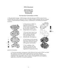
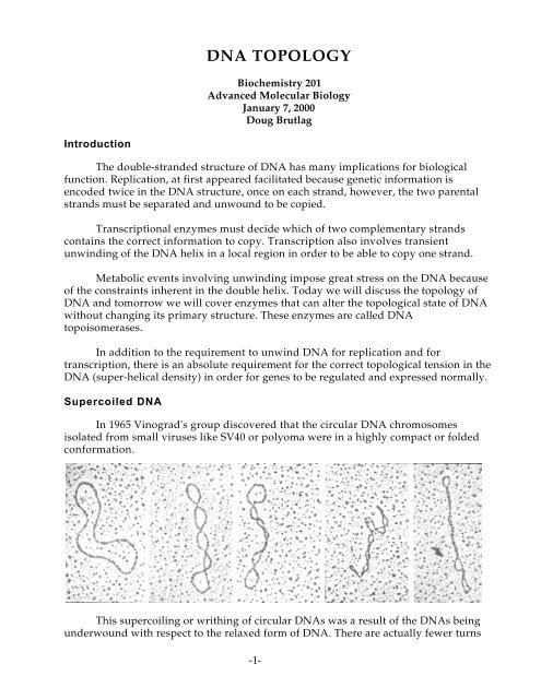
<strong>DNA</strong> TOPOLOGYBiochemistry 201Advanced Molecular BiologyJanuary 7, 2000Doug BrutlagIntroductionThe double-stranded structure of <strong>DNA</strong> has many implications for biologicalfunction. Replication, at first appeared facilitated because genetic information isencoded twice in the <strong>DNA</strong> structure, once on each strand, however, the two parentalstrands must be separated and unwound to be copied.Transcriptional enzymes must decide which of two complementary strandscontains the correct information to copy. Transcription also involves transientunwinding of the <strong>DNA</strong> helix in a local region in order to be able to copy one strand.Metabolic events involving unwinding impose great stress on the <strong>DNA</strong> becauseof the constraints inherent in the double helix. Today we will discuss the topology of<strong>DNA</strong> and tomorrow we will cover enzymes that can alter the topological state of <strong>DNA</strong>without changing its primary structure. These enzymes are called <strong>DNA</strong>topoisomerases.In addition to the requirement to unwind <strong>DNA</strong> for replication and fortranscription, there is an absolute requirement for the correct topological tension in the<strong>DNA</strong> (super-helical density) in order for genes to be regulated and expressed normally.Supercoiled <strong>DNA</strong>In 1965 Vinograd's group discovered that the circular <strong>DNA</strong> chromosomesisolated from small viruses like SV40 or polyoma were in a highly compact or foldedconformation.This supercoiling or writhing of circular <strong>DNA</strong>s was a result of the <strong>DNA</strong>s beingunderwound with respect to the relaxed form of <strong>DNA</strong>. There are actually fewer turns-1-
<strong>DNA</strong> <strong>Topology</strong>in the <strong>DNA</strong> helix than one would expect given the natural pitch of <strong>DNA</strong> in solution(10.4 base pairs per turn).When a linear <strong>DNA</strong> is free in solution it assumes a pitch which contains 10.4base pairs per turn. This is less tightly wound than the 10.0 base pairs per turn in theWatson and Crick B-form <strong>DNA</strong>.In order to understand the origin of supercoiling; imagine a linear <strong>DNA</strong> 5200base pairs in length. If the <strong>DNA</strong> were in the B-form one would expect the two strands ofthe helix to be wrapped around each other 500 times (5200 bp/10.4 bp/turn). Imagine alinear <strong>DNA</strong> in which the two ends become connected to form an open circle. This isreferred to as a relaxed circular <strong>DNA</strong>. On the other hand, if the linear <strong>DNA</strong> wereunwound 10%, say 50 turns, before its ends were joined, then the <strong>DNA</strong> moleculewould be under stress. When the molecule is free in solution it will coil about itself inspace as the two strands simultaneously twist about each other in order to return toequilibrium value of 10.4 base pairs per turn.<strong>DNA</strong> that is underwound is referred to as negatively supercoiled. The heliceswind about each other in a right handed path in space.<strong>DNA</strong> that is overwound also will relax and assume a supercoiled conformationbut this is referred to as a positively supercoiled <strong>DNA</strong> helix. Positively coiled <strong>DNA</strong> hasits <strong>DNA</strong> helices wound around each other in a left-handed path in space.Linking, Twisting and WrithingThe total number of times one strand of the <strong>DNA</strong> helix is linked with the otherin a covalently closed circular molecule is known as the linking number L k .1. The linking number is only defined for covalently closed <strong>DNA</strong> and its value isfixed as long as the molecule remains covalently closed.2. The linking number does not change whether the covalently closed circle isforced to lie in a plane in a stressed conformation or whether it is allowed tosupercoil about itself freely in space.-2-
<strong>DNA</strong> <strong>Topology</strong>3. The linking number L k of a circular <strong>DNA</strong> can only be changed by breaking aphosphodiester bond in one of the two strands, allowing the intact strand to passthrough the broken strand and then rejoining the broken strand.4. L k is always an integer since two strands must always be wound about eachother an integral number of times upon closure.The linking number of a covalently closed circular <strong>DNA</strong> can be resolved intotwo components called the twists T w and the writhes W r .L k = T w + W rThe twists Tw are the number of times that the two strands are twisted abouteach other while the writhes Wr is the number of times that the <strong>DNA</strong> helix is coiledabout itself in three-dimensional space.The twist and the writhe are not necessarily integers; indeed most often they arenot. It is just their sum, the linking number, that is an integer. If we use an SV40 <strong>DNA</strong>molecule for example, which is precisely 5243 base pairs in length, we would find that:L k = T w + W r480 = 5243/10.4 - 24.13480 = 504.13 - 24.13Its length and its pitch in solution determine the twist of <strong>DNA</strong>. [T w = Length (bp)/Pitch(bp/turn)]-3-
<strong>DNA</strong> <strong>Topology</strong>The twist and the linking number, determine the value of the writhe that forcesthe <strong>DNA</strong> to assume a contorted path is space. [Wr = L k - T w ]Unlike the Twist and the Linking number, the writhe of <strong>DNA</strong> only depends onthe path the helix axis takes in space, not on the fact that the <strong>DNA</strong> his two strands. Ifthe path of the <strong>DNA</strong> is in a plane, the Wr is always zero. Also if the path of the <strong>DNA</strong>helix were on the surface of a sphere (like the seams of a tennis ball or base ball) thenthe total Writhe can also be shown to be zero.Writhes can also come in different forms. If a <strong>DNA</strong> molecule wraps around itself, thenthe writhes are known as supertwists. If a <strong>DNA</strong> molecule wraps around something else-4-
<strong>DNA</strong> <strong>Topology</strong>(another molecule for instance) then the writhes are known as solenoidial writhe. Insolution, the writhes can isomerize between the two types.Measuring SupercoilsThe superhelical density of a circular <strong>DNA</strong> can be observed and measured inseveral ways, electron microscopy, sedimentation velocity, or by electrophoresis of<strong>DNA</strong> in a medium that provides frictional resistance.The first quantitativemethod for measuringsupercoiling was thesedimentation procedure.Since supercoiled moleculesare more compact theysediment faster in a centrifugethan when they are relaxed.A more elegant andsimpler modern method fordetermining the number ofsuperhelical turns in <strong>DNA</strong> isby electrophoresis in anagarose gel.Supercoiled <strong>DNA</strong> migrates muchmore rapidly than does a relaxed moleculeof the same length. <strong>DNA</strong> separates intodiscrete bands depending on the linkingnumber.Since <strong>DNA</strong>s resolved in this waydiffer from each other only in theirtopology, they are referred to as topologicalisomers, or topoisomers.Molecules that differ by one unit inlinking number can be separated byelectrophoresis in agarose due to thedifference in their writhe (that is due todifference in folding). The variation inlinking number is reflected in a differencein the writhe. The variation in writhe issubsequently reflected in the state ofcompaction of the <strong>DNA</strong> molecule.-5-
<strong>DNA</strong> <strong>Topology</strong>Neither of these methods bythemselves can distinguish negativelyand positively supercoiled <strong>DNA</strong>. Bothforms sediment and electrophoresemore rapidly than relaxed <strong>DNA</strong>.However by sedimenting orelectrophoresing supercoiled <strong>DNA</strong> inthe presence of an intercalating agentsuch as ethidium bromide orchloroquine, one can distinguishnegatively supercoiled <strong>DNA</strong> frompositively supercoiled <strong>DNA</strong>. Whennegatively supercoiled <strong>DNA</strong> binds anintercalating agent, the average pitch isreduced because the twist anglebetween adjacent base pairs on eitherside of the intercalating agent isreduced from 36° to as little as 10° of twist.This reduction of twist causes acompensatory increase in writhe in acovalently closed molecule. Hence amolecule that is initially negativelysupercoiled will become more relaxed and apositively supercoiled molecule willbecome more twisted. By electrophoresingor sedimenting supercoiled <strong>DNA</strong> inincreasing concentrations of anintercalating agent and knowing theaffinity of the agent for <strong>DNA</strong> it is possibleto calculate the writhe in the <strong>DNA</strong>.It is also possible to measure thewrithe and the sense of the write usingintercalating agents in 2 dimensional gelelectrophoresis experiments. By firstelectrophoresing <strong>DNA</strong> in one dimensionin the absence of intercalating agents asabove, and then adding an intercalatingagent to partially increase the writhe in apositive sense and electrophoresing the<strong>DNA</strong> in a second dimension, it is possibleto separate positive and negatively writhed<strong>DNA</strong> in the dimensions. It is also possible to get much better resolution of the highlytwisted forms of <strong>DNA</strong> found in natural supercoiled <strong>DNA</strong> samples.Measuring the Pitch of <strong>DNA</strong>Since the superhelical state of <strong>DNA</strong> is dependent on the pitch of <strong>DNA</strong> it becamecritical to have an accurate method to determine a <strong>DNA</strong>s pitch under different-6-
<strong>DNA</strong> <strong>Topology</strong>experimental conditions. Since agarose gels can separate molecule differing by a singletwist or writhe these were used to measure the change in pitch of <strong>DNA</strong> as a function ofboth temperature and sequence.By relaxing circular <strong>DNA</strong> molecules at various temperatures and thenelectrophoresing the <strong>DNA</strong>s all at a single temperature both Vinograd's group andWang's group determined that <strong>DNA</strong> tends to unwind by 0.012° per base pair per °centigrade as it is heated.While this does not seem like much change compared to the 36° rotation perbase pair in the <strong>DNA</strong> helix, in even a short <strong>DNA</strong> molecule 10,000 base pairs in length,this amounts to an unwinding of 120° (1/3 turn) per ° centigrade. Heating a 10-kbcircular <strong>DNA</strong> from 20° C to 35° C can reduce the overall twist by 5 full turns.Wang also used gelelectrophoresis to measurethe absolute pitch of <strong>DNA</strong>in solution. By using aseries of <strong>DNA</strong> moleculesthat differed from eachother by a few base pairs (11,25, 36 etc.) he was able tomeasure the amount oftwist contributed by theextra base pairs. Thesemeasurements confirmedthe X-ray crystal structuremeasurements of a pitch of<strong>DNA</strong> of 10.4 base pairs perturn of the helix (or 34.62°of twist per base pair). Healso confirmed thedependence of the pitch onthe sequence of <strong>DNA</strong>.Klug furtherconfirmed thesemeasurementsindependently using acompletely differentmethod. By adsorbing smallfragments of <strong>DNA</strong> to amica surface and thentreating the adsorbed <strong>DNA</strong> with <strong>DNA</strong>se I to nick the <strong>DNA</strong>, Klug showed that the <strong>DNA</strong>was accessible to nicking only on its side away from the mica surface and that thisaccessible region repeated every 10.6 base pairs, suggesting a pitch of that magnitude.-7-
<strong>DNA</strong> <strong>Topology</strong>ReferencesBauer, W. R., Crick, F. H. C. and White, J. H. (1980). Supercoiled <strong>DNA</strong>. ScientificAmerican 243, 118-133.Benham, C. J. (1985). Theoretical analysis of conformational equilibria in superhelical<strong>DNA</strong>. Annu. Rev. Biophys. Biophys. Chem. 14, 23-45.Benham, C. J. (1987). The influence of tertiary structural restraints on conformationaltransitions in superhelical <strong>DNA</strong>. Nucleic. Acids. Res. 15, 9985-9995.Crothers, D. M. (1998). <strong>DNA</strong> curvature and deformation in protein-<strong>DNA</strong> complexes: astep in the right direction [comment]. Proc Natl Acad Sci U S A, 95(26), 15163-15165.Cozzarelli, N. R. (1991). The structure and function of <strong>DNA</strong> supercoiling and catenanes.Harvey Lect, 87, 35-55.Crick, F. H. C., Wang, J. C. and Bauer, W. R. (1979). Is <strong>DNA</strong> really a double helix? J. Mol.Biol. 129, 449-457.El Hassan, M. A. and Calladine, C. R. (1998). Two distinct modes of protein-inducedbending in <strong>DNA</strong>. J Mol Biol, 282(2), 331-343.Giaever, G. N., Snyder, L., and Wang, J. C. (1988). <strong>DNA</strong> supercoiling in vivo. Biophys.Chem. 29, 7-15.Smith, G,R, (1981). <strong>DNA</strong> supercoiling: Another level for regulating gene expression.Cell 24, 599-600.Vologodskii, A. V. and Frank-Kamenetskii, M. D. (1992). Modeling supercoiled <strong>DNA</strong>.Methods Enzymol, 211, 467-80.Wang, J. C. (1974). Interactions between twisted <strong>DNA</strong>s and enzymes: The effects ofsuperhelical turns. J. Mol. Biol. 87, 797-816.Wang, J. C., and Giaever, G. N. (1988). Action at a distance along a <strong>DNA</strong>. Science 240,300-304.Wang, J. C. (1979). Helical repeat of <strong>DNA</strong> in solution. Proc. Natl. Acad. Sci.. USA 76, 200-203.Wasserman, S. A., and Cozzarelli, N. R. (1986). Biochemical topology: applications to<strong>DNA</strong> recombination and replication. Science 232, 951-960.-8-