Ootaxonomic Investigation of Five Lutzomyia Species ... - SciELO
Ootaxonomic Investigation of Five Lutzomyia Species ... - SciELO
Ootaxonomic Investigation of Five Lutzomyia Species ... - SciELO

Create successful ePaper yourself
Turn your PDF publications into a flip-book with our unique Google optimized e-Paper software.
Mem Inst Oswaldo Cruz, Rio de Janeiro, Vol. 96(2): 197-204, February 2001<br />
<strong>Ootaxonomic</strong> <strong>Investigation</strong> <strong>of</strong> <strong>Five</strong> <strong>Lutzomyia</strong> <strong>Species</strong><br />
(Diptera, Psychodidae) from Venezuela<br />
AM Fausto, MD Feliciangeli*, M Maroli**, M Mazzini<br />
197<br />
Dipartimento di Scienze Ambientali, Università della Tuscia, Via San Camillo de Lellis, 01100 Viterbo, Italy<br />
*Escuela de Medicina, Facultad de Ciencias de la Salud, Universidad de Carabobo, Núcleo Aragua, Maracay,<br />
Venezuela **Laboratorio di Parassitologia, Istituto Superiore di Sanità, Roma, Italy<br />
The eggshell fine structure <strong>of</strong> five sand fly species from Venezuela belonging to the genus <strong>Lutzomyia</strong><br />
(L. migonei, L. ovallesi, L. absonodonta, L. gomezi and L. panamensis) was examined by scanning<br />
electron microscopy. The chorionic sculpturing <strong>of</strong> L. migonei, L. ovallesi, L. absonodonta and L. gomezi<br />
was characterized by series <strong>of</strong> columns arranged in palisade to form sinuous ridges. In inter-ridge<br />
areas, the basal layer was covered with fibrous material. The outer chorion <strong>of</strong> L. panamensis had a<br />
pattern known as “mountain- or volcano-like”. The morphology <strong>of</strong> the posterior pole and aeropyle had<br />
a common structure in the five species, with some species-specific characters. The eggshell features <strong>of</strong><br />
the five species are compared with those <strong>of</strong> other phlebotomine sand flies.<br />
Key words: phlebotomine sand fly - ootaxonomy - eggshell - aeropyle - fine structure - scanning electron<br />
microscopy<br />
Sand fly taxonomy was declared to be “an indispensable<br />
basis for every work on sand fly biology<br />
and on their vector role” by the World Health<br />
Organization (WHO 1977). However, the taxonomic<br />
status <strong>of</strong> phlebotomines is much more obscure<br />
than that <strong>of</strong> other insects <strong>of</strong> medical significance,<br />
such as mosquitoes (Lane 1986), although<br />
many attempts have been made to find unequivocal<br />
characters to distinguish morphologically similar<br />
species and to propose stable classifications<br />
(Lewis et al. 1977, Artemiev 1991, Ashford 1991).<br />
Scanning electron microscope (SEM) studies<br />
<strong>of</strong> eggshell morphology have shown that differences<br />
between related species <strong>of</strong> insects are reliable<br />
taxonomic markers (Hinton 1981, Mazzini<br />
1987, Mazzini et al. 1993a). Ootaxonomy <strong>of</strong> various<br />
families <strong>of</strong> dipterans is in an advanced stage<br />
(for a review, see Margaritis & Mazzini 1998).<br />
With regard to sand flies, studies on eggshell morphology<br />
have been delayed by the difficulty <strong>of</strong> finding<br />
eggs in nature and <strong>of</strong> breeding certain species.<br />
The eggs <strong>of</strong> Old World sand flies have only been<br />
studied for a limited number <strong>of</strong> species (Irungo et<br />
al. 1985, Lane & El Sawaf 1986, Gebre-Michael<br />
This work has been funded by CDCHT, Universidad de<br />
Carabobo (FCS-91-044) and Italian MURST-ex40%.<br />
Corresponding author. Fax:+39-0761-357114. E-mail:<br />
fausto@unitus.it<br />
Reiceved 8 February 2000<br />
Accepted 15 August 2000<br />
& Lane 1991, Fausto et al. 1991, 1992, 1993, Rogo<br />
et al. 1992, Ghosh & Mukhopadhway 1996). These<br />
studies suggest that differences in egg sculpture <strong>of</strong><br />
Sergentomyia and Phlebotomus species are sufficient<br />
for separation <strong>of</strong> the two genera, hitherto<br />
based on other taxonomic characters (Perfil’iev<br />
1966). <strong>Ootaxonomic</strong> investigations have been carried<br />
out in many Neotropical species (about 10%<br />
<strong>of</strong> the about 400 known species) demonstrating the<br />
usefulness <strong>of</strong> chorionic ultrastructure in separating<br />
closely related species (Ward & Ready 1975,<br />
Zimmerman et al. 1977, Endris et al. 1987,<br />
Feliciangeli et al. 1993, Sierra et al. 1995, Perez &<br />
Ogusuku 1997).<br />
In the present study, the eggshell fine structure<br />
<strong>of</strong> five sand fly species belonging to the genus<br />
<strong>Lutzomyia</strong> - L. (Psychodopygus) panamensis, L.<br />
(<strong>Lutzomyia</strong>) gomezi, L. (<strong>Species</strong> Group<br />
Verrucarum) ovallesi, L. (<strong>Species</strong> Group Migonei)<br />
migonei, and L. (Micropygomyia) absonodonta -<br />
from Venezuela was examined by SEM. The first<br />
four species are known to be anthropophilic and<br />
have also been demonstrated or incriminated as<br />
vectors <strong>of</strong> Leishmania spp. in different Latin<br />
American countries. L. panamensis is regarded as<br />
a primary vector <strong>of</strong> cutaneous leishmaniasis (CL)<br />
in Venezuela (Rodriguez et al. 1999) and as a secondary<br />
vector in Panama, Guatemala and Colombia<br />
(Desjeux 1991). L. gomezi was reported to be<br />
naturally infected with Le. braziliensis in Panama<br />
(Johnson et al. 1993), Colombia (Young et al.<br />
1987), Ecuador (Gomez & Hashiguchi 1987) and<br />
Venezuela (Feliciangeli 1991, Feliciangeli et al.<br />
1994). L. ovallesi is recognized as a vector <strong>of</strong> CL
198 Ootaxonomy in <strong>Lutzomyia</strong> • AM Fausto et al.<br />
in Venezuela (Feliciangeli 1991, Feliciangeli et al.<br />
1994) and Guatemala (Rowton et al. 1992). It was<br />
found to be infected with a variant <strong>of</strong> Le.<br />
panamensis/guyanensis in Venezuela (Bonfante-<br />
Garrido et al. 1991) and with unidentified flagellates<br />
in Belize, Panama (Williams 1970) and Colombia<br />
(Young et al. 1987). L. migonei is a suspected<br />
vector <strong>of</strong> Le. braziliensis in the State <strong>of</strong><br />
Ceará, Brazil (Azevedo et al. 1990). The medical<br />
significance <strong>of</strong> L. absonodonta is unknown.<br />
Closely allied species in the subgenus Micropygomyia<br />
are known to feed on lizards.<br />
The chorionic patterns <strong>of</strong> the five species and<br />
other species belonging to different genera <strong>of</strong><br />
Phlebotominae were compared.<br />
MATERIALS AND METHODS<br />
Newly laid eggs were obtained from naturally<br />
blood-engorged sand flies <strong>of</strong> the following species:<br />
L. (<strong>Species</strong> Group Migonei) migonei, L. (<strong>Species</strong><br />
Group Verrucarum) ovallesi, L. (Micropygomyia)<br />
absonodonta, L. (<strong>Lutzomyia</strong>) gomezi, L.<br />
(Psychodopygus) panamensis.<br />
For scanning electron microscopy, the eggs were<br />
fixed for 1 h at 4°C in 4% paraformaldehyde and<br />
5% glutaraldehyde in 0.1 M cacodylate buffer at<br />
pH 7.2 (Karnovsky 1965), then rinsed overnight in<br />
cacodylate buffer, post-fixed in 1% osmium tetroxide<br />
for 1 h and dehydrated in a graded ethanol series.<br />
The material was dried by the critical point<br />
method using liquid CO 2 in a Balzers CPD 020 apparatus,<br />
attached to specimen holders, coated with<br />
gold in a Balzers Union MED 010 evaporator and<br />
observed with a 5200 Jeol JSM electron microscope.<br />
RESULTS<br />
Common characters <strong>of</strong> all eggs are elongated,<br />
cigar-like form with one side slightly flattened and<br />
both poles rounded (Figs 1, 5, 9, 14).<br />
The five species had specific chorionic sculpturing<br />
which enabled the species to be distiguished<br />
from each other.<br />
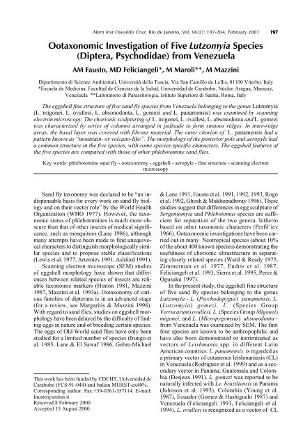
L. migonei (Figs 1-4) - The egg had a median<br />
width <strong>of</strong> about 80 µm and a length <strong>of</strong> about 280<br />
µm (Fig. 1). The eggshell surface was characterised<br />
by polygonal design, consisting <strong>of</strong> longitudinal<br />
ridges united by cross-ridges, which defined irregular<br />
rectangular areas with a major side parallel to<br />
the longitudinal axis <strong>of</strong> the egg (Fig. 3). Each ridge<br />
consisted <strong>of</strong> a single series <strong>of</strong> cylindrical columns,<br />
about 2 µm high, united at the top (Figs 3-4). In<br />
inter-ridge areas, the basal layer was covered in<br />
uniformly distributed fibrous material (Figs 3-4).<br />
The posterior pole was delimited by the terminal<br />
parts <strong>of</strong> a few chorionic ridges. Marked protuberances<br />
<strong>of</strong> different size were disposed around<br />
the aeropylar openings (Fig. 2).<br />
L. ovallesi (Figs 5-8) - The eggs were about<br />
270 µm in length and 80 µm in width (Fig. 5) with<br />
a complete basal layer covered with a compact coat<br />
<strong>of</strong> fine fibrous material (Figs 6, 7). The outer<br />
chorion had a series <strong>of</strong> sinuous longitudinal ridges<br />
with a few cross-ridges defining rectangular areas<br />
<strong>of</strong> different size (Fig. 6). The ridges consisted <strong>of</strong> a<br />
double series <strong>of</strong> columns (high about 0.5 µm)<br />
linked at the top (Fig. 7). Some <strong>of</strong> the chorionic<br />
ridges extended as far as the polar regions.<br />
The posterior pole consisted <strong>of</strong> a circular area<br />
delimited by the end part <strong>of</strong> chorionic ridges and<br />
divided into two semicircular areas by two short<br />
transverse ridges (Fig. 8). Each area had an aeropylar<br />
opening and several small conical protrusions.<br />
L. absonodonta (Figs 9-11) - The eggs were<br />
about 300 µm long and 75 µm wide (Fig. 9). The<br />
chorionic sculpture consisted <strong>of</strong> longitudinal columnar<br />
ridges defining elliptical areas <strong>of</strong> about 35-<br />
40 µm in length (Fig. 10). Each area was crossed<br />
by transverse fine ridges to form irregular quadrilaterals.<br />
As a result the egg surface had a reticular<br />
pattern. The basal layer <strong>of</strong> chorion between the<br />
ridges was covered in coarsely arranged fibrous<br />
material, that formed minute microvilli (Fig. 10).<br />
The borders <strong>of</strong> the posterior pole region were not<br />
well defined. This area was covered by several protuberances<br />
randomly distributed around the two<br />
aeropylar openings (Fig. 11).<br />
L. gomezi (Figs 12-13) - The surface sculpturing<br />
<strong>of</strong> eggs from Venezuelan females had a pattern<br />
<strong>of</strong> polygons formed by intersecting ridges consisting<br />
<strong>of</strong> columns arranged in palisade completely<br />
united at the top (Fig. 12). The areas enclosed by<br />
the ridges were four-sided with rounded corners<br />
and sides measuring about 15-20 µm (Fig. 12). The<br />
basal layer was covered with regularly arranged<br />
fibrous material. The posterior pole region was devoid<br />
<strong>of</strong> the structures characterising the rest <strong>of</strong> the<br />
egg surface and was delimited by the ends <strong>of</strong> the<br />
chorionic ridges and irregular circular ridges (Fig.<br />
13). Two non-columnar ridges partially divided the<br />
polar region into two semicircular areas in which<br />
small protrusions were observed beside each<br />
aeropylar opening.<br />
L. panamensis (Figs 14-17) - The egg had a<br />
median width <strong>of</strong> about 100 µm and a length <strong>of</strong> about<br />
340 µm (Fig. 14). The chorionic pattern was different<br />
from that <strong>of</strong> the other species, consisting <strong>of</strong> a<br />
uniform layer <strong>of</strong> numerous and tall mountains bearing<br />
volcano-like structures regularly disposed (Fig.<br />
15). These structures showed prominent irregular<br />
edges, enclosing a central depression in which holes<br />
<strong>of</strong> different sizes were visible (Fig. 16). The posterior<br />
pole was surrounded by a circle <strong>of</strong> short noncolumnar<br />
ridges. The aeropylar openings were surrounded<br />
by small uneven ridges (Fig. 17).
Mem Inst Oswaldo Cruz, Rio de Janeiro, Vol. 96(2), February 2001<br />
199<br />
Scanning electron microscopy micrographs <strong>of</strong> the outer chorionic sculpturing <strong>of</strong> <strong>Lutzomyia</strong> migonei. Fig. 1: whole egg. The<br />
eggshell surface has a polygonal pattern formed by longitudinal ridges and non-columnar cross ridges (Fig. 3). Each ridge<br />
consists <strong>of</strong> a single series <strong>of</strong> columns united at the top (Fig. 4). The basal layer is covered with uniformly arranged fibrous<br />
material (Fig. 4). The posterior pole, delimited by the terminal portion <strong>of</strong> chorionic ridges, shows protuberances and aeropylar<br />
openings (Fig. 2). Bar: Fig.1 = 20 µm; Figs 2-4 = 2 µm<br />
DISCUSSION<br />
The eggshell sculptures <strong>of</strong> the five species<br />
showed species-specific characters, useful for species<br />
identification. However, they had some features<br />
in common with eggs <strong>of</strong> other New and Old<br />
World sand fly species. Morphological categories,<br />
based on the chorionic patterns <strong>of</strong> <strong>Lutzomyia</strong> eggs,<br />
have been proposed by different authors. Ward and<br />
Ready (1975) proposed three categories: “volcanolike<br />
or mountain-like”, “polygonal”, and “parallel<br />
ridging”. The latter was divided by Endris et al.<br />
(1987) into “connected ridges” and “unconnected<br />
ridges”. With the recent discovery <strong>of</strong> other chorionic<br />
patterns, three new categories were added:<br />
“elliptical” (Feliciangeli et al. 1993), “verrucose”<br />
and “disperse” (Pérez & Ogusuku 1997). With regard<br />
to chorionic patterns described in the Old<br />
World species, Gebre-Michael and Lane (1991)<br />
divided the category “unconnected ridges” into the<br />
groups: “fragmented chained” and “complete<br />
chained”. The latter was defined as “reticular” by<br />
Fausto et al. (1992).<br />
The five species described in this paper can be<br />
grouped in the following categories: L. migonei<br />
“connected parallel ridges”; L. ovallesi “connected<br />
parallel ridges” (with few connections); L.<br />
absonodonta “reticular”; L. gomezi “polygonal”;<br />
L. panamensis “volcano-like or mountain-like”.
200 Ootaxonomy in <strong>Lutzomyia</strong> • AM Fausto et al.<br />
Most <strong>of</strong> patterns (L. migonei, L. ovallesi, L.<br />
absonodonta, and L. gomezi) have prominent<br />
ridges consisting <strong>of</strong> one or more series <strong>of</strong> columns.<br />
These patterns, common in New World species,<br />
are the only ones present in Old World species<br />
belonging to genera Phlebotomus and Sergentomyia<br />
(Fausto et al. 1992, 1993). However,<br />
Phlebotomus eggs show great variability in the arrangement<br />
<strong>of</strong> the columns, and the eggs <strong>of</strong><br />
Sergentomyia species are morphologically uniform,<br />
with only a polygonal pattern and none <strong>of</strong><br />
the fibrous material usually covers the basal layer<br />
between the ridges. These results are in line with<br />
comparative spermatology data, suggesting that the<br />
genus Sergentomyia followed a different evolutionary<br />
path to Phlebotomus and <strong>Lutzomyia</strong> (Dallai et<br />
al. 1984, Mazzini et al. 1993b, Fausto et al. 1995).<br />
The “volcano or mountain-like” pattern described<br />
in L. panamensis eggs has only been observed<br />
in species <strong>of</strong> the <strong>Lutzomyia</strong> genus. This particular<br />
pattern may have evolved in response to a<br />
damp microhabitat, i.e. swamp forest, in which<br />
these species are abundant (Ward & Ready 1975).<br />
As shown in Fig. 16, the prominent “volcano-like”<br />
structures protecting the aeropylar openings, are<br />
probably the basis for a well developed plastron<br />
and reflect its habitat preference. The chorionic<br />
sculpturing <strong>of</strong> L. panamensis from Panama<br />
(Zimmerman et al. 1977) is very similar to that <strong>of</strong><br />
L. panamensis from Venezuela, whereas two different<br />
chorionic patterns have been described for<br />
L. gomezi from Panama (Zimmerman et al. 1977)<br />
and Colombia (Sierra et al. 1995) (elongated hexagonal<br />
polygons) and Brazil (Ward & Ready 1975)<br />
Scanning electron microscopy micrographs <strong>of</strong> egg <strong>of</strong> <strong>Lutzomyia</strong> ovallesi. Fig.5: whole egg. The chorion shows sinuous longitudinal<br />
ridges with few cross-ridges forming rectangular areas (Fig. 6). The ridges consist <strong>of</strong> a double series <strong>of</strong> columns linked at<br />
the top (Fig. 7). Some <strong>of</strong> chorionic ridges reach the polar regions. The posterior pole is divided into two semicircular areas,<br />
having aeropylar openings and small conical protrusions, by two short transverse ridges (Fig. 8). Bar: Fig. 5 = 20 µm; Fig. 6 =<br />
5 µm; Figs 7, 8 = 2 µm
Mem Inst Oswaldo Cruz, Rio de Janeiro, Vol. 96(2), February 2001<br />
201<br />
(regular pentagonal polygons). We found that eggshell<br />
sculpture <strong>of</strong> the Venezuelan specimens was<br />
<strong>of</strong> the Brazilian type.<br />
Almost nothing is known about the breeding<br />
sites <strong>of</strong> the present and previous species described,<br />
and there is not yet enough data to attempt a taxo-<br />
Scanning electron microscopy micrographs <strong>of</strong> eggs <strong>of</strong> <strong>Lutzomyia</strong> absonodonta (Figs 9-11) and L. gomezi (Figs 12,13). Fig.9:<br />
whole egg. The chorionic sculpture has short longitudinal united by cross-ridges to form irregular quadrilaterals; the basal layer<br />
is covered with arranged coarsely fibrous material, forming minute microvilli (Fig. 10). The posterior pole is characterised by<br />
protuberances randomly distributed around the two aeropylar openings (arrows) (Fig. 11). Fig. 12: polygonal pattern due to<br />
intersecting ridges, in which most <strong>of</strong> polygons are quadrilaterals with rounded corners. An irregular circular ridge delimits the<br />
polar region where small protrusions can be observed beside two aeropylar openings (Fig.13). Bar: Fig. 9 = 20 µm; Figs 10, 11,<br />
13 = 2 µm; Fig. 12 = 5 µm
202 Ootaxonomy in <strong>Lutzomyia</strong> • AM Fausto et al.<br />
nomic and evolutionary interpretation. However,<br />
the similar exochorion pattern in L. gomezi from<br />
Panama and Colombia and the other pattern shared<br />
by L. gomezi from Venezuela and Brazil is <strong>of</strong> interest<br />
and may be related to the Andean barrier<br />
between them. L. gomezi may <strong>of</strong> course form a<br />
species complex (Feliciangeli 1997) and this should<br />
be clarified by isoenzymes and DNA fingerprinting<br />
<strong>of</strong> different populations.<br />
Comparison <strong>of</strong> the chorionic patterns <strong>of</strong> the<br />
present species and other <strong>Lutzomyia</strong> species (Ward<br />
& Ready 1975, Zimmerman et al. 1977, Endris et<br />
al. 1987, Feliciangeli et al. 1993) shows characters<br />
in most cases compatible with systematic position.<br />
<strong>Species</strong> <strong>of</strong> subgenus Psychodopygus have<br />
“volcano-like or mountain-like” patterns, like L.<br />
panamensis. L. ovallesi has similar chorionic sculpturing<br />
(“connected parallel ridges”) to L. evansi<br />
(“polygonal”) (Feliciangeli et al. 1993) and to other<br />
species belonging to the Verrucarum group. However,<br />
chorion sculpturing pattern <strong>of</strong> L. verrucarum<br />
from a Peruvian Andes valley combines “con-<br />
Scanning electron microscopy micrographs <strong>of</strong> an egg <strong>of</strong> <strong>Lutzomyia</strong> panamensis. Fig.14: whole egg. The chorionic pattern<br />
consists <strong>of</strong> a uniform layer <strong>of</strong> mountains with regular “volcano-like” structures (Fig. 15). Prominent irregular edges delimit a<br />
central depression in which holes <strong>of</strong> different sizes are visible (Fig. 16). The posterior pole, surrounded by a series <strong>of</strong> short noncolumnar<br />
ridges, shows aeropylar openings (arrows) and small uneven ridges (Fig. 17). Bar: Fig.14 = 20 µm; Fig. 15 = 5 µm;<br />
Figs 16, 17 = 2 µm
Mem Inst Oswaldo Cruz, Rio de Janeiro, Vol. 96(2), February 2001<br />
203<br />
nected ridges” pattern with “reticular” pattern<br />
(Pérez & Ogusuku 1997). Slight contrasts <strong>of</strong> eggs<br />
morphology described in L. verrucarum from different<br />
localities could be associated with geographical<br />
isolation <strong>of</strong> sand fly populations in different<br />
Andean valleys (Pérez & Ogusuku 1997). The eggs<br />
<strong>of</strong> L. venezuelensis, the only species <strong>of</strong> Micropygomyia<br />
subgenus previously studied<br />
(Feliciangeli et al. 1993), are in the “elliptical” category,<br />
and those <strong>of</strong> L. absonodonta, belonging to<br />
the same subgenus, in the “reticular” category.<br />
These two categories are very similar by virtue <strong>of</strong><br />
a common basal structure. Connected parallel<br />
ridges give L. migonei eggs a polygonal aspect<br />
similar to those described, using light microscopy,<br />
for the eggs <strong>of</strong> L. lenti and L. bahiensis (<strong>Species</strong><br />
Group Migonei) (Feliciangeli et al. 1993).<br />
The morphology <strong>of</strong> the posterior pole, described<br />
for the first time in Phlebotomus eggs<br />
(Fausto et al. 1992), has not yet been used much as<br />
an ootaxonomic character <strong>of</strong> sand fly eggs: most<br />
morphological descriptions <strong>of</strong> <strong>Lutzomyia</strong> eggs lack<br />
this structural detail. However, as reported for other<br />
species (Fausto et al. 1992, 1993, Pérez & Ogusuku<br />
1997), the posterior poles <strong>of</strong> the present five species<br />
show species-specific morphology, which<br />
could be important for sand fly ootaxonomy.<br />
However, more information, especially regarding<br />
the egg morphology <strong>of</strong> other genera, is needed<br />
to indicate phylogenetic relationships among the<br />
sand fly taxa.<br />
REFERENCES<br />
Artemiev MM 1991. A classification <strong>of</strong> the subfamily<br />
Phlebotominae. Parassitologia 33: 69-78.<br />
Ashford RW 1991. A new morphological character to<br />
distinguish Sergentomyia and Phlebotomus.<br />
Parassitologia 33: 79-83.<br />
Azevedo CR, Rangel EF, Queiroz RG 1990. <strong>Lutzomyia</strong><br />
migonei (França, 1920) naturally infected with<br />
perypilarian flagellates in Baturité, a focus <strong>of</strong> cutaneous<br />
leishmaniasis in Ceará State, Brazil. Mem Inst<br />
Oswaldo Cruz Rio de Janeiro 85: 479.<br />
Bonfante-Garrido R, Spinetti H, Cupillo E, Momen H,<br />
Grimaldi G 1991. <strong>Lutzomyia</strong> ovallesi (Diptera: Psychodidae)<br />
as a vector <strong>of</strong> cutaneous leishmaniasis in<br />
Venezuela. Parassitologia 33: 99-104.<br />
Dallai R, Baccetti B, Mazzini M, Sabatinelli G 1984.<br />
The spermatozoon <strong>of</strong> three species <strong>of</strong> Phlebotomus<br />
(Phlebotominae) and the acrosomal evolution in<br />
Nematoceran Dipterans. Int J Insect Morph Embryol<br />
13: 1-10.<br />
Desjeux P 1991. Information on the epidemiology and<br />
control <strong>of</strong> the leishmaniases by country and territory.<br />
WHO/LEISH/91.30.<br />
Endris RG, Young DG, Perkins PV 1987. Ultrastructural<br />
comparison <strong>of</strong> egg surface morphology <strong>of</strong> five<br />
<strong>Lutzomyia</strong> species (Diptera: Psychodidae). J Med<br />
Entomol 24: 412-415.<br />
Fausto AM, Maroli M, Mazzini M 1991. Ootaxonomy<br />
investigation <strong>of</strong> three sandfly species (Diptera, Psychodidae).<br />
Parassitologia 33: 225-228.<br />
Fausto AM, Maroli M, Mazzini M 1992. Ootaxonomy<br />
and eggshell structure <strong>of</strong> Phlebotomus sand flies.<br />
Med Vet Entomol 6: 201-208.<br />
Fausto AM, Maroli M, Mazzini M 1993. Scanning electron<br />
microscopical study <strong>of</strong> the eggshell <strong>of</strong> three<br />
species <strong>of</strong> Sergentomyia (Diptera, Psychodidae).<br />
Insect Sci Appl 14: 483-488.<br />
Fausto AM, Mazzini M, Maroli M, Feliciangeli D 1995.<br />
Spermatozoon <strong>of</strong> the sandfly <strong>Lutzomyia</strong> longipalpis<br />
(Lutz and Neiva) (Diptera, Psychodidae). Boll Zool<br />
62: 339-343.<br />
Feliciangeli MD 1991. Vectors <strong>of</strong> leishmaniasis in Venezuela.<br />
Parassitologia 33: 229-236.<br />
Feliciangeli MD 1997. Hourly activity <strong>of</strong> <strong>Lutzomyia</strong><br />
ovallesi and L. gomezi (Diptera: Psychodidae), vectors<br />
<strong>of</strong> cutaneous leishmaniasis in northcentral Venezuela.<br />
J Med Entomol 34: 110-115.<br />
Feliciangeli MD, Castejon OC, Limongi J 1993. Egg<br />
surface ultrastructure <strong>of</strong> eight New World<br />
phlebotomine sandfly species (Diptera: Psychodidae).<br />
J Med Entomol 30: 651-656.<br />
Feliciangeli MD, Rodriguez N, Bravo A, Arias F,<br />
Guzman B 1994. Vectors <strong>of</strong> cutaneous leishmaniasis<br />
in north-central Venezuela. Med Vet Entomol 8:<br />
317-324.<br />
Gebre-Michael T, Lane RP 1991. Scanning electron<br />
microscopy <strong>of</strong> eggs <strong>of</strong> Phlebotomus (Synphlebotomus)<br />
martini and P. (Syn.) celiae (Diptera:<br />
Phlebotominae). Parassitologia 33: 261-266.<br />
Ghosh KN, Mukhopadhway JA 1996. A comparison <strong>of</strong><br />
chorionic sculpturing <strong>of</strong> four Indian phlebotomine<br />
sand flies (Diptera: Psychodidae) by scanning electron<br />
microscopy. Parasitology 3: 61-68.<br />
Gomez E, Hashiguchi Y 1987. Natural infection <strong>of</strong> sand<br />
flies with Leishmania promastigotes. In Y<br />
Hashiguchi, Studies on New World Leishmaniasis<br />
and its Transmission, with Particular Reference to<br />
Ecuador, Kyowa Printing & Co, Kochi, Japan, p.<br />
70-78.<br />
Hinton HE 1981. Biology <strong>of</strong> Insect Eggs, Vols I-III,<br />
Pergamon Press, Oxford, 1125 pp.<br />
Irungu LW, Mutinga MJ, Kokward ED 1985. Chorionic<br />
sculpturing <strong>of</strong> eggs <strong>of</strong> some Kenyan phlebotomine<br />
sand flies. Insect Sci Appl 7: 45-48.<br />
Johnson PT, McConnell E, Hertig M 1993. Natural infection<br />
<strong>of</strong> leptomonad flagellates in Panamanian<br />
Phlebotomus sand flies. Exp Parasitol 14: 107-122.<br />
Karnovsky MJ 1965. A formaldehyde-glutaraldehyde<br />
fixative <strong>of</strong> high osmolality for use in electron microscopy.<br />
J Cell Biol 27: 137A-138A.<br />
Lane RP 1986. Recent advances in the systematics <strong>of</strong><br />
phlebotomine sand flies. Insect Sci Appl 7: 225-230.<br />
Lane RP, El Sawaf B 1986. The immature stages <strong>of</strong><br />
Phlebotomus langeroni (Diptera: Psychodidae). J<br />
Med Entomol 23: 263-268.<br />
Lewis DJ, Young DG, Fairchild GB, Minter DM 1977.<br />
Proposal for a stable classification <strong>of</strong> the phlebotomine<br />
sand flies (Diptera: Psychodidae). Syst<br />
Entomol 2: 319-332.
204 Ootaxonomy in <strong>Lutzomyia</strong> • AM Fausto et al.<br />
Margaritis LH, Mazzini M 1998. Structure <strong>of</strong> the egg.<br />
In Microscopic Anatomy <strong>of</strong> Invertebrates, Vol. 11C:<br />
Insecta, Wiley-Liss, New York, p. 995-1037.<br />
Mazzini M 1987. An overview <strong>of</strong> the egg structure in<br />
Orthopteroid insects. In B Baccetti, Evolutionary<br />
Biology <strong>of</strong> Orthopteroid Insects, Vol. II, Ellis<br />
Horwood Ltd., Chichester, England, p. 358-372.<br />
Mazzini M, Carcupino M, Fausto AM 1993a. Egg<br />
chorion architecture in stick insects. Int J Insect<br />
Morph Embryol 22: 391-415.<br />
Mazzini M, Fausto AM, Maroli M 1993b. Fine structure<br />
<strong>of</strong> spermatozoon <strong>of</strong> the sandfly Sergentomyia<br />
minuta (Diptera, Psychodidae). Boll Zool 59: 343-<br />
347.<br />
Perez EJ, Ogusuku E 1997. Chorion patterns on eggs <strong>of</strong><br />
<strong>Lutzomyia</strong> sand flies from the Peruvian Andes. Med<br />
Vet Entomol 11: 127-133.<br />
Perfil’ev PP 1966. Phlebotomidae (sandflies). In Fauna<br />
<strong>of</strong> the U.S.S.R., 382 pp. (English translation, Jerusalem,<br />
362 pp., 1968).<br />
Rodriguez N, Aguilar CM, Barrios MA, Barker DC 1999.<br />
Detection <strong>of</strong> Leishmania braziliensis in naturally infected<br />
individual sand flies by the polymerase chain<br />
reaction. Trans R Soc Trop Med Hyg 93: 47-49.<br />
Rogo LM, Kokwaro ED, Mutinga MJ, Khamala CP<br />
1992. Differentiation <strong>of</strong> vector species <strong>of</strong><br />
phlebotominae (Diptera: Psychodidae) in Kenya by<br />
chorionic sculpturing <strong>of</strong> their eggs. J Med Entomol<br />
29: 1042-1044.<br />
Rowton ED, de Mata M, Rizzo N, Porter CH, Navin<br />
TR 1992. Isolation <strong>of</strong> Leishmania braziliensis from<br />
<strong>Lutzomyia</strong> ovallesi (Diptera: Psychodidae) in Guatemala.<br />
Am J Tropl Med Hyg 46: 465-468.<br />
Sierra D, Uribe S, Velez ID 1995. Egg surface ultrastructure<br />
<strong>of</strong> New World phlebotomine sandfly<br />
<strong>Lutzomyia</strong> gomezi (Diptera: Psychodidae), Second<br />
International Symposium on Phebotomine Sand<br />
Flies, Mérida, Venezuela, p. 96.<br />
Ward RD, Ready PA 1975. Chorionic sculpturing in<br />
some sandfly eggs (Diptera, Psychodidae). J Entomol<br />
50: 127-134.<br />
WHO-World Health Organization 1977. Scientific working<br />
group on leishmaniasis. Report <strong>of</strong> first meeting,<br />
TDR/ LEISH-SWG(1)/77.3, Geneve.<br />
Williams P 1970. Phlebotomine sand flies and leishmaniasis<br />
in British Honduras (Belize). Trans R Soc Trop<br />
Med Hyg 64: 317-364.<br />
Young DG, Morales A, Kreutzer D, Alexander B,<br />
Corredor A, Tesh R, Ferro de Carrasquilla C, De<br />
Rodriguez C 1987. Isolation <strong>of</strong> Leishmania<br />
braziliensis (Kinetoplastida: Trypanosomatidae)<br />
from cryopreserved Columbian sand flies (Diptera:<br />
Psychodidae). J Med Entomol 24: 588-589.<br />
Zimmerman JH, Newson HD, Hooper GR, Christensen<br />
HA 1977. A comparison <strong>of</strong> egg surface structure <strong>of</strong><br />
six anthropophilic phlebotomine sand flies<br />
(<strong>Lutzomyia</strong>) with the scanning electron microscope<br />
(Diptera: Psychodidae). J Med Entomol 13: 574-579.