Sonochemical synthesis of nanocrystalline LaFeO 3 - ResearchGate
Sonochemical synthesis of nanocrystalline LaFeO 3 - ResearchGate
Sonochemical synthesis of nanocrystalline LaFeO 3 - ResearchGate

You also want an ePaper? Increase the reach of your titles
YUMPU automatically turns print PDFs into web optimized ePapers that Google loves.
Mettler Toledo TGA/SDTA851 instrument attached to a mass<br />
spectrometer (Balzers Instruments), over the temperature range<br />
<strong>of</strong> 30–900 uC (heating rate y10 uC min 21 ). Energy-dispersive<br />
analytical X-ray (EDAX) spectra were measured on a JEOL-<br />
JSM-840 scanning microscope. Mössbauer effect spectroscopy<br />
(MES) studies were performed using a 57 Co:Rh source (50mCi)<br />
and a conventional constant acceleration Mössbauer drive.<br />
Room-temperature magnetic properties were measured using a<br />
vibrating sample magnetometer (VSM).<br />
Preparation procedures<br />
Stage 1: preparation <strong>of</strong> La 2 O(CO 3 ) 2 ?1.4?H 2 O. The <strong>synthesis</strong><br />
<strong>of</strong> lanthanum carbonate particles was carried out with the aid<br />
<strong>of</strong> ultrasound radiation and the process followed is similar to<br />
that as reported by Jeevanandam et al. 18 120 ml <strong>of</strong> the<br />
experimental solution was irradiated for 3 h with high intensity<br />
ultrasound radiation by employing a direct immersion titanium<br />
horn (Sonics and Materials, 20 kHz, 600 W). Calorimetry was<br />
used to estimate the electro-acoustic or energy transfer<br />
efficiency <strong>of</strong> the transducer to the solution, in the present<br />
experiments. Based on the calorimetry experiments, the<br />
transfer efficiency <strong>of</strong> the ultrasonic energy from the transducer<br />
to the reactor solution was estimated as 54% (at 65%<br />
amplitude), with the assumption that all the ultrasonic<br />
energy was converted to heat in the reactor. Based on these<br />
calorimetry measurements, the power intensity <strong>of</strong> the system<br />
was calculated as 29.7 W cm 22 . The titanium horn tip was<br />
inserted into the solution to a depth <strong>of</strong> 1 cm. The temperature<br />
during the experiment increased to a maximum <strong>of</strong> 85 uC due to<br />
ultrasound passage, as measured by an iron–constantan<br />
thermocouple. After the precipitation process was complete,<br />
the precipitates were separated from the solution by centrifugation.<br />
The recovered precipitates were then washed several<br />
times with doubly distilled water and ethanol and then dried<br />
under vacuum at room temperature.<br />
The role <strong>of</strong> urea in the homogeneous precipitation <strong>of</strong><br />
lanthanum carbonate particles is that it decomposes by<br />
hydrolysis homogeneously in the solution upon heating,<br />
producing CO 22 3 , OH 2 and NH 1 4 ions in solution. This<br />
increase in the pH <strong>of</strong> the medium is suitable for the formation<br />
<strong>of</strong> lanthanum carbonate particles. Measurement <strong>of</strong> the pH<br />
before and after the ultrasound irradiation treatment revealed<br />
that the pH <strong>of</strong> the solution increased from 5.6 to 6.4. Applying<br />
ultrasound radiation during the reaction might accelerate the<br />
homogeneous precipitation process. In addition to this, it<br />
might also be beneficial in controlling the particle size and the<br />
shape.<br />
Stage 2: preparation <strong>of</strong> amorphous Fe 2 O 3 from Fe(CO) 5 and<br />
in situ dispersion <strong>of</strong> the obtained product on La 2 O(CO 3 ) 2 ?1.4?-<br />
H 2 O. This step utilizes the principle <strong>of</strong> obtaining pure<br />
amorphous materials from suitable precursors by means <strong>of</strong><br />
sonication. 19,20 Suslick et al. have already prepared pure<br />
amorphous iron, 19 amorphous cobalt, amorphous Fe/Co<br />
alloy 21 and amorphous molybdenum carbide, 22 using ultrasound.<br />
Following the same principle, the present approach<br />
involves generating amorphous Fe 2 O 3 from iron pentacarbonyl<br />
by ultrasound after which the resultant amorphous Fe 2 O 3 was<br />
dispersed in situ on lanthanum carbonate, again with the<br />
assistance <strong>of</strong> ultrasound. The process involves taking stoichiometric<br />
amounts <strong>of</strong> La 2 O(CO 3 ) 2 ?1.4?H 2 O and Fe(CO) 5 in<br />
decalin and irradiating with ultrasound (the method followed<br />
is similar to stage 1). The reaction was carried out in an<br />
atmosphere <strong>of</strong> air at 0 uC for 4 h, after which the obtained<br />
product was thoroughly washed with pentane, centrifuged, and<br />
dried in vacuum at room temperature. The product thus<br />
obtained will hereafter be referred to as the precursor. This<br />
precursor was calcined at 800 uC for 24 h in air to obtain the<br />
lanthanum ferrite fine powder.<br />
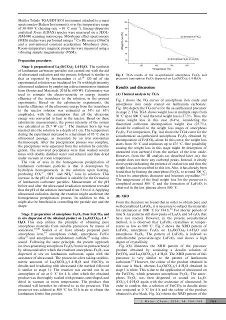
Fig. 1 TGA results <strong>of</strong> the as-synthesized amorphous Fe 2 O 3 and<br />
precursor (amorphous Fe 2 O 3 dispersed on La 2 O(CO 3 ) 2 ?1.4?H 2 O).<br />
Results and discussion<br />
(A) Thermal analysis by TGA<br />
Fig. 1 shows the TG curves <strong>of</strong> amorphous iron oxide and<br />
amorphous iron oxide coated on lanthanum carbonate.<br />
Fig. 1(b) depicts the TG curve for the as-synthesized precursor<br />
in stage 2. This TGA shows weight loss in multiple steps from<br />
30 uC up to 800 uC and the total weight loss is 27.3%. Thus, the<br />
excess weight loss in this case (8.4%), considering the<br />
theoretical carbonate decomposition weight loss (25.7%),<br />
should be confined to the weight loss stages <strong>of</strong> amorphous<br />
Fe 2 O 3 . For comparison, Fig. 1(a) shows the TGA curve for the<br />
sonochemical as-synthesized amorphous Fe 2 O 3 obtained by<br />
decomposition <strong>of</strong> Fe(CO) 5 alone. In this curve, the weight loss<br />
starts from 30 uC and continues up to 475 uC. One possibility<br />
causing the weight loss in this stage might be desorption <strong>of</strong><br />
unreacted iron carbonyl from the surface <strong>of</strong> the iron oxide.<br />
However, from the IR analysis (as described later on), the<br />
sample does not show any carbonyl peaks. Instead, it clearly<br />
shows peaks indicating the presence <strong>of</strong> oxalate ion and thus the<br />
weight loss can be ascribed to this ion. Also, it has already been<br />
found that by heating the amorphous Fe 2 O 3 , to around 300 uC,<br />
it loses its amorphous character and becomes crystalline. 20,23<br />
The temperature <strong>of</strong> the final weight loss for the precursor is<br />
completed around 800 uC and the formation <strong>of</strong> <strong>LaFeO</strong> 3 is<br />
observed in the last plateau above 800 uC.<br />
(B) XRD<br />
From the literature we found that in order to obtain pure and<br />
well-crystallized <strong>LaFeO</strong> 3 , it is necessary to subject the materials<br />
for calcination at 1000 uC for 184 h. 24 For shorter periods <strong>of</strong><br />
time X-ray patterns still show peaks <strong>of</strong> La 2 O 3 and a-Fe 2 O 3 that<br />
have not reacted. However, in the present sonochemical<br />
method, it is observed that <strong>LaFeO</strong> 3 crystallises at temperatures<br />
as low as 800 uC. Fig. 2 shows the XRD pattern <strong>of</strong><br />
<strong>LaFeO</strong> 3 , amorphous Fe 2 O 3 on La 2 O(CO 3 ) 2 ?1.4?H 2 O and<br />
amorphous Fe 2 O 3 . The pattern <strong>of</strong> <strong>LaFeO</strong> 3 is indexed as<br />
orthorhombic perovskite-type <strong>LaFeO</strong> 3 and shows a high<br />
degree <strong>of</strong> crystallinity.<br />
Fig. 2(b) illustrates the XRD pattern <strong>of</strong> the precursor<br />
product obtained by sonicating a decalin solution <strong>of</strong><br />
Fe(CO) 5 and La 2 O(CO 3 ) 2 ?1.4?H 2 O. The XRD pattern <strong>of</strong> this<br />
precursor is very similar to the pattern <strong>of</strong> lanthanum<br />
carbonate. 18 However, the colour <strong>of</strong> the product obtained in<br />
this case is black, whereas La 2 O(CO 3 ) 2 ?1.4?H 2 O obtained in<br />
stage 1 is white. This is due to the application <strong>of</strong> ultrasound on<br />
the Fe(CO) 5 , which generates amorphous Fe 2 O 3 . The amorphous<br />
Fe 2 O 3 was then dispersed or coated on La 2 O-<br />
(CO 3 ) 2 ?1.4?H 2 O again with the assistance <strong>of</strong> ultrasound. In<br />
order to confirm this, a solution <strong>of</strong> Fe(CO) 5 in decalin alone<br />
was sonicated at 0 uC for 4 h and the colour <strong>of</strong> the product<br />
obtained is also black. Fig. 2(a) shows the XRD pattern <strong>of</strong> the<br />
J. Mater. Chem., 2004, 14, 764–769 765