Sonochemical synthesis of nanocrystalline LaFeO 3 - ResearchGate
Sonochemical synthesis of nanocrystalline LaFeO 3 - ResearchGate
Sonochemical synthesis of nanocrystalline LaFeO 3 - ResearchGate

Create successful ePaper yourself
Turn your PDF publications into a flip-book with our unique Google optimized e-Paper software.
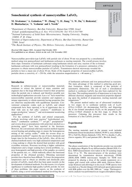
ARTICLE<br />
<strong>Sonochemical</strong> <strong>synthesis</strong> <strong>of</strong> <strong>nanocrystalline</strong> <strong>LaFeO</strong> 3<br />
M. Sivakumar, a A. Gedanken,* a W. Zhong, b Y. H. Jiang, b Y. W. Du, b I. Brukental, c<br />
D. Bhattacharya, c Y. Yeshurun c and I. Nowik d<br />
a Department <strong>of</strong> Chemistry, Bar-Ilan University, Ramat-Gan 52900, Israel.<br />
E-mail: gedanken@mail.biu.ac.il; Fax: 972-3-5351250; Tel: 972-3-5317769<br />
b National Laboratory <strong>of</strong> Solid State Microstructures, Nanjing University, Nanjing<br />
210093, P. R. China<br />
c Institute <strong>of</strong> Superconductivity, Department <strong>of</strong> Physics, Bar-Ilan University, Ramat-Gan<br />
52900, Israel<br />
d The Racah Institute <strong>of</strong> Physics, The Hebrew University, Jerusalem 91904, Israel<br />
Journal <strong>of</strong><br />
Materials<br />
Chemistry<br />
www.rsc.org/materials<br />
Received 28th August 2003, Accepted 22nd October 2003<br />
First published as an Advance Article on the web 11th November 2003<br />
Nanocrystalline perovskite-type <strong>LaFeO</strong> 3 with particle size <strong>of</strong> about 30 nm was prepared by a sonochemical<br />
method using iron pentacarbonyl and lanthanum carbonate as starting materials. The overall process involves<br />
three steps: formation <strong>of</strong> lanthanum carbonate using lanthanum nitrate and urea; reaction <strong>of</strong> the so-formed<br />
lanthanum carbonate with iron pentacarbonyl resulting in the formation <strong>of</strong> a precursor; calcination <strong>of</strong> the<br />
precursor to obtain <strong>nanocrystalline</strong> particles <strong>of</strong> <strong>LaFeO</strong> 3 . Transmission electron microscopy revealed the<br />
particles to have a mean size <strong>of</strong> about 30 nm. Study <strong>of</strong> the magnetic properties <strong>of</strong> <strong>nanocrystalline</strong> <strong>LaFeO</strong> 3<br />
particles shows a coercivity <strong>of</strong> y250 Oe, while the saturation magnetization is y40 memu g 21 .<br />
DOI: 10.1039/b310110j<br />
Introduction<br />
Research in submicroscopic or <strong>nanocrystalline</strong> materials<br />
continues to attract the interest <strong>of</strong> many scientists and<br />
engineers due to the large differences found in their properties<br />
when the particle size is reduced, and therefore possible new<br />
technological applications can arise. Lau et al. 1 have stated that<br />
<strong>nanocrystalline</strong> materials provide scientists and engineers a<br />
unique opportunity to obtain materials having properties that<br />
are otherwise unachievable with equilibrium materials. Conventional<br />
composite oxides such as <strong>LaFeO</strong> 3 and related<br />
compounds have been reported to be <strong>of</strong> importance due to<br />
their wide uses in fuel cells, 2 catalysts, 3,4 membranes in<br />
syngas production, 5 sensors, 6,7 and environmental monitoring<br />
applications. 8<br />
For the <strong>synthesis</strong> <strong>of</strong> <strong>LaFeO</strong> 3 and related compounds,<br />
methods involving solid state reaction, 9 hydrothermal <strong>synthesis</strong>,<br />
10 combustion <strong>synthesis</strong>, 11,12 sol–gel, 13 precipitation 14<br />
and reverse drop co-precipitation with poly(vinyl alcohol) as a<br />
protecting agent 15 have been reported in the literature.<br />
However, the conventional solid state reaction method requires<br />
several heating and grinding steps to ensure the homogeneous<br />
mixing <strong>of</strong> the various oxides, 16 while the reverse drop coprecipitation<br />
method 15 requires the use <strong>of</strong> more chemicals and<br />
longer time for the formation <strong>of</strong> the <strong>LaFeO</strong> 3 . Also, all the wet<br />
chemical methods need a very high calcination temperature and<br />
a long soaking to obtain the desired final powders with good<br />
crystalline structure. Processes involving a higher calcination<br />
temperature and a longer soaking have several problems, e.g.,<br />
poor homogeneity and high porosity <strong>of</strong> the samples, no control<br />
<strong>of</strong> the particle size, etc., which indirectly affects the functional<br />
properties <strong>of</strong> the obtained powders. Obtaining nanosized<br />
powders have therefore been difficult. To lower the preparation<br />
temperature and to obtain an ultrafine and chemically pure<br />
powder <strong>of</strong> <strong>LaFeO</strong> 3 , much effort has been spent recently on the<br />
development <strong>of</strong> chemical methods. For this reason, it is <strong>of</strong> great<br />
significance to <strong>synthesis</strong>e high quality <strong>LaFeO</strong> 3 powder. In view<br />
<strong>of</strong> these problems, this paper reports on the <strong>synthesis</strong> <strong>of</strong><br />
<strong>LaFeO</strong> 3 using a novel sonochemical method involving the use<br />
<strong>of</strong> lanthanum carbonate and iron pentacarbonyl as reactants.<br />
The main advantage <strong>of</strong> this method is achieving good product<br />
formation, which is composed <strong>of</strong> fine particles <strong>of</strong><br />
nanometric dimensions. The use <strong>of</strong> such a sonochemical<br />
method to synthesize <strong>LaFeO</strong> 3 has also been explored for the<br />
first time. The resulting material is <strong>of</strong> importance as it may have<br />
different electrical, magnetic and structural properties depending<br />
upon the method <strong>of</strong> preparation, starting materials and<br />
temperature. 17<br />
The present method makes use <strong>of</strong> ultrasound irradiation<br />
in two stages: (i) to synthesize uniform rods <strong>of</strong> La 2 O-<br />
(CO 3 ) 2 ?1.4?H 2 O, (ii) decomposing Fe(CO) 5 and dispersing<br />
the in situ formed amorphous Fe 2 O 3 on the prepared La 2 O-<br />
(CO 3 ) 2 ?1.4?H 2 O obtained in the first stage, to obtain the<br />
precursor. Calcining this precursor then results in the<br />
formation <strong>of</strong> a fine <strong>LaFeO</strong> 3 powder.<br />
Experimental<br />
Materials<br />
The starting materials used in the present work included<br />
lanthanum nitrate hexahydrate (Aldrich, 99.99%), urea (Aldrich,<br />
98%), Fe(CO) 5 (Strem chemicals, USA, 99.5%), decalin, C 10 H 18<br />
(Acros organics, 98%) and pentane (96%, Bio-lab, Israel).<br />
Characterisation<br />
The phase constitution <strong>of</strong> the hexaferrite powder was recorded<br />
by employing a Rigaku X-ray diffractometer (Model-2028, Co-<br />
Ka). XRD measurements were taken in the 2h scanning range<br />
from 15 to 70u. The microstructures <strong>of</strong> the products were<br />
determined by transmission electron microscopy (TEM)<br />
(JEOL-JEM 100SX microscope). Samples for TEM were<br />
prepared by placing a drop <strong>of</strong> the sample suspension on a<br />
copper grid coated with carbon (400 mesh, Electron Microscopy<br />
Sciences) and allowing it to dry in air. FTIR spectra were<br />
recorded on a Nicolet (Impact 410) infrared spectrophotometer<br />
with KBr pellets over the 400–3000 cm 21 range. Thermogravimetric<br />
analysis was performed in an oxygen atmosphere using a<br />
764 J. Mater. Chem., 2004, 14, 764–769 This journal is ß The Royal Society <strong>of</strong> Chemistry 2004
Mettler Toledo TGA/SDTA851 instrument attached to a mass<br />
spectrometer (Balzers Instruments), over the temperature range<br />
<strong>of</strong> 30–900 uC (heating rate y10 uC min 21 ). Energy-dispersive<br />
analytical X-ray (EDAX) spectra were measured on a JEOL-<br />
JSM-840 scanning microscope. Mössbauer effect spectroscopy<br />
(MES) studies were performed using a 57 Co:Rh source (50mCi)<br />
and a conventional constant acceleration Mössbauer drive.<br />
Room-temperature magnetic properties were measured using a<br />
vibrating sample magnetometer (VSM).<br />
Preparation procedures<br />
Stage 1: preparation <strong>of</strong> La 2 O(CO 3 ) 2 ?1.4?H 2 O. The <strong>synthesis</strong><br />
<strong>of</strong> lanthanum carbonate particles was carried out with the aid<br />
<strong>of</strong> ultrasound radiation and the process followed is similar to<br />
that as reported by Jeevanandam et al. 18 120 ml <strong>of</strong> the<br />
experimental solution was irradiated for 3 h with high intensity<br />
ultrasound radiation by employing a direct immersion titanium<br />
horn (Sonics and Materials, 20 kHz, 600 W). Calorimetry was<br />
used to estimate the electro-acoustic or energy transfer<br />
efficiency <strong>of</strong> the transducer to the solution, in the present<br />
experiments. Based on the calorimetry experiments, the<br />
transfer efficiency <strong>of</strong> the ultrasonic energy from the transducer<br />
to the reactor solution was estimated as 54% (at 65%<br />
amplitude), with the assumption that all the ultrasonic<br />
energy was converted to heat in the reactor. Based on these<br />
calorimetry measurements, the power intensity <strong>of</strong> the system<br />
was calculated as 29.7 W cm 22 . The titanium horn tip was<br />
inserted into the solution to a depth <strong>of</strong> 1 cm. The temperature<br />
during the experiment increased to a maximum <strong>of</strong> 85 uC due to<br />
ultrasound passage, as measured by an iron–constantan<br />
thermocouple. After the precipitation process was complete,<br />
the precipitates were separated from the solution by centrifugation.<br />
The recovered precipitates were then washed several<br />
times with doubly distilled water and ethanol and then dried<br />
under vacuum at room temperature.<br />
The role <strong>of</strong> urea in the homogeneous precipitation <strong>of</strong><br />
lanthanum carbonate particles is that it decomposes by<br />
hydrolysis homogeneously in the solution upon heating,<br />
producing CO 22 3 , OH 2 and NH 1 4 ions in solution. This<br />
increase in the pH <strong>of</strong> the medium is suitable for the formation<br />
<strong>of</strong> lanthanum carbonate particles. Measurement <strong>of</strong> the pH<br />
before and after the ultrasound irradiation treatment revealed<br />
that the pH <strong>of</strong> the solution increased from 5.6 to 6.4. Applying<br />
ultrasound radiation during the reaction might accelerate the<br />
homogeneous precipitation process. In addition to this, it<br />
might also be beneficial in controlling the particle size and the<br />
shape.<br />
Stage 2: preparation <strong>of</strong> amorphous Fe 2 O 3 from Fe(CO) 5 and<br />
in situ dispersion <strong>of</strong> the obtained product on La 2 O(CO 3 ) 2 ?1.4?-<br />
H 2 O. This step utilizes the principle <strong>of</strong> obtaining pure<br />
amorphous materials from suitable precursors by means <strong>of</strong><br />
sonication. 19,20 Suslick et al. have already prepared pure<br />
amorphous iron, 19 amorphous cobalt, amorphous Fe/Co<br />
alloy 21 and amorphous molybdenum carbide, 22 using ultrasound.<br />
Following the same principle, the present approach<br />
involves generating amorphous Fe 2 O 3 from iron pentacarbonyl<br />
by ultrasound after which the resultant amorphous Fe 2 O 3 was<br />
dispersed in situ on lanthanum carbonate, again with the<br />
assistance <strong>of</strong> ultrasound. The process involves taking stoichiometric<br />
amounts <strong>of</strong> La 2 O(CO 3 ) 2 ?1.4?H 2 O and Fe(CO) 5 in<br />
decalin and irradiating with ultrasound (the method followed<br />
is similar to stage 1). The reaction was carried out in an<br />
atmosphere <strong>of</strong> air at 0 uC for 4 h, after which the obtained<br />
product was thoroughly washed with pentane, centrifuged, and<br />
dried in vacuum at room temperature. The product thus<br />
obtained will hereafter be referred to as the precursor. This<br />
precursor was calcined at 800 uC for 24 h in air to obtain the<br />
lanthanum ferrite fine powder.<br />
Fig. 1 TGA results <strong>of</strong> the as-synthesized amorphous Fe 2 O 3 and<br />
precursor (amorphous Fe 2 O 3 dispersed on La 2 O(CO 3 ) 2 ?1.4?H 2 O).<br />
Results and discussion<br />
(A) Thermal analysis by TGA<br />
Fig. 1 shows the TG curves <strong>of</strong> amorphous iron oxide and<br />
amorphous iron oxide coated on lanthanum carbonate.<br />
Fig. 1(b) depicts the TG curve for the as-synthesized precursor<br />
in stage 2. This TGA shows weight loss in multiple steps from<br />
30 uC up to 800 uC and the total weight loss is 27.3%. Thus, the<br />
excess weight loss in this case (8.4%), considering the<br />
theoretical carbonate decomposition weight loss (25.7%),<br />
should be confined to the weight loss stages <strong>of</strong> amorphous<br />
Fe 2 O 3 . For comparison, Fig. 1(a) shows the TGA curve for the<br />
sonochemical as-synthesized amorphous Fe 2 O 3 obtained by<br />
decomposition <strong>of</strong> Fe(CO) 5 alone. In this curve, the weight loss<br />
starts from 30 uC and continues up to 475 uC. One possibility<br />
causing the weight loss in this stage might be desorption <strong>of</strong><br />
unreacted iron carbonyl from the surface <strong>of</strong> the iron oxide.<br />
However, from the IR analysis (as described later on), the<br />
sample does not show any carbonyl peaks. Instead, it clearly<br />
shows peaks indicating the presence <strong>of</strong> oxalate ion and thus the<br />
weight loss can be ascribed to this ion. Also, it has already been<br />
found that by heating the amorphous Fe 2 O 3 , to around 300 uC,<br />
it loses its amorphous character and becomes crystalline. 20,23<br />
The temperature <strong>of</strong> the final weight loss for the precursor is<br />
completed around 800 uC and the formation <strong>of</strong> <strong>LaFeO</strong> 3 is<br />
observed in the last plateau above 800 uC.<br />
(B) XRD<br />
From the literature we found that in order to obtain pure and<br />
well-crystallized <strong>LaFeO</strong> 3 , it is necessary to subject the materials<br />
for calcination at 1000 uC for 184 h. 24 For shorter periods <strong>of</strong><br />
time X-ray patterns still show peaks <strong>of</strong> La 2 O 3 and a-Fe 2 O 3 that<br />
have not reacted. However, in the present sonochemical<br />
method, it is observed that <strong>LaFeO</strong> 3 crystallises at temperatures<br />
as low as 800 uC. Fig. 2 shows the XRD pattern <strong>of</strong><br />
<strong>LaFeO</strong> 3 , amorphous Fe 2 O 3 on La 2 O(CO 3 ) 2 ?1.4?H 2 O and<br />
amorphous Fe 2 O 3 . The pattern <strong>of</strong> <strong>LaFeO</strong> 3 is indexed as<br />
orthorhombic perovskite-type <strong>LaFeO</strong> 3 and shows a high<br />
degree <strong>of</strong> crystallinity.<br />
Fig. 2(b) illustrates the XRD pattern <strong>of</strong> the precursor<br />
product obtained by sonicating a decalin solution <strong>of</strong><br />
Fe(CO) 5 and La 2 O(CO 3 ) 2 ?1.4?H 2 O. The XRD pattern <strong>of</strong> this<br />
precursor is very similar to the pattern <strong>of</strong> lanthanum<br />
carbonate. 18 However, the colour <strong>of</strong> the product obtained in<br />
this case is black, whereas La 2 O(CO 3 ) 2 ?1.4?H 2 O obtained in<br />
stage 1 is white. This is due to the application <strong>of</strong> ultrasound on<br />
the Fe(CO) 5 , which generates amorphous Fe 2 O 3 . The amorphous<br />
Fe 2 O 3 was then dispersed or coated on La 2 O-<br />
(CO 3 ) 2 ?1.4?H 2 O again with the assistance <strong>of</strong> ultrasound. In<br />
order to confirm this, a solution <strong>of</strong> Fe(CO) 5 in decalin alone<br />
was sonicated at 0 uC for 4 h and the colour <strong>of</strong> the product<br />
obtained is also black. Fig. 2(a) shows the XRD pattern <strong>of</strong> the<br />
J. Mater. Chem., 2004, 14, 764–769 765
Fig. 2 Powder X-Ray diffraction patterns <strong>of</strong> (a) amorphous Fe 2 O 3 ,<br />
(b) amorphous Fe 2 O 3 dispersed on La 2 O(CO 3 ) 2 ?1.4?H 2 Oand(c)<strong>LaFeO</strong> 3<br />
obtained by calcining the precursor at 800 uC in air for 24 h.<br />
resultant product obtained during this process. It can be seen<br />
from this pattern that the Fe 2 O 3 obtained in this process is<br />
amorphous. The absence <strong>of</strong> this amorphous background due to<br />
Fe 2 O 3 in Fig. 2(b) may be due to the dominance <strong>of</strong> crystalline<br />
La 2 O(CO 3 ) 2 ?1.4?H 2 O peaks. Fig. 2(c) shows the XRD pattern<br />
<strong>of</strong> <strong>LaFeO</strong> 3 obtained by calcining the precursor sample at 800 uC<br />
in air atmosphere for 24 h. The diffraction peaks match those<br />
reported for standard <strong>LaFeO</strong> 3 (JCPDS file No. 37-1493).<br />
Heating the precursor at a rate <strong>of</strong> 8 uC min 21 to 800 uC resulted<br />
in a well-crystallized ferrite phase. This was indicated by the<br />
appearance <strong>of</strong> the most intense reflections: (121), (202) and<br />
(240) peaks at 2h values <strong>of</strong> 38.28, 54.78 and 68.43u, respectively,<br />
as shown in Fig. 2(c). Thus, the formation <strong>of</strong> <strong>LaFeO</strong> 3 is<br />
completed at 800 uC. The formation temperature for <strong>LaFeO</strong> 3<br />
by the sonochemically prepared precursor is clearly lower than<br />
that observed in the conventional solid-state reaction utilizing<br />
La 2 O(CO 3 ) 2 ?1.4?H 2 O and Fe 2 O 3 (y1000 uC for 184 h) for<br />
forming a single phase <strong>of</strong> <strong>LaFeO</strong> 24 3 . Thus, the formation<br />
temperature <strong>of</strong> <strong>LaFeO</strong> 3 by this process compares reasonably<br />
well with many other chemistry based processing routes.<br />
(C) IR<br />
The IR spectra <strong>of</strong> the amorphous iron oxide coated on lanthanum<br />
carbonate, amorphous iron oxide (for comparison) and<br />
calcined <strong>nanocrystalline</strong> <strong>LaFeO</strong> 3 powder in the wavenumber<br />
range from 3000 to 400 cm 21 are shown in Fig. 3(a)–(c). The IR<br />
spectrum <strong>of</strong> Fig. 3(b) provides evidence for the presence <strong>of</strong><br />
carbonate ions <strong>of</strong> La 2 O(CO 3 ) 2 ?1.4H 2 O in the precursor; it also<br />
shows a broad band at ca. 1400cm 21 , which is assigned to the<br />
bending vibrational mode <strong>of</strong> bound water molecules. 18 The<br />
absorption band at ca.1481cm 21 is attributed to the n 3 mode <strong>of</strong><br />
the CO 22 3 ion while the other bands at ca. 1071, 850 and 725 cm 21<br />
have been assigned to the n 1 , n 2 and n 4 modes <strong>of</strong> the carbonate<br />
ions, respectively. 18 The above observed bands clearly indicate the<br />
presence <strong>of</strong> carbonate ions in the precursor. However, this<br />
spectrum does not show the peaks for amorphous Fe 2 O 3 which<br />
might be due to the relatively high intensity peaks <strong>of</strong> lanthanum<br />
carbonate. The IR spectrum <strong>of</strong> the amorphous Fe 2 O 3 synthesized<br />
by ultrasound alone (for comparison) shows absorption bands at<br />
473, 1424 and 1550 cm 21 (Fig. 3(a)). Also, the peaks observed at<br />
1424 and 1550 cm 21 support the presence <strong>of</strong> oxalate ion.<br />
In addition, the peak centered at 473 cm 21 indicates the presence<br />
<strong>of</strong> amorphous iron oxide. 25 Fig. 3(c) shows the IR spectrum <strong>of</strong><br />
<strong>LaFeO</strong> 3 . This spectrum shows well-established strong absorption<br />
bands at y570 and y430 cm 21 in the powder calcined at<br />
Fig. 3 IR spectra <strong>of</strong> (a) amorphous Fe 2 O 3 , (b) amorphous Fe 2 O 3<br />
dispersed on La 2 O(CO 3 ) 2 ?1.4H 2 O and (c) <strong>LaFeO</strong> 3 obtained by<br />
calcining the precursor at 800 uC in air for 24 h.<br />
800 uC indicating the formation <strong>of</strong> lanthanum ferrite. The<br />
570 cm 21 band is attributed to the Fe–O stretching vibration<br />
(n 1 mode), and the 430 cm 21 band corresponds to the O–Fe–O<br />
deformation vibration (n 2 mode). 26 Comparing, Fig. 3(c) with<br />
Fig. 3(a) and (b), we can see after calcination that the<br />
characteristic bands <strong>of</strong> La 2 O- (CO 3 ) 2 ?1.4H 2 O and iron oxide<br />
vanish at this calcination temperature and only Fe–O stretching<br />
vibration bands are found in the <strong>synthesis</strong>ed powder. The above<br />
interpretation <strong>of</strong> the IR spectral results is based on the assumption<br />
that the synthesized carbonate, Fe 2 O 3 , precursor, and lanthanum<br />
ferrite are essentially pure materials. These results agree with the<br />
XRD phase-analysis findings.<br />
(D) EDAX<br />
Elemental analysis using EDAX indicates that the product <strong>of</strong><br />
calcination obtained from the precursor has a La:Fe ratio <strong>of</strong><br />
1:1 (within the range 0.98–1.01) indicating the equal presence<br />
<strong>of</strong> La and Fe in the sample. Also, a similar analysis for the<br />
amorphous Fe 2 O 3 obtained from Fe(CO) 5 shows the presence<br />
<strong>of</strong> Fe and O in a ratio ranging from 1:1.5 to 1:2. The higher<br />
ratio is due to the strong adsorption <strong>of</strong> oxygen as has been<br />
reported by Cao et al. 23 No other elements were detected in the<br />
EDAX <strong>of</strong> the amorphous compound.<br />
(E) Mössbauer studies<br />
As confirmation <strong>of</strong> the phase purity <strong>of</strong> the prepared samples,<br />
Fig. 4 shows the 57 Fe Mössbauer spectrum measured at 27 uC<br />
Fig. 4 Mössbauer spectrum <strong>of</strong> the <strong>LaFeO</strong> 3 sample obtained by<br />
calcining the precursor at 800 uC in air for 24 h.<br />
766 J. Mater. Chem., 2004, 14, 764–769
<strong>of</strong> the <strong>LaFeO</strong> 3 powder calcined at 800 uC for 24 h in air. A<br />
theoretical spectrum (the solid line) least-squared fitted to the<br />
experimental points, yields the following hyperfine interaction<br />
parameters: isomer shift relative to iron metal: 0.30(3) mm 21 ,<br />
Quadrupole interaction Q/2 ~ 20.06(3) mm 21 , and magnetic<br />
hyperfine field 521(2) kOe. These parameters agree perfectly<br />
with those <strong>of</strong> bulk <strong>LaFeO</strong> 3 . 27<br />
(F) Permeability spectra<br />
A useful route to investigate the mechanism <strong>of</strong> domain wall<br />
motions and domain rotations is to measure the complex<br />
permeability (m* ~ m’2 im@) as a function <strong>of</strong> frequency, i.e., the<br />
so-called magnetic spectrum. In addition, by studying such a<br />
magnetic spectrum, the effective magnetic anisotropy field, as<br />
well as the damping mechanism <strong>of</strong> domain wall displacements,<br />
can be checked. In general, the dispersions caused by wall<br />
displacements occur in a radio frequency range (below<br />
10 MHz), and those caused by domain rotations <strong>of</strong>ten<br />
appear in the microwave range (100 MHz to 10 GHz). This<br />
methodology has been extensively used in ferrites. 28,29 Thus, a<br />
similar study has been carried out for the <strong>LaFeO</strong> 3 powder<br />
prepared by the present ultrasonic method and calcination at<br />
800 uC for 24 h in air.<br />
The frequency dependence <strong>of</strong> the complex permeability (real<br />
part m’ and imaginary part m@) is shown in Fig. 5. For<br />
permeability measurements, the nanosized powder sample was<br />
pressed to a ring with a typical size <strong>of</strong> 13 mm OD, 7 mm ID and<br />
1 mm thickness. The pressed ring has a lower density than the<br />
sintered one. For polycrystalline ferrites, the permeability is<br />
related to the magnetizing mechanisms: spin ratation and<br />
domain wall motion. According to theory, in single domain<br />
grains there are no domain wall motions. Therefore, m 0 (static<br />
permeability, i.e. m’ with frequency limit to zero) is only caused<br />
by reversible domain rotations and follows the relation m 0 ~<br />
1 1 8pM s /3H a , where H a is the effective magnetic anisotropy<br />
field. Because M s <strong>of</strong> <strong>LaFeO</strong> 3 is very small, the complex<br />
permeability is small. In addition, the nonmagnetic impurities<br />
such as boundaries and cavities from the low-density sample<br />
may cause the permeability to decline further. In the<br />
corresponding bulk sample, there is domain wall motion,<br />
whereas, in nanosized samples, there are only reversible<br />
domain rotations. The m 0 contributed by domain rotations is<br />
generally smaller than that by domain wall displacements. It<br />
shoud be noted that the imaginary part m@ <strong>of</strong> <strong>nanocrystalline</strong><br />
perovskite-type <strong>LaFeO</strong> 3 is very small, which may be important<br />
for further <strong>nanocrystalline</strong> dielectric materials development.<br />
Fig. 6 (a) Transmission electron micrograph <strong>of</strong> amorphous Fe 2 O 3<br />
dispersed on La 2 O(CO 3 ) 2 ?1.4H 2 O and (b) transmission electron<br />
micrograph <strong>of</strong> <strong>LaFeO</strong> 3 obtained by calcining the precursor at 800 uC<br />
in air for 24 h.<br />
(G) TEM<br />
Fig. 6(a) depicts a TEM image <strong>of</strong> the amorphous Fe 2 O 3<br />
powder dispersed on the hexagonal La 2 O(CO 3 ) 2 ?1.4H 2 O rods<br />
(average diameter and length are 1.0 and 0.15 mm, respectively).<br />
It is clearly seen that there is no crystalline formation <strong>of</strong><br />
Fe 2 O 3 in this powder. It can also be observed that this<br />
amorphous Fe 2 O 3 is an agglomerate <strong>of</strong> small particles<br />
aggregated in a spongelike form. The micrograph in Fig. 6(b)<br />
indicates the morphology <strong>of</strong> the lanthanum ferrite sample<br />
obtained after treating the precursor at 800 uC. It can be<br />
observed from this micrograph that after calcination, the<br />
particles have an average diameter <strong>of</strong> about 30 nm, which<br />
further confirms the nanometer dimensions <strong>of</strong> the particles. No<br />
remaining La 2 O(CO 3 ) 2 ?1.4H 2 O and Fe 2 O 3 could be observed<br />
in the micrograph in spite <strong>of</strong> the low heating treatment<br />
temperature. This result agrees with the results <strong>of</strong> XRD and IR.<br />
Fig. 5 Plots <strong>of</strong> complex permeability as a function <strong>of</strong> frequency for a<br />
polycrystalline <strong>LaFeO</strong> 3 sample at room temperature.<br />
(H) Magnetic properties <strong>of</strong> <strong>nanocrystalline</strong> <strong>LaFeO</strong> 3<br />
Fig. 7 shows a magnetic hysteresis loop <strong>of</strong> <strong>nanocrystalline</strong><br />
<strong>LaFeO</strong> 3 obtained by the present sonochemical method. We<br />
have measured the magnetic properties <strong>of</strong> the <strong>nanocrystalline</strong><br />
J. Mater. Chem., 2004, 14, 764–769 767
(specifically, amorphous Fe 2 O 3 ) also contributes to the<br />
lowering <strong>of</strong> calcination temperature. It can be seen from the<br />
work <strong>of</strong> Vazquez-Vazquez et al. 24 that by using conventional<br />
lanthanum and iron nitrates, the mean particle size <strong>of</strong> the<br />
ferrite obtained is about 120 nm, at a calcination temperature<br />
<strong>of</strong> 800 uC. In addition, formation <strong>of</strong> the secondary phases <strong>of</strong><br />
La(OH) 3 , a-Fe 2 O 3 and ferrihydrite have also been noted at this<br />
temperature. It has also been found that a temperature <strong>of</strong><br />
1000 uC for 184 h is necessary to obtain pure and wellcrystallized<br />
<strong>LaFeO</strong> 3 . However, in the present case, by applying<br />
a calcination temperature <strong>of</strong> 800 uC for 24 h, the mean particle<br />
size <strong>of</strong> the obtained ferrite is 30 nm and no secondary phases<br />
are observed, which might be due to the highly reactive<br />
amorphous Fe 2 O 3 generated by ultrasound already present in<br />
the system.<br />
Fig. 7 The magnetization vs. applied field (measured at room temperature)<br />
pattern for the ultrafine <strong>LaFeO</strong> 3 system prepared by a<br />
sonochemical technique followed by calcination at 800 uC in air for 24 h.<br />
<strong>LaFeO</strong> 3 at room temperature using a vibrating sample<br />
magnetometer (VSM). From the hysteresis plot, we can<br />
observe hysteresis but no clear saturation <strong>of</strong> magnetization<br />
within the field limit <strong>of</strong> y16000 Oe. Normally, the <strong>LaFeO</strong> 3<br />
system is either weakly ferromagnetic (WFM) or antiferromagnetic<br />
(AFM) with a Néel temperature T N y 477 uC. 30 For<br />
a 30 nm <strong>LaFeO</strong> 3 particle size system, Qi et al. 31 reported a<br />
coercive field <strong>of</strong> y90 G and a saturation magnetization M s <strong>of</strong><br />
y2.75 emu g 21 at room temperature. In our case, the<br />
corresponding values are y250 Oe and y40 memu g 21 ,<br />
respectively. This reflects a further increase in the magnetocrystalline<br />
anisotropy, which is expected in the <strong>nanocrystalline</strong><br />
system. The squareness ratio M r /M s in our case is y0.055,<br />
which is quite small, compared to the value (y0.135)<br />
reported. 31 All these results point out to the presence <strong>of</strong> a<br />
large number <strong>of</strong> fine particles in our system. The average<br />
particle size <strong>of</strong> our system is y30 nm.<br />
In the conventional solid-state reaction <strong>of</strong> Fe 2 O 3 with<br />
lanthanum carbonate to form the lanthanum ferrite, the<br />
major limitation is the diffusion factor. In the present<br />
sonochemical method this diffusion resistance may be overcome,<br />
as there is an in situ generation <strong>of</strong> amorphous Fe 2 O 3<br />
from Fe(CO) 5 by ultrasound, followed by its dispersion or<br />
deposition on lanthanum carbonate, again assisted by ultrasound.<br />
This process might facilitate the diffusion, as well as the<br />
reaction, <strong>of</strong> highly reactive lanthanum ion (La 31 ) with the<br />
amorphous Fe 2 O 3 , resulting in the completion <strong>of</strong> the phase<br />
transition <strong>of</strong> the precursor to the <strong>LaFeO</strong> 3 ferrite at a lower<br />
calcination temperature <strong>of</strong> 800 uC, employed in the present<br />
method.<br />
The amorphous nature <strong>of</strong> Fe 2 O 3 synthesized by ultrasound<br />
might also play a key role in increasing its reactivity with<br />
lanthanum carbonate, resulting in the formation <strong>of</strong> a ferrite<br />
phase at the lower calcination temperature. The importance <strong>of</strong><br />
amorphicity in increasing the reactivity has already been<br />
discussed. 32 Bellisent et al. 21 have clearly shown that sonochemically<br />
prepared amorphous Fe, Co and Fe–CO nanomaterials<br />
are better catalysts than the corresponding crystalline<br />
nanomaterials prepared by other methods. In addition, Yee<br />
et al. 33 have functionalized amorphous Fe 2 O 3 nanoparticles<br />
with alkanesulfonic and octadecanephosphonic acids. This<br />
work further supports the fact that it is possible to form a<br />
coating <strong>of</strong> organic molecules more easily on amorphous Fe 2 O 3 .<br />
In addition to the above factors, the size <strong>of</strong> the reactants<br />
Conclusions<br />
In the present investigation, <strong>LaFeO</strong> 3 has been synthesized from<br />
lanthanum carbonate and iron pentacarbonyl via a sonochemical<br />
method for the first time. The sonochemically synthesized<br />
<strong>LaFeO</strong> 3 is a <strong>nanocrystalline</strong> material with high purity and good<br />
homogeneity. It was obtained with lower processing temperatures<br />
and shorter annealing times. The obtained nan<strong>of</strong>errite<br />
powder is expected to be used in applications for functional<br />
ceramics (sensors, SOFCs, electrodes, or even magnetic and<br />
ferroelectric applications).<br />
Acknowledgements<br />
We thank The Ministry <strong>of</strong> Science, Sport and Culture for a<br />
Materials Science grant through the Sino–Israeli program in<br />
Materials Science. We also thank Ms Louise Braverman for<br />
editorial assistance.<br />
References<br />
1 M. L. Lau, H. G. Jiang, R. J. Perez, J. Juarezislas and<br />
E. J. Lavernia, Nanostruct. Mater., 1996, 7, 847.<br />
2 N. Q. Minh, J. Am. Ceram. Soc., 1993, 76, 563.<br />
3 A. Delmastro, D. Mazza, S. Ronchetti, M. Vallino, R. Spinicci,<br />
P. Brovetto and M. Salis, Mater. Sci. Eng. B, 2001, 79, 140.<br />
4 S. L. Bai, X. X. Fu, J. Z. Wang, Q. H. Yang, Y. H. Sun and<br />
S. L. Zeng, Chin. J. Appl. Chem., 2000, 17, 343.<br />
5 Q. Ming, M. D. Nersesyan, A. Wagner, J. Ritchie,<br />
J. T. Richardson, D. Luss, A. J. Jacobson and Y. L. Yang,<br />
Solid State Ionics, 1999, 122, 113.<br />
6 T. Arakawa, H. Kurachi and J. Shiokawa, J. Mater. Sci., 1985, 20,<br />
1207.<br />
7 Y. Shimizu, M. Shimabukuro, H. Arai and T. Seiyama, Chem.<br />
Lett., 1985, 163, 917.<br />
8 G. Martinelli, M. C. Carotta, M. Ferroni, Y. Sadaoka and<br />
E. Traversa, Sens. Actuators B, 1999, 55, 99.<br />
9 B. I. Ita, Ph.D Thesis, University <strong>of</strong> Calabar, Nigeria, 1998.<br />
10 W. J. Zheng, R. H. Liu, D. K. Peng and G. Y. Meng, Mater. Lett.,<br />
2000, 43, 19.<br />
11 S. S. Manoharan and K. C. Patil, J. Solid State Chem., 1993, 102,<br />
267.<br />
12 A. Chakraborty, P. S. Devi and H. S. Maiti, J. Mater. Res., 1995,<br />
10, 918.<br />
13 J. Xiao, G. Y. Hong, D. C. Yu, X. T. Dong, D. J. Wang, L. Jiang<br />
and T. J. Li, Acta Chim. Sin., 1994, 52, 784.<br />
14 N. Pandya, R. G. Kulkarni and P. H. Parsania, Mater. Res. Bull.,<br />
1990, 25, 1073.<br />
15 X. Li, H. B. Zhang and M. Y. Zhao, Mater. Chem. Phys., 1994, 37,<br />
132.<br />
16 P. Ravindranathan, S. Komarneni and R. Roy, J. Mater. Sci.<br />
Lett., 1993, 12, 369.<br />
17 B. I. Ita, U. J. Ekpe and U. J. Ibok, Global J. Pure Appl. Chem.,in<br />
press.<br />
18 P. Jeevanandam, Y. Koltypin, O. Palchik and A. Gedanken,<br />
J. Mater. Chem., 2001, 11, 869.<br />
19 K. S. Suslick, S. B. Choe, A. A. Cichowlas and M. W. Grinstaff,<br />
Nature, 1991, 353, 414.<br />
768 J. Mater. Chem., 2004, 14, 764–769
20 X. Cao, Y. Koltypin, R. Prozorov, G. Kataby and A. Gedanken,<br />
J. Mater. Chem., 1997, 7, 2447.<br />
21 R. Bellissent, G. Galli, T. Hyeon, S. Magazu, D. Majolino,<br />
P. Migliardo and K. S. Suslick, Phys. Scr., 1995, T57, 79.<br />
22 T. H. Hyeon, M. M. Fang and K. S. Suslick, J. Am. Chem. Soc.,<br />
1996, 118, 5492.<br />
23 X. Cao, R. Prozorov, Y. Koltypin, G. Kataby and A. Gedanken,<br />
J. Mater. Res., 1997, 12, 402.<br />
24 C. Vazquez-Vazquez, P. Kogerler, M. A. Lopez-Quintela,<br />
R. D. Sanchez and J. Rivas, J. Mater. Res., 1998, 13, 451.<br />
25 B. Grzeta, M. Ristic, I. Nowik and S. Music, J. Alloys Compd.,<br />
2002, 344, 304.<br />
26 G. V. S. Rao, C. N. R. Rao and J. R. Ferraro, Appl. Spectrosc.,<br />
1970, 24, 436.<br />
27 M. Eibschut, S. Shtrikma and D. Treves, Phys. Rev., 1967, 156,<br />
562.<br />
28 G. T. Rado, R. W. Wright and W. H. Emerson, Phys. Rev., 1950,<br />
80, 273.<br />
29 G. T. Rado, R. W. Wright, W. H. Emerson and A. Terris, Phys.<br />
Rev., 1952, 88, 909.<br />
30 Y. N. Venevtsev and V. V. Gagulin, Ferroelectrics, 1994, 162, 23.<br />
31 X. W. Qi, J. Zhou, Z. X. Yue, Z. L. Gui and L. T. Li, Mater.<br />
Chem. Phys., 2003, 78, 25.<br />
32 K. S. Suslick, T. W. Hyeon and M. M. Fang, Chem. Mater., 1996,<br />
8, 2172.<br />
33 C. Yee, G. Kataby, A. Ulman, T. Prozorov, H. White, A. King,<br />
M. Rafailovich, J. Sokolov and A. Gedanken, Langmuir, 1999, 15,<br />
7111.<br />
J. Mater. Chem., 2004, 14, 764–769 769