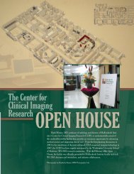
The Center for Clinical Imaging Research - Mallinckrodt Institute of ...
The Center for Clinical Imaging Research - Mallinckrodt Institute of ...
The Center for Clinical Imaging Research - Mallinckrodt Institute of ...

You also want an ePaper? Increase the reach of your titles
YUMPU automatically turns print PDFs into web optimized ePapers that Google loves.
<strong>The</strong> <strong>Center</strong> <strong>for</strong><br />
<strong>Clinical</strong> <strong>Imaging</strong><br />
<strong>Research</strong><br />
A COMPREHENSIVE IMAGING CENTER DEDICATED TO RESEARCH<br />
by Candace O’Connor<br />
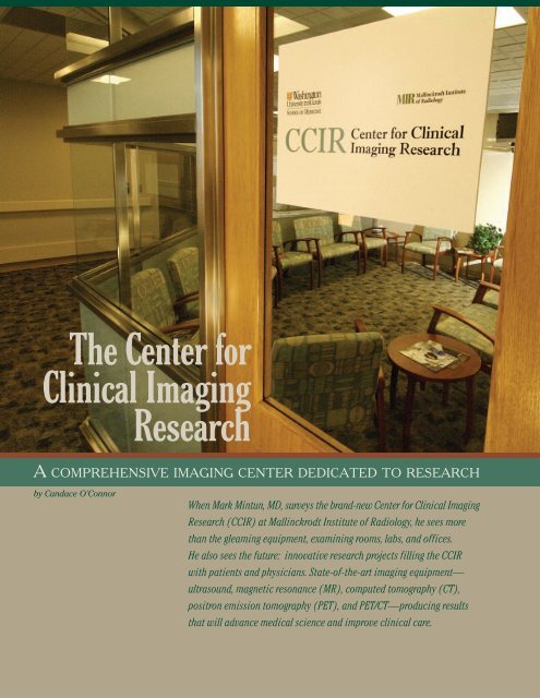
When Mark Mintun, MD, surveys the brand-new <strong>Center</strong> <strong>for</strong> <strong>Clinical</strong> <strong>Imaging</strong><br />
<strong>Research</strong> (CCIR) at <strong>Mallinckrodt</strong> <strong>Institute</strong> <strong>of</strong> Radiology, he sees more<br />
than the gleaming equipment, examining rooms, labs, and <strong>of</strong>fices.<br />
He also sees the future: innovative research projects filling the CCIR<br />
with patients and physicians. State-<strong>of</strong>-the-art imaging equipment—<br />
ultrasound, magnetic resonance (MR), computed tomography (CT),<br />
positron emission tomography (PET), and PET/CT—producing results<br />
that will advance medical science and improve clinical care.
<strong>The</strong> <strong>Center</strong> <strong>for</strong><br />
<strong>Clinical</strong> <strong>Imaging</strong><br />
<strong>Research</strong><br />
He points out a well-appointed<br />
lab that will test blood samples<br />
and a processing room where<br />
scientists will view images at<br />
top-<strong>of</strong>-the-line workstations. One <strong>of</strong><br />
the side rooms will store carts that<br />
hold researchers’ supplies used <strong>for</strong><br />
the imaging studies. Though it is<br />
nearly empty now, the “room will<br />
be lined with carts,”<br />
predicts Mintun, the<br />
CCIR director, with<br />
enthusiasm. “<strong>The</strong>y will<br />
need to have parking<br />
spots inside!”<br />
For now, the CCIR<br />
is just getting started.<br />
<strong>The</strong> 8,800-square-foot<br />
facility, on the tenth<br />
floor <strong>of</strong> the Barnes-<br />
Jewish Hospital West<br />
Mark Mintun, MD Pavilion and near the<br />
Neurosurgery Intensive Care Unit<br />
(ICU), has been open since the<br />
end <strong>of</strong> June. While the staff is still<br />
adding some finishing touches, the<br />
opening fanfare is yet to come,<br />
with a gala reception scheduled<br />
<strong>for</strong> later this year.<br />
Yet this young CCIR already is<br />
setting the standard <strong>for</strong> radiology<br />
research nationwide because <strong>of</strong> its<br />
meticulously planned layout and<br />
extraordinary location. Not only<br />
are neurosurgery patients just<br />
steps away, but the Surgery ICU is<br />
two floors down, so that inpatients<br />
can be transported <strong>for</strong> studies with<br />
little fuss or risk. In other medical<br />
centers, imaging research facilities<br />
are <strong>of</strong>ten blocks away.<br />
“A comprehensive imaging<br />
center and a research-dedicated<br />
imaging center, right in the middle<br />
<strong>of</strong> a hospital—we don’t know <strong>of</strong><br />
any other medical group that has<br />
done this,” says Mintun, a pr<strong>of</strong>essor<br />
<strong>of</strong> radiology who also holds joint<br />
appointments in psychiatry, neurobiology,<br />
and biomedical engineering.<br />
“It is a huge advantage to have<br />
a state-<strong>of</strong>-the-art imaging facility<br />
that allows us to routinely conduct<br />
clinical research involving inpatients<br />
as well as outpatients,” adds Gilbert<br />
Jost, MD, director <strong>of</strong> <strong>Mallinckrodt</strong><br />
<strong>Institute</strong> and chair <strong>of</strong> Washington<br />
University’s Department <strong>of</strong> Radiology.<br />
THE CCIR TAKES SHAPE<br />
In 2003, Mintun began discussing<br />
the idea <strong>of</strong> such a center with Jost<br />
and colleagues Marcus Raichle, MD,<br />
then cochief <strong>of</strong> the Division <strong>of</strong><br />
Radiological Sciences; William<br />
Powers, MD, a renowned neurologist<br />
at Washington University; and<br />
Barry Siegel, MD, chief <strong>of</strong> the Division<br />
<strong>of</strong> Nuclear Medicine. <strong>The</strong>y knew<br />
that a <strong>Mallinckrodt</strong> <strong>Institute</strong>-owned,<br />
clinical radiology area inside<br />
Barnes-Jewish Hospital would<br />
soon be vacant as equipment was<br />
being moved to the <strong>Center</strong> <strong>for</strong><br />
Advanced <strong>Imaging</strong>. What would be<br />
the best use <strong>for</strong> that empty space?<br />
<strong>The</strong> group was inspired by a<br />
well-used PET scanner inside the<br />
neuro-ICU, a valuable tool <strong>for</strong><br />
researchers because <strong>of</strong> its close<br />
proximity to patients. “Because <strong>of</strong><br />
its location, that PET scanner has<br />
greatly advanced the understanding<br />
<strong>of</strong> what goes on in traumatic brain<br />
injury and stroke, and the scanner<br />
also has been used <strong>for</strong> research<br />
studies <strong>of</strong> depression,” says<br />
Mintun, who has conducted some<br />
<strong>of</strong> his own studies on that machine.<br />
In 2005, <strong>Mallinckrodt</strong> <strong>Institute</strong><br />
signed an agreement with Siemens<br />
Medical Solutions: <strong>The</strong> medical<br />
equipment manufacturer would<br />
provide its most advanced, Food<br />
and Drug Administration-approved<br />
imaging technology, and the <strong>Institute</strong><br />
would use it <strong>for</strong> cutting-edge therapy<br />
research. <strong>The</strong>y would “push this<br />
equipment to the limit,” says Mintun,<br />
“and find out what it can do.”<br />
BUILDING THE CCIR<br />
Designing and building the<br />
CCIR required much <strong>of</strong> Mintun’s<br />
time <strong>for</strong> three years. Once the<br />
architects had sketches in place,<br />
focus groups (including cardiac,<br />
cancer, and brain researchers)<br />
spent hours discussing how<br />
many scanners to acquire or what<br />
special-use rooms to construct.<br />
Real-time, 3-D/4-D ultrasound technology<br />
has advantages over the conventional 2-D<br />
method, including shorter exam times; data<br />
manipulation <strong>for</strong> obtaining more in<strong>for</strong>mation<br />
<strong>for</strong> treatment planning, chemotherapy,<br />
radiotherapy, and surgery; and tracking<br />
volume changes with treatment.<br />
In the CCIR’s radiopharmacy, Linda Becker,<br />
CNMT, prepares radioactive doses used <strong>for</strong><br />
PET and PET/CT studies.
Jennifer Halliday, a lab assistant, is preparing a<br />
cardiac PET study. In the background are Jo Ann Marsala,<br />
RN, clinical research nurse coordinator, and Todd Cade,<br />
PT, PhD, assistant pr<strong>of</strong>essor in physical therapy.<br />
This imaging equipment<br />
entailed special construction<br />
requirements. “Since this facility<br />
was being built inside a working<br />
hospital,” Mintun says, “we had to<br />
be highly aware <strong>of</strong> safety issues,<br />
such as lead shielding to contain<br />
the radiation and steel shielding <strong>for</strong><br />
the magnetic fields <strong>of</strong> the scanners.”<br />
Other needs also posed challenges.<br />
In one room, pneumatic<br />
tubing was installed to transport<br />
radiotracers (used in PET imaging)<br />
directly from the three cyclotrons<br />
housed at <strong>Mallinckrodt</strong> <strong>Institute</strong>.<br />
Throughout the CCIR area, the<br />
planners had to devise ways to<br />
receive ill—sometimes bedridden—<br />
patients and to ensure their<br />
continuous care.<br />
IMAGING GOALS<br />
Behind all the planning <strong>for</strong> the<br />
physical plant is a carefully developed<br />
concept <strong>for</strong> the CCIR’s imaging<br />
work. While research in animal<br />
models can be invasive, human<br />
studies must be as noninvasive as<br />
possible. “<strong>Imaging</strong> provides a<br />
dramatic way <strong>of</strong> taking measurements<br />
in living people with a<br />
minimum <strong>of</strong> invasive procedures,”<br />
says Mintun.<br />
According to Mintun, imaging<br />
has blossomed over the past<br />
decade. CT scanners are at least<br />
100 times faster than they once<br />
were, and MR scanners now provide<br />
high-resolution images. PET<br />
scanners, in conjunction with new<br />
tracers, allow scientists to see cell<br />
activity that previously could only<br />
be captured by removing the<br />
targeted tissue and examining it<br />
under a microscope.<br />
IT TOOK A VILLAGE…<br />
<strong>The</strong> concept <strong>of</strong> the CCIR became a<br />
reality as a result <strong>of</strong> the collaborative ef<strong>for</strong>ts<br />
<strong>of</strong> several people and groups, including<br />
the following:<br />
• <strong>The</strong> CCIR staff<br />
• Electronic Radiology Laboratory<br />
(ERL): Fred Prior, PhD, research<br />
associate pr<strong>of</strong>essor and ERL director;<br />
Lawrence Tarbox, PhD, research assistant<br />
pr<strong>of</strong>essor; Stephen Moore, MS,<br />
research assistant pr<strong>of</strong>essor<br />
• <strong>Clinical</strong> <strong>Research</strong> Laboratory: Shelly<br />
Meese, clinical research development<br />
coordinator; Robin Haverman, manager,<br />
research operations<br />
• Gilbert Jost, MD, chair, Department <strong>of</strong><br />
Radiology, and director, <strong>Mallinckrodt</strong><br />
<strong>Institute</strong><br />
FOCAL SPOT, SUMMER/FALL 2007 3
Michael Harrod, CNMT, in the control room <strong>for</strong> the<br />
positron emission tomography (PET) scanner, which<br />
provides 2-D and 3-D acquisitions.<br />
Betsy Thomas, RN, is shown in the nursing area, where<br />
study patients will complete medical <strong>for</strong>ms and study<br />
questionnaires, have blood drawn, and, depending on<br />
the study, undergo an electrocardiogram <strong>of</strong> the heart<br />
or receive intravenous fluids.<br />
<strong>The</strong> CCIR will focus on three<br />
major imaging applications:<br />
• A place where physicians can<br />
conduct the final clinical tests <strong>of</strong><br />
an imaging application that is<br />
nearly ready to <strong>for</strong> patient use<br />
• Help scientists better understand<br />
a disease or its treatment at a<br />
basic, biological level by examining<br />
its biomarkers—“Perhaps there<br />
are treatments <strong>for</strong> Parkinson’s<br />
disease that don’t work perfectly,”<br />
Mintun explains. “<strong>The</strong>n,<br />
researchers hypothesize that<br />
the disease could actually be<br />
reversed by making the dead<br />
brain cells grow back. Be<strong>for</strong>e<br />
those researchers start a clinical<br />
trial, they must know if imaging<br />
can see these cells being <strong>for</strong>med.”<br />
• Promote work at a more fundamental<br />
level. How did a disease<br />
occur in the first place? What is<br />
the normal development <strong>of</strong> the<br />
brain in childhood? What is<br />
normal aging and what causes<br />
the brain to decline? Is it a<br />
gradual process or a series <strong>of</strong><br />
small insults that add up to<br />
dramatic change?<br />
CCIR’S NEW TECHNOLOGY<br />
Already, two major studies<br />
have moved into the CCIR:<br />
• an ongoing protocol measuring<br />
the metabolism <strong>of</strong> the heart<br />
• a study looking at amyloid imaging<br />
in Alzheimer’s disease.<br />
As scientists write grants and<br />
acquire regulatory approvals, additional<br />
clinical trials will be added.<br />
This research will take place<br />
using the latest equipment. In one<br />
room is the Antares ® ultrasound<br />
machine, with adapters <strong>for</strong> vascular,<br />
body, and cardiac imaging. Down<br />
the hall are two Siemens MR scanners:<br />
an imposing 3-Tesla scanner,<br />
<strong>of</strong>ten used <strong>for</strong> brain studies<br />
because <strong>of</strong> its high resolution; and<br />
a smaller 1.5-Tesla unit that allows<br />
fewer artifacts on the imaging field.<br />
Both have Total <strong>Imaging</strong> Matrix<br />
capability, in which coils positioned<br />
around the patient allow the<br />
machine to acquire a large amount<br />
<strong>of</strong> data simultaneously.<br />
In another lab is a new PET/CT<br />
scanner that is highly effective in<br />
overlapping biochemistry and<br />
anatomy in one image. According<br />
to Mintun, most clinical PET<br />
scanners have PET/CT capability<br />
because <strong>of</strong> the diagnostic advantages<br />
they <strong>of</strong>fer, especially <strong>for</strong> patients<br />
with cancer. For example, with<br />
PET/CT, physicians can now pinpoint<br />
a suspicious spot in the<br />
abdomen that might be a small,<br />
cancerous lymph node.<br />
“Not only can PET/CT provide<br />
cancer evaluation, especially how<br />
the cancer has<br />
responded to therapy,”<br />
says Mintun,<br />
“but it also has the<br />
promise <strong>of</strong> showing<br />
biochemical<br />
changes in the<br />
tumor long be<strong>for</strong>e<br />
the anatomical<br />
changes are seen.”<br />
Another new<br />
piece <strong>of</strong> equipment<br />
is a Siemens Definition<br />
® CT unit that<br />
contains two fully<br />
functioning CT<br />
scanners. <strong>The</strong>y can<br />
Dott Wallace,<br />
special projects administrator<br />
operate simultaneously to collect<br />
data twice as fast, or they can work<br />
independently to gather different<br />
types <strong>of</strong> data.<br />
20 MALLINCKRODT INSTITUTE OF RADIOLOGY
Ruth Holdener, RT, with the SOMATOM Definition computed<br />
tomography scanner: uses two X-ray sources and<br />
a two-detector system simultaneously, has a 64-sliceper-rotation<br />
acquisition.<br />
MAKING WORK EASIER<br />
<strong>Research</strong>ers already have been<br />
contacting Mintun, saying that the<br />
CCIR will make their work much<br />
easier. When researchers need two<br />
imaging methods <strong>for</strong> a study, they<br />
will find both in the same location.<br />
If they need to combine imaging plus<br />
heart monitoring or imaging plus a<br />
computerized assessment, those<br />
are possible in the CCIR as well.<br />
<strong>The</strong> work <strong>of</strong> the <strong>Institute</strong>’s<br />
Electronic Radiology Laboratory<br />
(ERL) also will facilitate research.<br />
Thanks to the ERL’s innovative<br />
programming, the CCIR will have<br />
web-based scheduling in which a<br />
patient’s protocol is immediately<br />
available. <strong>The</strong> images <strong>of</strong> that patient,<br />
with all personal identification<br />
removed, are downloaded into the<br />
computer system, archived with a<br />
protocol number, and routed to any<br />
computer specified by the scientist.<br />
And the <strong>Clinical</strong> <strong>Research</strong><br />
Laboratory staff is available to<br />
help investigators design protocols<br />
properly, making sure they have the<br />
correct regulatory approvals and<br />
research coordination. “<strong>The</strong> technical<br />
aspects <strong>of</strong> imaging<br />
are complicated and<br />
daunting <strong>for</strong> many<br />
investigators,” says<br />
Mintun. “We want to<br />
make sure they can<br />
take advantage <strong>of</strong><br />
our expertise.”<br />
Altogether, Mintun<br />
says, he is thrilled with<br />
the start the CCIR has<br />
made. “For years, it<br />
was something we<br />
were planning to do<br />
and then working on.<br />
Now it is very exciting<br />
to actually walk around<br />
the center and to see<br />
what is happening.” MIR<br />
Editor’s note: Learn<br />
more about the CCIR at<br />
http://ccir.wustl.edu.<br />
<strong>The</strong> Magnetom Trio, a 3-Tesla<br />
magnetic resonance imaging<br />
(MRI) unit, scans up to 32 coil<br />
channels simultaneously. <strong>The</strong><br />
image on the monitor was<br />
obtained by a procedure called<br />
tractography, which uses diffusion<br />
tensor imaging to study bundles<br />
<strong>of</strong> fiber tracts in the brain.<br />
CCIR STAFF<br />
Administration<br />
• Mark Mintun, MD, director<br />
• Jeffrey Brown, MD, associate director<br />
• John Kotyk, PhD, associate director<br />
• Marion Harris, administrator, Division <strong>of</strong><br />
<strong>Research</strong> Development<br />
• Dott Wallace, special project administrator<br />
• Robin Link, administrative coordinator<br />
<strong>Clinical</strong><br />
• Betsy Thomas, RN, nurse specialist<br />
• Ruth Holdener, RT, research<br />
patient coordinator<br />
In<strong>for</strong>mation technology<br />
• Matthew House, systems manager<br />
• Steve Baldwin, data analyst<br />
• Wiemen Shen, senior programmer analyst<br />
Magnetic resonance imaging<br />
• Glenn Foster, RT, supervisor<br />
• Scott Love, RT, technologist<br />
• Mark Nolte, RT, technologist<br />
Positron emission tomography<br />
• Michael White, CNMT, supervisor<br />
• Linda Becker, CNMT, technologist<br />
• Michael Harrod, CNMT, technologist<br />
Computed tomography<br />
• Christopher Allen, RT, technologist