Erbil Akbudak
Erbil Akbudak
Erbil Akbudak
- No tags were found...
You also want an ePaper? Increase the reach of your titles
YUMPU automatically turns print PDFs into web optimized ePapers that Google loves.
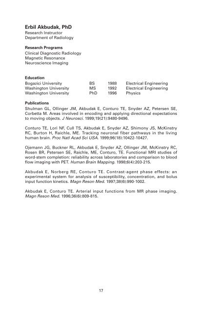
<strong>Erbil</strong> <strong>Akbudak</strong>, PhDResearch InstructorDepartment of RadiologyResearch ProgramsClinical Diagnostic RadiologyMagnetic ResonanceNeuroscience ImagingEducationBogazici University BS 1988 Electrical EngineeringWashington University MS 1992 Electrical EngineeringWashington University PhD 1996 PhysicsPublicationsShulman GL, Ollinger JM, <strong>Akbudak</strong> E, Conturo TE, Snyder AZ, Petersen SE,Corbetta M. Areas involved in encoding and applying directional expectationsto moving objects. J Neurosci. 1999;19(21):9480-9496.Conturo TE, Lori NF, Cull TS, <strong>Akbudak</strong> E, Snyder AZ, Shimony JS, McKinstryRC, Burton H, Raichle, ME. Tracking neuronal fiber pathways in the livinghuman brain. Proc Natl Acad Sci USA. 1999;96(18):10422-10427.Ojemann JG, Buckner RL, <strong>Akbudak</strong> E, Snyder AZ, Ollinger JM, McKinstry RC,Rosen BR, Petersen SE, Raichle, ME, Conturo, TE. Functional MRI studies ofword-stem completion: reliability across laboratories and comparison to bloodflow imaging with PET. Human Brain Mapping. 1998;6(4):203-215.<strong>Akbudak</strong> E, Norberg RE, Conturo TE. Contrast-agent phase effects: anexperimental system for analysis of susceptibility, concentration, and bolusinput function kinetics. Magn Reson Med. 1997;38(6):990-1002.<strong>Akbudak</strong> E, Conturo TE. Arterial input functions from MR phase imaging.Magn Reson Med. 1996;36(6):809-815.17
<strong>Erbil</strong> <strong>Akbudak</strong>, PhDResearch InstructorDepartment of RadiologyAs part of the neuroimaging MR group, Dr. <strong>Akbudak</strong> is interested in functionaland structural mapping of the human brain and the spinal cord usingmagnetic resonance (MR) techniques.The properties (magnitude and phase) of the magnetic resonance signaloriginating from the water molecules in a biosystem are sensitive to theenvironment (eg, tissue, blood, contrast agent) as well as their kinetic behavior(eg, flow rate, diffusion). By tuning, manipulating, and optimizing timingparameters as well as external inputs (eg, RF pulse, field gradient), severaltechniques have been and are being developed and put to use.Functional magnetic resonance imaging (fMRI), a sensory and cognitivemapping of the normal human brain, has become a standard tool in theneuroimaging laboratory. FMRI relies on intrinsic MR water signal changescaused by the decoupling between blood flow and oxygen consumption. Therecent acquisition of two state-of-the-art Siemens MR scanners (Sonata 1.5Tand Allegra 3T head-only), in addition to the whole-body 1.5T Vision scanneralready in place, will greatly enhance this research.Additionally, two echoplanar-based fast-imaging techniques are showingclinical potential: A diffusion-sensitive MR-based technique is being developedto map white-matter fiber tracts as well as to provide a basis for earlydetection of stroke. Secondly, a contrast agent-based quantitative mapping ofcerebral blood volume and cerebral blood flow will aid in understanding thehuman brain’s hemodynamics as well as distinguishing the brain’s pathologiesin the human brain.Specifically, Dr. <strong>Akbudak</strong>’s MR research focuses on the complex relationshipbetween the nature of the biosystem and its reflection on the characteristics ofthe MR data acquired—whether it be susceptibility diffusion, T1 and T2,relaxation mechanisms, etc. This in turn aids in clinical findings and thegathering of new information on how the brain works.East BuildingCampus Box 8225Phone: (314) 362-6376Fax: (314) 362-6110E-mail: erbil@npg.wustl.edu18


