Analyses of the Temporomandibular Disc - Prague Medical Report
Analyses of the Temporomandibular Disc - Prague Medical Report
Analyses of the Temporomandibular Disc - Prague Medical Report

You also want an ePaper? Increase the reach of your titles
YUMPU automatically turns print PDFs into web optimized ePapers that Google loves.
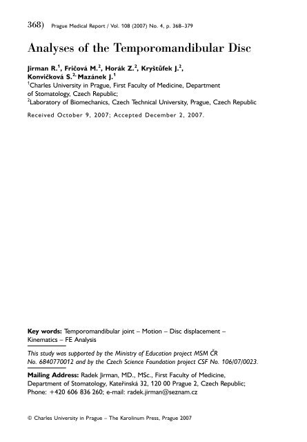
368) <strong>Prague</strong> <strong>Medical</strong> <strong>Report</strong> / Vol. 108 (2007) No. 4, p. 368–379<br />
<strong>Analyses</strong> <strong>of</strong> <strong>the</strong> <strong>Temporomandibular</strong> <strong>Disc</strong><br />
Jirman R. 1 , Fričová M. 2 , Horák Z. 2 , Kryštůfek J. 2 ,<br />
Konvičková S. 2, Mazánek J. 1<br />
1 Charles University in <strong>Prague</strong>, First Faculty <strong>of</strong> Medicine, Department<br />
<strong>of</strong> Stomatology, Czech Republic;<br />
2 Laboratory <strong>of</strong> Biomechanics, Czech Technical University, <strong>Prague</strong>, Czech Republic<br />
Received October 9, 2007; Accepted December 2, 2007.<br />
Key words: <strong>Temporomandibular</strong> joint – Motion – <strong>Disc</strong> displacement –<br />
Kinematics – FE Analysis<br />
This study was supported by <strong>the</strong> Ministry <strong>of</strong> Education project MSM ČR<br />
No. 6840770012 and by <strong>the</strong> Czech Science Foundation project CSF No. 106/07/0023.<br />
Mailing Address: Radek Jirman, MD., MSc., First Faculty <strong>of</strong> Medicine,<br />
Department <strong>of</strong> Stomatology, Kateřinská 32, 120 00 <strong>Prague</strong> 2, Czech Republic;<br />
Phone: +420 606 836 260; e-mail: radek.jirman@seznam.cz<br />
Jirman R.; Fričová M.; Horák Z.; Kryštůfek J.; Konvičková S.; Mazánek J.<br />
© Charles University in <strong>Prague</strong> – The Karolinum Press, <strong>Prague</strong> 2007
<strong>Prague</strong> <strong>Medical</strong> <strong>Report</strong> / Vol. 108 (2007) No. 4, p. 368–379<br />
369)<br />
Abstract: This project is <strong>the</strong> beginning <strong>of</strong> a large research work with a goal to<br />
develop a new total replacement <strong>of</strong> temporomandibular (TM) joint. First aim <strong>of</strong><br />
this work was to determine <strong>the</strong> relative displacement <strong>of</strong> <strong>the</strong> TM disc and <strong>the</strong><br />
mandible during mouth opening. The movement <strong>of</strong> <strong>the</strong> TM disc was studied using<br />
a magnetic resonance imaging. Sagittal static images in revolved sections <strong>of</strong> <strong>the</strong> TM<br />
joint were obtained in various positions <strong>of</strong> jaw opening from 0 to 50 mm. The<br />
results provided a description <strong>of</strong> <strong>the</strong> TM disc displacements as a function <strong>of</strong> jaw<br />
opening. The displacements <strong>of</strong> <strong>the</strong> mandible and TM disc were about 16 mm and<br />
10 mm respectively at mouth opening <strong>of</strong> 50 mm, maximum rotation <strong>of</strong> <strong>the</strong><br />
mandible was 34ş. The results <strong>of</strong> <strong>the</strong>se measurements can be used for clinical<br />
diagnostics and also <strong>the</strong>y were used as inputs for <strong>the</strong> follows finite element analysis<br />
(FEA). Second aim <strong>of</strong> this work was to create stress and strain analysis <strong>of</strong> TM joint<br />
using non-linear FEA. Complex <strong>of</strong> TM joint consists <strong>of</strong> mandibular disc, half skull<br />
and half mandible during normal jaw opening. The results illustrate <strong>the</strong> stress<br />
distributions in <strong>the</strong> TMJ during a normal jaw opening.<br />
Introduction<br />
Problem <strong>of</strong> face and jaw disorders is very extensive and complex, and it requires<br />
much effort to elaborate and implement new curative procedures and renew <strong>the</strong><br />
function and shape <strong>of</strong> <strong>the</strong> or<strong>of</strong>acial area with respect to <strong>the</strong> patient. It is estimated<br />
that 20% <strong>of</strong> <strong>the</strong> population suffers from temporomandibular disorders (TMD).<br />
Several studies have reported disc perforations and degeneration <strong>of</strong> <strong>the</strong> articulating<br />
surfaces [1]. Any TM disc motion derangement during mouth opening will<br />
significantly influence TMD. From <strong>the</strong> anatomical and biomechanical point <strong>of</strong> view,<br />
<strong>the</strong> temporomandibular joint (TMJ) is a sophisticated bicondylar articulatory<br />
complex that places high demands on neuromuscular control with a frequency <strong>of</strong><br />
motion indicated up to 2000 periods per day. This makes <strong>the</strong> TMJ one <strong>of</strong> <strong>the</strong> most<br />
frequently exerted joints in <strong>the</strong> human body, which in conjunction with <strong>the</strong><br />
individual uniqueness <strong>of</strong> this joint place high demands on its design and reliability.<br />
This project is <strong>the</strong> beginning <strong>of</strong> a large research work with <strong>the</strong> goal to develop a<br />
new total replacement <strong>of</strong> TMJ. <strong>Temporomandibular</strong> joint replacement is very<br />
sophisticated and with individual approach to patient. For successful development<br />
<strong>of</strong> <strong>the</strong> TMJ replacement it is very important to know several conditions. First <strong>of</strong><br />
<strong>the</strong>m is to understand and to know how to describe <strong>the</strong> relative motion <strong>of</strong> <strong>the</strong><br />
system <strong>of</strong> TMJ parts. It is <strong>the</strong>refore necessary to perform experimental<br />
measurements in order to observe this physiological movement. Condylar and TM<br />
disc displacements are useful in understanding <strong>the</strong> aetiology <strong>of</strong> <strong>the</strong> disease and in<br />
evaluating <strong>the</strong> treatment. The movement <strong>of</strong> <strong>the</strong> TM disc during jaw opening and<br />
closure is very sophisticated. It was difficult to depict its motion exactly, because<br />
<strong>the</strong> disc shape changes significantly during <strong>the</strong> motion. The number <strong>of</strong><br />
experimental studies is limited, because it is difficult to insert experimental<br />
devices, such as strain gauges, into <strong>the</strong> joint without causing damage to its tissues<br />
<strong>Analyses</strong> <strong>of</strong> <strong>the</strong> <strong>Temporomandibular</strong> <strong>Disc</strong>
370) <strong>Prague</strong> <strong>Medical</strong> <strong>Report</strong> / Vol. 108 (2007) No. 4, p. 368–379<br />
and influencing <strong>the</strong>ir mechanical behaviour. In several studies, condylar motion has<br />
been studied directly with <strong>the</strong> use <strong>of</strong> MRI [2, 3, 4], videoanalysis [5], or ultrasound<br />
imaging [6]. The data obtained from this experimental measurement during jaw<br />
opening was measured only for a maximum distance between central lower and<br />
upper incisors <strong>of</strong> 17 mm. In fact, <strong>the</strong> maximum comfortable mouth opening is<br />
greater (approx. 30–50 mm), and moreover, <strong>the</strong>re were no references about TM<br />
disc displacement. Experimental and numerical studies concerning jaw movement<br />
and <strong>the</strong> distribution <strong>of</strong> <strong>the</strong> loads in <strong>the</strong> TMJ have been performed in many models<br />
[7, 8, 9, 10]. However, <strong>the</strong> TM disc is crucial for proper TMJ function, and it has<br />
been omitted or simplified in many numerical models and experiments [9, 11, 12].<br />
Second necessary condition for successful development <strong>of</strong> <strong>the</strong> TMJ replacement is<br />
to know distribution <strong>of</strong> <strong>the</strong> loads in <strong>the</strong> TMJ. Ma<strong>the</strong>matical models <strong>of</strong> <strong>the</strong> human<br />
masticatory system including <strong>the</strong> TMJ have been demonstrated to be a powerful<br />
tool to predict <strong>the</strong> loads acting on this joint [7, 8, 9, 10, 12, 13, 14, 15, 16].<br />
However, many studies have oversimplified <strong>the</strong> geometry and behaviour <strong>of</strong> <strong>the</strong><br />
TM disc. Therefore, <strong>the</strong> tissue deformations and <strong>the</strong> distribution <strong>of</strong> loads inside <strong>the</strong><br />
joint could not be analyzed. Numerical modelling can provide with fur<strong>the</strong>r <strong>of</strong> <strong>the</strong><br />
understanding physiology <strong>of</strong> <strong>the</strong> joint but also pathogenesis <strong>of</strong> <strong>the</strong> joint diseases.<br />
Application <strong>of</strong> <strong>the</strong> finite element stress analysis technique to <strong>the</strong> biomechanical<br />
investigation <strong>of</strong> <strong>the</strong> TMJ is very suitable. Finite element analyses <strong>of</strong> TMJ were<br />
created by many authors [7, 10, 12, 13, 15], but in this analysis was used some<br />
simplification. Problems <strong>of</strong> all FE analyses are definition <strong>of</strong> <strong>the</strong> muscle forces,<br />
movement <strong>of</strong> <strong>the</strong> TM disc during jaw opening and definition <strong>of</strong> TM disc material<br />
properties. Some analyses were created as 2D contact problems or separated<br />
parts were analyzed. Results <strong>of</strong> <strong>the</strong>se analyses were affected by <strong>the</strong> boundary<br />
conditions and complexity <strong>of</strong> <strong>the</strong> models. Therefore it was necessary to create<br />
complex three-dimensional model <strong>of</strong> TMJ as global system <strong>of</strong> <strong>the</strong> skull, mandible,<br />
TM disc, ligaments and muscles. Contacts interactions were defined between <strong>the</strong><br />
TM disc and <strong>the</strong> skull and between <strong>the</strong> TM disc and <strong>the</strong> mandible.<br />
Material and Methods<br />
Kinematics <strong>of</strong> <strong>the</strong> TM disc<br />
The experimental measurements were obtained on one volunteer (male, 25 years<br />
old) – only one TMJ with no abnormalities. Our measurement method is based on<br />
[2]. Imaging was carried out at <strong>the</strong> Radiodiagnostic clinic <strong>of</strong> <strong>the</strong> 1st <strong>Medical</strong> Faculty<br />
at Charles University. The measuring device was Intera 1.5 T (Philips), and <strong>the</strong><br />
subject was scanned into 3 mm thick slices with <strong>the</strong> FFE dynamic scan sequence in<br />
<strong>the</strong> image plane <strong>of</strong> <strong>the</strong> slices. The head was fixed to avoid major displacements;<br />
smaller displacements such as swallowing and position discomfort <strong>of</strong> <strong>the</strong> head<br />
were corrected using <strong>the</strong> geometric transformation described below. The image<br />
plane was rotated from a mid-sagittal plane <strong>of</strong> 30ş in <strong>the</strong> direction <strong>of</strong> <strong>the</strong> jaw<br />
(Figure 1). Image plane sections <strong>of</strong> TMJ were scanned in <strong>the</strong> specified location <strong>of</strong><br />
Jirman R.; Fričová M.; Horák Z.; Kryštůfek J.; Konvičková S.; Mazánek J.
<strong>Prague</strong> <strong>Medical</strong> <strong>Report</strong> / Vol. 108 (2007) No. 4, p. 368–379<br />
371)<br />
jaw opening from 0 to 50 mm (distance between central lower and upper incisors)<br />
using a customised 1-directional “spreader” device. We used a Vernier calliper<br />
design and proposed an appropriate shape for convenient mouth opening. Six<br />
static mouth opening positions were obtained, starting with <strong>the</strong> maximum jaw<br />
opening (50 mm) and proceeding to closure (0 mm), and an MR image <strong>of</strong> <strong>the</strong> TMJ<br />
was taken for each position. It was necessary to minimize <strong>the</strong> imaging time over<br />
which <strong>the</strong> patient must keep <strong>the</strong> jaw open. This can be uncomfortable for <strong>the</strong><br />
patient in <strong>the</strong> supine position, as saliva collects in <strong>the</strong> back <strong>of</strong> <strong>the</strong> pharynx [3, 17].<br />
The contours <strong>of</strong> <strong>the</strong> TMJ components: fossa mandibularis and tuberculum<br />
articulare on <strong>the</strong> skull, <strong>the</strong> mandible condyle and <strong>the</strong> TM disc were taken from <strong>the</strong><br />
MR images. These contours were traced in one slice (our chosen image plane) by<br />
parametric curves defined by <strong>the</strong> number <strong>of</strong> points for each part <strong>of</strong> <strong>the</strong> joint. We<br />
assigned two points at each end <strong>of</strong> <strong>the</strong> curve; <strong>the</strong>re were set points A, B and C, D<br />
on <strong>the</strong> skull and on <strong>the</strong> mandible condyle, respectively (Figure 2). One reference<br />
point (marker) was assigned for <strong>the</strong> fossa mandibularis – point E, and also for <strong>the</strong><br />
mandibular condyle – point F (Figure 2). The positioning <strong>of</strong> <strong>the</strong> reference point<br />
was hard to define due to <strong>the</strong> broken shapes <strong>of</strong> <strong>the</strong> biological structures. We<br />
determined <strong>the</strong> points as intersection <strong>of</strong> normals (centre <strong>of</strong> curvature) on <strong>the</strong><br />
above mentioned contours (points E and F related to skull and mandible,<br />
respectively, Figure 2). It was more difficult to decide how to deal with <strong>the</strong> disc<br />
reference points, because <strong>the</strong> TM disc changed its shape significantly during <strong>the</strong><br />
motion. The only solution that we found was to define markers for each mouth<br />
opening in <strong>the</strong> location <strong>of</strong> <strong>the</strong> muscle and ligament conjunction, points G, H<br />
respectively, because <strong>the</strong>se particular points could be explicitly defined.<br />
The displacements for each part <strong>of</strong> <strong>the</strong> TMJ were obtained from <strong>the</strong> sequence <strong>of</strong><br />
<strong>the</strong> contour transformation (trajectories <strong>of</strong> all points are shown in Figure 3). First,<br />
<strong>the</strong> transformation described <strong>the</strong> position <strong>of</strong> all reference points into <strong>the</strong> local<br />
coordinate system <strong>of</strong> <strong>the</strong> skull (index S). A global (index G) Cartesian coordinate<br />
system (CCS) was chosen to coincide with <strong>the</strong> coordinate system in <strong>the</strong> MR image<br />
Figure 1 – MR image <strong>of</strong> <strong>the</strong><br />
TMJ at <strong>the</strong> mid-opening<br />
<strong>of</strong> <strong>the</strong> mandible with initial<br />
point and defined CCS; image<br />
plane with <strong>the</strong> highlighted<br />
condyle – C, fossa<br />
mandibularis – FM and <strong>the</strong><br />
TM disc – D (on <strong>the</strong> left),<br />
rotation <strong>of</strong> image plane<br />
from mid-sagittal plane<br />
(on <strong>the</strong> right).<br />
<strong>Analyses</strong> <strong>of</strong> <strong>the</strong> <strong>Temporomandibular</strong> <strong>Disc</strong>
372) <strong>Prague</strong> <strong>Medical</strong> <strong>Report</strong> / Vol. 108 (2007) No. 4, p. 368–379<br />
(Figure 1). The first MR image (assigned as <strong>the</strong> n-th index) was established as a<br />
reference image. Second, all markers were transformed from <strong>the</strong> local coordinates<br />
(from <strong>the</strong> above-mentioned transformation) into <strong>the</strong> global coordinate system for<br />
all mouth opening positions; <strong>the</strong> temporal bone points A, B and E were identical<br />
for all positions. This was essential, because although <strong>the</strong> head was fixed, <strong>the</strong> skull<br />
might move towards <strong>the</strong> skin during <strong>the</strong> examination. The third was <strong>the</strong><br />
transformation <strong>of</strong> point F into <strong>the</strong> coordinate system from <strong>the</strong> global CCS to <strong>the</strong><br />
local CCS <strong>of</strong> <strong>the</strong> mandible (index M). Finally, point F (local CCS <strong>of</strong> <strong>the</strong> mandible)<br />
was translated and rotated into <strong>the</strong> global coordinate system for all mouth<br />
opening positions. The four transformations can be described by <strong>the</strong> following<br />
instructions [18]:<br />
1. Coordinate transformation <strong>of</strong> reference points B, C, D, E, G and H from <strong>the</strong><br />
global (upper index G) Cartesian coordinate system to local CCS <strong>of</strong> <strong>the</strong> skull<br />
(upper index S) respectively for i=1, 2,.., n, when n is position number <strong>of</strong> jaw<br />
opening (in our case n=6) and r indicates extended radius vector.<br />
GGGGGGSSSSSS<br />
r Bi<br />
r, Ci<br />
r, Di<br />
r, Ei<br />
r, Gi<br />
r, Hi<br />
® r Bi<br />
r, Ci<br />
r, Di<br />
r, Ei<br />
r, Gi<br />
r,<br />
Hi<br />
2. Correction <strong>of</strong> points B, C, D, E, G and H position in global CCS respectively for<br />
i=1, 2,..., n, when n is <strong>the</strong> number <strong>of</strong> <strong>the</strong> jaw opening position.<br />
SSSSSSGGGGGG<br />
r Bi<br />
r, Ci<br />
r, Di<br />
r, Ei<br />
r, Gi<br />
r,® r BNi<br />
r, CNi<br />
r, DNi<br />
r, ENi<br />
r, GNi<br />
r,<br />
HiHNi<br />
A<br />
B<br />
C<br />
Figure 2 – Contours <strong>of</strong> TMJ part with assigned<br />
reference points from A to H for maximum mouth<br />
opening (on <strong>the</strong> left); defined coordinates systems,<br />
index G – global CCS, index S – local CCS <strong>of</strong> <strong>the</strong><br />
skull and index M – local CCS <strong>of</strong> <strong>the</strong> mandible (on<br />
<strong>the</strong> right).<br />
Figure 3 – Scheme <strong>of</strong> <strong>the</strong> reference point motion<br />
(index 1 mouth is closing, index 6 mouth is opened<br />
to <strong>the</strong> maximum): (a) scheme <strong>of</strong> <strong>the</strong> mandibular<br />
condyle movement, (b) image detail <strong>of</strong> <strong>the</strong> TM disc<br />
reference point movement, (c) coordinate system for<br />
graphic interpretation <strong>of</strong> <strong>the</strong> reference point motion.<br />
Jirman R.; Fričová M.; Horák Z.; Kryštůfek J.; Konvičková S.; Mazánek J.
<strong>Prague</strong> <strong>Medical</strong> <strong>Report</strong> / Vol. 108 (2007) No. 4, p. 368–379<br />
373)<br />
3. Coordinate transformation <strong>of</strong> point F from global (index G) CCS to local CCS <strong>of</strong><br />
mandible (index M)<br />
GM<br />
r Fn<br />
® r Fn<br />
.<br />
4. Computation <strong>of</strong> ano<strong>the</strong>r position (i=1, 2,..., n-1) <strong>of</strong> jaw opening was done in<br />
global CCS.<br />
FE analysis <strong>of</strong> <strong>the</strong> TM joint<br />
The geometry <strong>of</strong> <strong>the</strong> model was obtained from <strong>the</strong> head <strong>of</strong> embalmed male<br />
cadaver, showing no abnormalities, using a CT and MRI scans. Muscles and<br />
ligaments were additionally modelled in to <strong>the</strong> CAD program according to <strong>the</strong><br />
anatomical knowledge. Only half <strong>of</strong> <strong>the</strong> skull and mandible were used for<br />
simplification <strong>of</strong> <strong>the</strong> FE analysis. Geometrical model <strong>of</strong> <strong>the</strong> TMJ is shown in<br />
(Figure 4). Model <strong>of</strong> <strong>the</strong> TMJ was exported from <strong>the</strong> CAD program in to <strong>the</strong><br />
automated mesh generator NETGEN, where were generated four-noded<br />
tetrahedral elements <strong>of</strong> <strong>the</strong> skull and mandible. TM disc was meshed by eightnoded<br />
brick elements in to <strong>the</strong> TrueGrid ® mesh program. Finally muscles were<br />
defined by connector elements. This special element allows various definitions <strong>of</strong><br />
<strong>the</strong> material properties, loading forces and <strong>the</strong> element behaviour. Connector<br />
element type axial was used for this application. This type provides a connection<br />
between two nodes where <strong>the</strong> relative displacement is along <strong>the</strong> line separating<br />
<strong>the</strong> two nodes.<br />
It models discrete physical connections such as axial springs, axial dashpots, or<br />
node-to-node (gap-like) contact. The axial connection does not constrain any<br />
component <strong>of</strong> relative motion. The available component <strong>of</strong> relative motion u acts<br />
along <strong>the</strong> line connecting <strong>the</strong> two nodes, measures <strong>the</strong> change in distance<br />
separating <strong>the</strong> two nodes. Muscles represented by connector elements were<br />
linked with mandible and skull by distributed coupling constraints. This<br />
Figure 4 – Geometrical model <strong>of</strong> <strong>the</strong> skull and mandible. All muscles and ligaments are shown.<br />
<strong>Analyses</strong> <strong>of</strong> <strong>the</strong> <strong>Temporomandibular</strong> <strong>Disc</strong>
374) <strong>Prague</strong> <strong>Medical</strong> <strong>Report</strong> / Vol. 108 (2007) No. 4, p. 368–379<br />
represented muscle insertion in to <strong>the</strong> bone. The coupling constraint provides<br />
coupling between a reference node and a group <strong>of</strong> nodes. Distributing coupling<br />
constrains <strong>the</strong> motion <strong>of</strong> <strong>the</strong> coupling nodes to <strong>the</strong> translation and rotation <strong>of</strong> <strong>the</strong><br />
reference node. This constraint is enforced in an average sense in a way that<br />
enables control <strong>of</strong> <strong>the</strong> transmission <strong>of</strong> loads through weight factors at <strong>the</strong> coupling<br />
nodes. The constraint distributes loads such that <strong>the</strong> resultants <strong>of</strong> <strong>the</strong> forces at <strong>the</strong><br />
coupling nodes are equivalent to <strong>the</strong> forces and moments at <strong>the</strong> reference node. FE<br />
model <strong>of</strong> <strong>the</strong> TMJ is shown in (Figure 5).<br />
FE analysis was defined as a nonlinear contact task and solved in ABAQUS 6.6<br />
(Hibbit, Karlsson, Sorensen, Inc., Providence, RI). Contact interactions were defined<br />
between <strong>the</strong> TM disc and <strong>the</strong> skull and between <strong>the</strong> TM disc and <strong>the</strong> mandible. All<br />
contacts were defined as surface to surface slide contact. The coefficient <strong>of</strong> friction<br />
for articular surfaces is unknown. It was estimated that <strong>the</strong> coefficient <strong>of</strong> friction<br />
must be smaller than 0.15 because <strong>of</strong> <strong>the</strong> existence <strong>of</strong> <strong>the</strong> synovial fluid. Coefficient<br />
<strong>of</strong> friction was estimated 0.08. The FE model consisted <strong>of</strong> 180 eight-noded<br />
elements, 54578 four-noded tetrahedral elements, and 48 connector elements.<br />
Total number <strong>of</strong> nodes was 16665. Data on <strong>the</strong> material properties <strong>of</strong> all TMJ parts<br />
were taken from published data. The bone <strong>of</strong> <strong>the</strong> skull and mandible were<br />
considered to be isotropic and linear elastic. Tooth and ligaments were also defined<br />
as isotropic and linear elastic. Because range <strong>of</strong> magnitude TM disc material<br />
properties in published data is big, Young’s modulus and Poisson’s ratio were<br />
estimated. TM disc was also defined as homogenous and isotropic. All material<br />
properties assigned to <strong>the</strong> structural elements are listed in (Table 1) [12, 19].<br />
Advantage <strong>of</strong> using connector element was <strong>the</strong> possibilities apply resultant forces<br />
directly. Geometrical parameters are completely defined by <strong>the</strong> muscle insertion in<br />
<strong>the</strong> bone. Normal jaw clenching was used for this analysis and forces were applied<br />
in connector elements. All forces were assumed to be symmetrical and had equal<br />
Figure 5 – FE model <strong>of</strong> skull and mandible.<br />
All muscles (red lines), all ligaments (green)<br />
and TM disc (blue) in detail were shown.<br />
Jirman R.; Fričová M.; Horák Z.; Kryštůfek J.; Konvičková S.; Mazánek J.
<strong>Prague</strong> <strong>Medical</strong> <strong>Report</strong> / Vol. 108 (2007) No. 4, p. 368–379<br />
375)<br />
magnitude on <strong>the</strong> right and left side <strong>of</strong> <strong>the</strong> mandible. Magnitudes <strong>of</strong> all applied<br />
forces were taken from published data [3, 11, 20]. Symmetry boundary conditions<br />
were applied on <strong>the</strong> sagittal surfaces <strong>of</strong> <strong>the</strong> skull and mandible. Base <strong>of</strong> <strong>the</strong> skull<br />
was constrained and tooth displacement in <strong>the</strong> z-direction was constrained for<br />
normal jaw clenching<br />
Results<br />
Results <strong>of</strong> <strong>the</strong> experimental measurements showed <strong>the</strong> motion <strong>of</strong> <strong>the</strong> TM disc and<br />
<strong>the</strong> mandible markers during mouth opening. Figure 3 shows an example <strong>of</strong> <strong>the</strong><br />
opening movement path <strong>of</strong> <strong>the</strong> reference points. We observed <strong>the</strong> relative motion<br />
<strong>of</strong> <strong>the</strong> TM disc and <strong>the</strong> mandible in <strong>the</strong> x-axis and in <strong>the</strong> y-axis and also <strong>the</strong><br />
rotation depending on <strong>the</strong> mouth opening (Figure 6, Table 3). Regression <strong>of</strong> <strong>the</strong><br />
measured points coordinates, depending on mouth opening, represents a third- or<br />
second-order polynomial curve (Figure 6). A correlation coefficients range from<br />
R 2 =0.97 to R 2 =0.99. The movement <strong>of</strong> <strong>the</strong> TM disc is shown in Fig. 6 and <strong>the</strong><br />
reference points G, H displacements are described in Table 3. which show <strong>the</strong><br />
TM disc behaviour during jaw opening. The motion <strong>of</strong> <strong>the</strong> mandibular condyle was<br />
Table 1 – Material properties<strong>of</strong> <strong>the</strong> TMJ components<br />
Material Young’s modulus [MPa] Poisson’s ratio [–]<br />
Bone 16300 0.31<br />
Ligaments 1200 0.28<br />
Tooth 19000 0.30<br />
TM <strong>Disc</strong> 16 0.45<br />
Table 2 – Muscle forces corresponding with maximal jaw clenching<br />
muscles<br />
Lateral pterygoid Medial pterygoid Temporalis Masseter<br />
Force [N] 378.0 191.4 528.6 340.0<br />
Table 3 – Values <strong>of</strong> displacement <strong>of</strong> all reference points F, G, H in <strong>the</strong><br />
specified directory and rotation <strong>of</strong> mandibular reference point F<br />
Mandible TM disc TM disc<br />
m [mm] " [°] Point F [mm] Point G [mm] Point H [mm]<br />
x y x y x y<br />
10 0.00 0.00 0.00 0.00 0.00 0.00 0.00<br />
10 9.46 –6.01 –2.82 1.53 –1.31 –0.43 –3.45<br />
20 14.75 –7.69 –3.33 1.20 –1.93 –1.40 –4.37<br />
30 20.53 –11.26 –2.86 –2.80 –1.13 –3.55 –4.86<br />
40 27.10 –13.51 –2.53 –4.17 0.66 –6.79 –6.38<br />
50 34.39 –15.81 –1.10 –9.83 1.85 –6.03 –6.82<br />
<strong>Analyses</strong> <strong>of</strong> <strong>the</strong> <strong>Temporomandibular</strong> <strong>Disc</strong>
376) <strong>Prague</strong> <strong>Medical</strong> <strong>Report</strong> / Vol. 108 (2007) No. 4, p. 368–379<br />
Figure 6 – Displacement <strong>of</strong><br />
reference points on <strong>the</strong> TM<br />
disc and <strong>the</strong> mandibular<br />
condyle (from top): motion <strong>of</strong><br />
mandible reference point F;<br />
relative motion <strong>of</strong> <strong>the</strong> TM<br />
disc reference points G<br />
and H; rotation <strong>of</strong> mandible<br />
about reference point F.<br />
Jirman R.; Fričová M.; Horák Z.; Kryštůfek J.; Konvičková S.; Mazánek J.
<strong>Prague</strong> <strong>Medical</strong> <strong>Report</strong> / Vol. 108 (2007) No. 4, p. 368–379<br />
377)<br />
investigated in two directions and one angle. The initial position was established<br />
for a closed mouth, where <strong>the</strong> angle was considered as 0°, so that <strong>the</strong> displayed<br />
angle was absolute. Figure 6. and Table 3. show that <strong>the</strong> maximum displacement in<br />
<strong>the</strong> x-direction was 15.81 mm, and in <strong>the</strong> y-direction it was 3.33 mm. So <strong>the</strong><br />
maximum translational displacement was 15.85 mm and <strong>the</strong> maximum rotation <strong>of</strong><br />
<strong>the</strong> mandibular condyle was 34.39° (Figure 6, Table 3).<br />
Results <strong>of</strong> <strong>the</strong> FE analysis illustrate <strong>the</strong> stress distributions in <strong>the</strong> TMJ during a<br />
normal jaw clenching. The stress distributions from this model are given in<br />
(Figure 7 and Figure 8). High von-Misses stresses were seen at <strong>the</strong> mandible near<br />
<strong>the</strong> processus coronoideus. The maximum von Misses stress was about 5.5<br />
[MPa]. High contact pressures were seen at <strong>the</strong> contact surface on <strong>the</strong> TM disc,<br />
contact was defined between TM disc and mandible. The maximum contact<br />
pressure was about 6.3 [MPa]. Maximal von Misses stress on <strong>the</strong> skull were<br />
located on <strong>the</strong> arcus zygomaticus, where is connected m. Masseter with <strong>the</strong> skull.<br />
The maximum von Misses stress was about 3 [MPa]. The forces in <strong>the</strong> ligaments<br />
were quite small. The maximum ligament forces were located in <strong>the</strong> joint capsule<br />
(0.3 N).<br />
A B C<br />
Figure 7 – Distribution <strong>of</strong> <strong>the</strong> von Misses stresses [MPa] in <strong>the</strong> skull (a) and mandible (b) and distribution <strong>of</strong><br />
<strong>the</strong> contact pressures [MPa] in <strong>the</strong> TM disc (c).<br />
A<br />
B<br />
Figure 8 – Distribution<br />
<strong>of</strong> <strong>the</strong> von Misses stresses<br />
[MPa] in <strong>the</strong><br />
temporomandibular joint.<br />
View <strong>of</strong> <strong>the</strong> frontal plane<br />
(a) and sagittal plane (b).<br />
<strong>Analyses</strong> <strong>of</strong> <strong>the</strong> <strong>Temporomandibular</strong> <strong>Disc</strong>
378) <strong>Prague</strong> <strong>Medical</strong> <strong>Report</strong> / Vol. 108 (2007) No. 4, p. 368–379<br />
<strong>Disc</strong>ussion<br />
The TM disc plays a crucial role in TMJ motion; it is <strong>the</strong>refore necessary to know its<br />
mechanical behaviour as well as its movement. The displacement <strong>of</strong> <strong>the</strong> mandibular<br />
condyle was described in previous publication [2] for maximum mouth opening <strong>of</strong><br />
17 mm. This is not <strong>the</strong> maximum comfortable mouth opening, so it was necessary<br />
to broaden <strong>the</strong>se measurements. In addition, <strong>the</strong>re was no investigation <strong>of</strong> TM disc<br />
movement. The results for mouth opening <strong>of</strong> 17 mm have been used for computing<br />
FEA [10], but <strong>the</strong> results <strong>of</strong> this analysis must be insufficient. Our measurement<br />
method is based on [2], but we obtained a more complex description <strong>of</strong> <strong>the</strong><br />
mandible and especially <strong>of</strong> TM disc behaviour. From our study we obtained <strong>the</strong><br />
displacement <strong>of</strong> <strong>the</strong> TM disc and <strong>of</strong> <strong>the</strong> mandible condyle in relation to mouth<br />
opening. The maximum mouth opening was 50 mm; this distance was defined<br />
between lower and upper incisors. This study shows that <strong>the</strong> condyle first moves<br />
down and forward (downward and anteriorly) and <strong>the</strong>n rotates open. This motion<br />
is shown in Figure 6. MRI is a non-invasive method which can be used easily and<br />
efficiently for visualization <strong>of</strong> s<strong>of</strong>t tissues. Adjusting <strong>the</strong> MRI device was crucial.<br />
There are many scanning techniques, but in most <strong>of</strong> <strong>the</strong>m <strong>the</strong> disc is not easily<br />
observed. Next, <strong>the</strong>re can be significant loss due to slice thickness. Several<br />
experiments with MR specialists led to a proper scanning sequence. Finally, <strong>the</strong><br />
precision <strong>of</strong> <strong>the</strong> results is also limited by <strong>the</strong> quality <strong>of</strong> <strong>the</strong> MR images and by <strong>the</strong><br />
accuracy <strong>of</strong> <strong>the</strong> contours <strong>of</strong> <strong>the</strong> TMJ part. All parts were shaped according to <strong>the</strong>ir<br />
anatomical geometry in cooperation with an experienced dentist. One <strong>of</strong> <strong>the</strong> main<br />
disadvantages was that <strong>the</strong> individual quasi-static images that were analyzed in this<br />
initial study did not capture everything that was truly seen when examining <strong>the</strong> TMJ<br />
in motion. In [2] <strong>the</strong> motion <strong>of</strong> <strong>the</strong> mandibular condyle was studied for <strong>the</strong><br />
reference point defined on <strong>the</strong> condyle, which does not seem to be precise. In our<br />
approach, <strong>the</strong> centre <strong>of</strong> <strong>the</strong> curvature should be <strong>the</strong> more exact description <strong>of</strong> <strong>the</strong><br />
reference point. Ano<strong>the</strong>r disadvantage <strong>of</strong> <strong>the</strong> approach is that <strong>the</strong>re were just two<br />
dimensions. Improvement into a three-dimensional scheme will surely be one <strong>of</strong> <strong>the</strong><br />
first future developments. The spatial conception needs to be more accurate due to<br />
<strong>the</strong> extremely complex shape <strong>of</strong> <strong>the</strong> TM disc. A fur<strong>the</strong>r disadvantage was that only<br />
one subject on one side <strong>of</strong> <strong>the</strong> joint was examined. A study is already in progress to<br />
overcome this disadvantage. We intend to supplement some statistical evaluation to<br />
enhance <strong>the</strong> statements presented here. The study provided a three-dimensional<br />
model <strong>of</strong> <strong>the</strong> temporomandibular joint. The presented model, as far as we know, is<br />
<strong>the</strong> first complete three-dimensional model. All parts <strong>of</strong> <strong>the</strong> TMJ were shaped<br />
according to <strong>the</strong>ir anatomical geometry which was sampled with high resolution.<br />
This study verified applied model in <strong>the</strong> real situation during a normal jaw clenching.<br />
Stresses in <strong>the</strong> joint components (disc, mandible, skull and ligaments) with a normal<br />
jaw clenching have been analyzed. Results <strong>of</strong> this analysis shown, that <strong>the</strong> numerical<br />
simulation <strong>of</strong> this geometrically complicated joint is possible by <strong>the</strong> FEM. Compared<br />
with <strong>the</strong> results taken from published data [7, 8, 13], <strong>the</strong> stress distribution in <strong>the</strong><br />
Jirman R.; Fričová M.; Horák Z.; Kryštůfek J.; Konvičková S.; Mazánek J.
<strong>Prague</strong> <strong>Medical</strong> <strong>Report</strong> / Vol. 108 (2007) No. 4, p. 368–379<br />
379)<br />
TM disc are corresponding. In this model simple material definitions were used, but<br />
this pilot study was made for model verification. Next simplification was ligaments<br />
simulation by <strong>the</strong> connector elements. Finally <strong>the</strong> model didn’t include deformable<br />
cartilage layers on <strong>the</strong> articulating surfaces <strong>of</strong> mandible and skull. All <strong>of</strong> <strong>the</strong>se<br />
simplifications will be changed in <strong>the</strong> next generation <strong>of</strong> <strong>the</strong> TMJ model.<br />
References<br />
1. JERGENSON M., BARTON J: The occurrence <strong>of</strong> TMJ disc perforations in an aging population. Journal <strong>of</strong><br />
Dental Research 77: 264, 1998.<br />
2. CHEN, J., BUCKWALTER, K.: Displacement Analysis <strong>of</strong> <strong>the</strong> <strong>Temporomandibular</strong> Condyle from Magnetic<br />
Resonance Images. Journal <strong>of</strong> Biomechanics 26: 1455–1462, 1993.<br />
3. DRACE J. E., ENZMANN D. R.: Defining <strong>the</strong> Normal <strong>Temporomandibular</strong> Joint: Closed-, Partially Open-,<br />
and Open-Mouth MR Imaging <strong>of</strong> Asymptomatic Subjects. Radiology 177: 67–71, 1990.<br />
4. PETEROVA V., JIRMAN R., MAZANEK J., SEIDL Z.: The Examination <strong>of</strong> <strong>the</strong> <strong>Temporomandibular</strong> Joint<br />
on 1,5T Magnetic Resonance. <strong>Prague</strong> <strong>Medical</strong> <strong>Report</strong> 105: 29–34, 2005.<br />
5. YATABE M., ZWIJNENBURG A., MEGENS C. C. E. J., NAEIJE M.: Movements <strong>of</strong> <strong>the</strong> Mandibular<br />
Condyle Kinematic Center during Jaw Opening and Closing. Journal <strong>of</strong> Dental Research 76: 714–719, 1997.<br />
6. BRAUN, S., HICKEN, J.S.: Ultrasound imaging <strong>of</strong> condylar motion: a preliminary report. Angle<br />
Orthodontist 70: 383–386, 2000.<br />
7. BEEK M., KOOLSTRA J. H.: Three-dimensional finite element analysis <strong>of</strong> <strong>the</strong> human temporomandibular<br />
joint disc. Journal <strong>of</strong> Biomechanics 33: 307–316, 2000.<br />
8. CHOI A. H., BEN-NISSAN B., CONWAY R. C.: Three-dimensional modelling and finite element analysis<br />
<strong>of</strong> <strong>the</strong> human mandible during occlusion. Australian Dental Journal 50: 42–48, 2005.<br />
9. DONZELLI P. S., GALLO L. M., SPILKER L. R.: Biphasic finite element simulation <strong>of</strong> <strong>the</strong> TMJ disc from in<br />
vivo kinematic and geometric measurement. Journal <strong>of</strong> Biomechanics 37: 42–48, 2004.<br />
10. PEREZ DEL PALOMAR A., DOBLARE M.: 3D Finite Element Simulation <strong>of</strong> <strong>the</strong> Opening Movement <strong>of</strong> <strong>the</strong><br />
Mandible in Healthy and Pathologic Situations. Journal <strong>of</strong> Biomechanical Engineering 128: 242–249, 2006.<br />
11. BEEK M., AARNTS M. P., KOOLSTRA J.H.: Dynamic properties <strong>of</strong> <strong>the</strong> human temporomandibular joint<br />
disc. Journal <strong>of</strong> Dental Research 80: 876–880, 2001.<br />
12. PEREZ DEL PALOMAR A, DOBLARE M.: The effect <strong>of</strong> collagen reinforcement in <strong>the</strong> behaviour<br />
<strong>of</strong> <strong>the</strong> temporomandibular joint disc. Journal <strong>of</strong> Biomechanics 39: 1075–1085, 2006.<br />
13. CHEN J., XU L. A.: Finite Element Analysis <strong>of</strong> <strong>the</strong> Human <strong>Temporomandibular</strong> Joint. Journal. <strong>of</strong><br />
Biomechanical Engineering 116: 401–407, 1994.<br />
14. HATTORI Y., SATOH C., SEKI S., WATANABE Y., OGINO Y., WATANABE M.: Occlusal and TMJ Loads<br />
in Subjects with Experimentally Shortened Dental Arches. Journal. <strong>of</strong> Dental Research 82: 532–536, 2003.<br />
15. MAY B., SAHA S., SALTZMAN. M.: Three-dimensional ma<strong>the</strong>matical model <strong>of</strong> temporomandibular joint<br />
loading. Clinical Biomechanics 16: 489–495, 2001.<br />
16. SELLERS W. I., CROMPTON R. H.: Using sensitivity analysis to validate <strong>the</strong> predictions <strong>of</strong><br />
a biomechanical model <strong>of</strong> bite forces. Annals <strong>of</strong> Anatomy 186: 89–95, 2004.<br />
17. KATZBERG R. W.: <strong>Temporomandibular</strong> Joint Imaging. Radiology 170: 297–307, 1989.<br />
18. STEJSKAL V., VALASEK M.: Kinematics and dynamics <strong>of</strong> machinery. Marcel Dekker Inc., New York (NY),<br />
USA, 1996.<br />
19. TANAKA E., VAN EIJDEN T.: Biomechanical Behaviour <strong>of</strong> <strong>the</strong> <strong>Temporomandibular</strong> Joint <strong>Disc</strong>.<br />
Crit. Rev. Oral Biol. Med 14: 138–150, 2003.<br />
20. NICKEL J. C., IWASAKI L.R., WALKER R. D., MCLACHLAN K. R., MCCALL W. D.: Human Masticatory<br />
Muscle Forces during Static Biting. Journal <strong>of</strong> Dental Research 82: 212–217, 2003.<br />
<strong>Analyses</strong> <strong>of</strong> <strong>the</strong> <strong>Temporomandibular</strong> <strong>Disc</strong>