(PDF). - Prague Medical Report
(PDF). - Prague Medical Report
(PDF). - Prague Medical Report

Create successful ePaper yourself
Turn your PDF publications into a flip-book with our unique Google optimized e-Paper software.
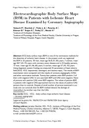
<strong>Prague</strong> <strong>Medical</strong> <strong>Report</strong> / Vol. 113 (2012) No. 4, p. 303–308<br />
303)<br />
Persistent Spontaneous Pneumothorax<br />
for Four Years: A Case <strong>Report</strong><br />
Mizuno Y., Iwata H., Shirahashi K., Matsui M., Takemura H.<br />
Department of General and Cardiothoracic Surgery, Graduate School of Medicine,<br />
Gifu University, Gifu, Japan<br />
Received February 26, 2012; Accepted October 10, 2012.<br />
Key words: Pneumothorax – VATS lobectomy – Minimally invasive surgery –<br />
Pleural decortication<br />
Abstract: Pneumothorax, defined as the presence of air in the pleural space, is<br />
usually classified as spontaneous or traumatic; it is unusual for pneumothorax<br />
to be categorized as being acute or chronic. Even if conservative treatment is<br />
chosen, the pneumothorax is cured when air in the pleural space dissolves into<br />
the venous blood. A 50-years-old Japanese man with no prior medical history<br />
was referred to our department with a right pneumothorax and two rightsided<br />
pulmonary nodules on chest X-ray and CT. The chest radiographs of past<br />
mass screening which was taken four years ago showed right pneumothorax<br />
and right-sided pulmonary nodules. From then, all chest radiograph and chest<br />
computed tomography showed right pneumothorax and pulmonary nodules. But<br />
he underwent no medical interventions. We designed to perform an operation for<br />
a treatment of right pneumothorax and the diagnosis of pulmonary tumors. We<br />
underwent right upper lobectomy and pleural decortication under video assisted<br />
thoracic surgery. We obtained pathological diagnosis of inflammatory pseudotumor<br />
and surrounding atelectasis. He was cured from pneumothorax and pulmonary<br />
tumors. A unique case of spontaneous pneumothorax presenting with a pleural<br />
air space that was confirmed by chest radiographs and computed tomography<br />
examinations over a 4-year period is reported.<br />
Mailing Address: Yoshimasa Mizuno, MD., PhD., Department of General and<br />
Cardiothoracic Surgery, Graduate School of Medicine, Gifu University, 1-1 Yanagido,<br />
Gifu, 501-1194, Japan; Phone: +81 58 230 6325; Fax: +81 58 230 6326; e-mail:<br />
mizunoyoshidasa@yahoo.co.jp<br />
© Charles University in <strong>Prague</strong> – Karolinum Press, <strong>Prague</strong> 2012<br />
PMR 2012 04 2670.indd 303 12.11.12 7:10
304)<br />
<strong>Prague</strong> <strong>Medical</strong> <strong>Report</strong> / Vol. 113 (2012) No. 4, p. 303–308<br />
Introduction<br />
Pneumothorax is classified into spontaneous and traumatic (Noppen and De<br />
Keukeleire, 2008). Spontaneous pneumothorax occurs without recognized lung<br />
disease (primary spontaneous pneumothorax), due to an underlying lung disease<br />
(secondary spontaneous pneumothorax), or in conjunction with menstruation<br />
(catamenial pneumothorax) (Noppen and De Keukeleire, 2008). Spontaneous<br />
pneumothorax is a disease that usually develops acutely with ipsilateral chest pain<br />
and mild dyspnea (Noppen and De Keukeleire, 2008). Because air in the pleural<br />
space dissolves into the capillary blood, if no new gas enters the pleural space, the<br />
intrapleural gas pressure tends to decrease and the lung re-expands (Cormier and<br />
Provencher, 2009). The amount of air in the pleural cavity decreases progressively,<br />
and, finally, the pleural space disappears. Therefore, in theory, pneumothorax cannot<br />
be present for a long time, and spontaneous pneumothorax is not usually classified<br />
as an acute or chronic disease. A unique case of spontaneous pneumothorax<br />
detected by chest radiographs and computed tomography (CT) examinations<br />
continuously over a 4-year period is reported.<br />
Case report<br />
A 50-years-old man with no prior history of cancer, infection, or any relevant<br />
occupation was referred to our department after his family doctor had found a<br />
right pneumothorax and two right-sided pulmonary nodules on chest X-ray and<br />
CT: a 44-mm-diameter mass in the right upper lobe and a 36-mm-diameter mass in<br />
the right lower lobe. The patient had a smoking history (current smoker: 30 packs/<br />
year), but no emphysematous change was detectable on chest CT and respiratory<br />
function testing (FEV 1.0 72.1%). He had no symptoms, and physical examination was<br />
unremarkable. There were no specific abnormalities on laboratory examination,<br />
including tumor markers or physiological testing, except for mild elevation of<br />
C-reactive protein (1.37 mg/dl). When the chest radiographs and CTs of past<br />
mass screening and medical examinations were examined, right pneumothorax and<br />
right-sided pulmonary nodules were detectable on the chest radiograph taken four<br />
years earlier and on the chest CT taken three years earlier. After that, all chest<br />
radiographs (8 times) and CTs (5 times) (Figure 1) showed a right pneumothorax<br />
of various degrees and right-sided pulmonary masses that grew gradually. Though<br />
he was diagnosed as having a right pneumothorax, he underwent no treatment<br />
because he had no symptoms, and the pneumothorax on chest radiographs and<br />
chest CTs was small over the 4-year period. Regarding the pulmonary nodules,<br />
though he had been recommended to undergo the detailed examinations by<br />
his family doctor, he had been rejecting. Most recently, 18F-fluorodeoxyglucose<br />
positron emission tomography (18F-FDG PET)/CT and bronchofiberscopy were<br />
performed by his family doctor. 18F-FDG PET/CT showed no areas of increased<br />
18F-FDG uptake, including the pulmonary masses. Bronchofiberscopy revealed no<br />
malignant findings. With a diagnosis of right pneumothorax and pulmonary tumors,<br />
Mizuno Y.; Iwata H.; Shirahashi K.; Matsui M.; Takemura H.<br />
PMR 2012 04 2670.indd 304 12.11.12 7:10
<strong>Prague</strong> <strong>Medical</strong> <strong>Report</strong> / Vol. 113 (2012) No. 4, p. 303–308<br />
305)<br />
Figure 1 – The findings of chest computed tomography (CT).<br />
Chest CTs before pulmonary resection show pneumothorax<br />
with a pulmonary nodule in the right upper lobe. The chest<br />
CT after surgery shows no pleural air space.<br />
A<br />
B<br />
Figure 2 – The intraoperative findings.<br />
A: The pulmonary nodule located in the right upper lobe (RUL) is white in colour. Inflammatory change and<br />
moderate thickness of the visceral pleura on the periphery of the white pulmonary nodule are detectable.<br />
A serous pleural effusion is present.<br />
B: There is little inflammatory reaction in the visceral pleura of the right middle lobe (RML) and the right lower<br />
lobe (RLL). There are scattered, moderate inflammatory lesions of the parietal pleura, and many of them are<br />
seen in the area of the pulmonary apex.<br />
Persistent Spontaneous Pneumothorax<br />
PMR 2012 04 2670.indd 305 12.11.12 7:10
306)<br />
<strong>Prague</strong> <strong>Medical</strong> <strong>Report</strong> / Vol. 113 (2012) No. 4, p. 303–308<br />
an operation to treat the pneumothorax and obtain a pathological diagnosis of the<br />
pulmonary tumors was performed.<br />
The intrathoracic findings are shown in Figure 2. There were no intrathoracic<br />
adhesions. The visceral pleura of the right upper lobe had inflammatory changes,<br />
and white masses in the right upper and lower lobes were easily detectable. The<br />
right upper lobe was collapsed and unexpandable, even under positive airway<br />
pressure. The visceral pleura of the right middle lobe and the right lower lobe<br />
except for the white lesion and its periphery had little inflammatory change.<br />
Therefore, the lobes were expandable easily under positive airway pressure.<br />
There was a small serous pleural effusion, and fluid was collected for general and<br />
acid-fast bacterial cultures. Moderate inflammatory lesions of the parietal pleura<br />
were scattered in the area of the pulmonary apex. A right upper lobectomy was<br />
performed by video-assisted thoracoscopic surgery (VATS). The intraoperative<br />
pathological diagnosis of the resected specimen showed no malignancy. The white<br />
mass consisted of a hypocellular fibrotic lesion, the peripheral region of which<br />
was composed of inflammatory thickening of the visceral pleura and collapsed<br />
alveoli. The patient was diagnosed as having an inflammatory pseudotumor<br />
and surrounding atelectasis. Since the right lower lobe mass appeared to be<br />
Figure 3 – The pathological findings.<br />
A: The mass is a hypocellular fibrotic lesion, and the<br />
peripheral visceral pleura of the mass also show<br />
hypocellular fibrotic change.<br />
B: There are few cells in the tumor on high-power<br />
examination.<br />
C: The visceral pleura of the right upper lobe (RUL),<br />
which is far from the tumor, shows mild inflammatory<br />
change and thickening.<br />
Mizuno Y.; Iwata H.; Shirahashi K.; Matsui M.; Takemura H.<br />
PMR 2012 04 2670.indd 306 12.11.12 7:10
<strong>Prague</strong> <strong>Medical</strong> <strong>Report</strong> / Vol. 113 (2012) No. 4, p. 303–308<br />
307)<br />
the same lesion, pleural decortication was performed, along with biopsy of the<br />
parietal pleura. The patient’s clinical course has been uneventful. The bacterial<br />
cultures showed no growth. The final pathological diagnosis was inflammatory<br />
pseudotumors with surrounding atelectasis and pleuritis (Figure 3). Chest CT<br />
2 months after the operation showed no residual pleural cavity (Figure 1). The<br />
pulmonary nodule in the right lower lobe gradually decreased in size on the chest<br />
radiographs.<br />
Discussion<br />
Spontaneous pneumothorax is classified into primary spontaneous pneumothorax<br />
and secondary spontaneous pneumothorax (Noppen and De Keukeleire, 2008;<br />
MacDuff et al., 2010). Flowcharts for managing spontaneous pneumothorax<br />
have been described for both primary and secondary cases (Noppen and De<br />
Keukeleire, 2008; MacDuff et al., 2010). Many therapeutic options are available for<br />
treating spontaneous pneumothorax. If patients are not dyspneic and have a small<br />
pneumothorax on chest radiographs, observation is a therapeutic option (Noppen<br />
and De Keukeleire, 2008; MacDuff et al., 2010). Since gases are resorbed into the<br />
venous blood and no new gases enter the pleural space, the amount of air<br />
in the pleural cavity decreases progressively and the lung re-expands (Cormier<br />
and Provencher, 2009). It has been reported that the average rate of lung<br />
re-expansion with conservative treatment for spontaneous pneumothorax was<br />
1.25 to 2.2% per day (Kircher and Swartzel, 1954; Kelly et al., 2006). Therefore, it<br />
is unusual for pneumothorax to persist continuously for a long time. Conversely,<br />
unexpandable lung is reported to be a mechanical complication resulting in the<br />
inability of the lung to expand to the chest wall (Huggins et al., 2010). Huggins<br />
et al. (2010) proposed that unexpandable lung be divided into trapped lung and<br />
lung entrapment. Trapped lung and lung entrapment were defined as the sequels<br />
of remote pleural space inflammation resulting in the development of a mature,<br />
fibrous membrane that impedes lung expansion during fluid removal and a<br />
complication of active pleural inflammation, malignancy, or hemothorax, respectively<br />
(Huggins et al., 2010). Since the present case did not have obvious past history of<br />
inflammatory disease of the lung or pleura, it did not strictly fulfil the definition<br />
of unexpandable lung proposed by Huggins et al. (2010). However, the operative<br />
and histological findings showed pleural inflammation, it may be appropriate<br />
that the present case was a subtype of trapped lung. We hypothesized that the<br />
pneumothorax was caused by a pleural laceration due to hyperstraining of the<br />
visceral pleura when the lung re-expanded, and the lung collapse and inflammatory<br />
reaction induced closure and healing of the pleural wound. We considered that the<br />
repetition of these processes caused the condition of persistent pneumothorax.<br />
This case was very unique that all clinical process developed spontaneously and<br />
fulfilled the criteria as follows: (1) continuously detectable pneumothorax on<br />
all chest radiographs and CTs; (2) no obvious past history of pleural diseases;<br />
Persistent Spontaneous Pneumothorax<br />
PMR 2012 04 2670.indd 307 12.11.12 7:10
308)<br />
<strong>Prague</strong> <strong>Medical</strong> <strong>Report</strong> / Vol. 113 (2012) No. 4, p. 303–308<br />
(3) no past history of thoracic trauma or medical intervention that could cause<br />
pneumothorax; and (4) gas-producing bacteria were not detected on pleural<br />
effusion cultures. Because of small pleural air space, he received conservative<br />
treatment. The inflammatory pseudotumor increased in the clinical course, it was<br />
necessary for him to undergo right upper lobectomy.<br />
Conclusion<br />
A surgical intervention should be performed for diagnosis and treatment when<br />
persistent pneumothorax and mass lesions existed even if patients had small<br />
pleural air space and fulfilled the above-mentioned criteria.<br />
References<br />
Cormier, Y., Provencher, S. (2009) Resorption of gases from the pleural space. In: General Thoracic Surgery, 7 th<br />
Ed., eds. Shields, T. W., LoCicero, J., Reed, C. E., Feins, R. H., pp. 735–737, Lippincott Williams and Wilkins,<br />
Philadelphia.<br />
Huggins, J. T., Doelken, P., Sahn, S. A. (2010) The unexpandable lung. F1000 Medicine <strong>Report</strong>s 2, 77.<br />
Kelly, A. M., Loy, J., Tsang, A. Y., Graham, C. A. (2006) Estimating the rate of re-expansion of spontaneous<br />
pneumothorax by a formula derived from computed tomography volumetry studies. Emerg. Med. J. 23,<br />
780–782.<br />
Kircher, L. T. Jr., Swartzel, R. L. (1954) Spontaneous pneumothorax and its treatment. JAMA 155, 24–29.<br />
MacDuff, A., Arnold, A., Harvey, J.; BTS Pleural Disease Guideline Group (2010) Management of spontaneous<br />
pneumothorax: British Thoracic Society Pleural Disease Guideline 2010. Thorax 65, 18–31 (Suppl. 2).<br />
Noppen, M., De Keukeleire, T. (2008) Pneumothorax. Respiration 76, 121–127.<br />
Mizuno Y.; Iwata H.; Shirahashi K.; Matsui M.; Takemura H.<br />
PMR 2012 04 2670.indd 308 12.11.12 7:10