Quintessence Journals - Sandra Kalil Bussadori
Quintessence Journals - Sandra Kalil Bussadori
Quintessence Journals - Sandra Kalil Bussadori

You also want an ePaper? Increase the reach of your titles
YUMPU automatically turns print PDFs into web optimized ePapers that Google loves.
Copyright<br />
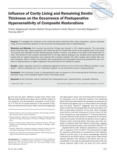
Influence of Cavity Lining and Remaining Dentin<br />
Thickness on the Occurrence of Postoperative<br />
Hypersensitivity of Composite Restorations<br />
Not for Publication<br />
<strong>Quintessence</strong><br />
for<br />
Not<br />
by<br />
Florian Wegehaupt a /Herbert Betke b /Nicole Solloch c /Ulrike Musch a,b /Annette Wiegand a,b /<br />
Thomas Attin d,e<br />
Purpose: To investigate the influence of the remaining dentin thickness after cavity preparation, calcium hydroxide<br />
lining, and two restorative systems on the occurrence of postoperative pain or hypersensitivity.<br />
Materials and Methods: One hundred twenty-three fillings were placed in 123 healthy patients. The remaining<br />
dentin thickness after caries excavation was measured with the Prepometer device at the deepest area of the cavity.<br />
The cavities were allocated to three different groups (shallow, medium, and deep) on the basis of the Prepometer results.<br />
The decision to use a calcium hydroxide liner or not was made by tossing a coin. Cavities which were to be later<br />
treated with an indirect restoration were restored with a buildup material. The other cavities were treated with a hybrid<br />
composite. After 6 months, the patients were re-examined and interviewed concerning postoperative pain incidents<br />
or hypersensitivity. A logistic regression was performed for the statistical analysis.<br />
Results: Logistic regression showed no statistically significant influence of any of the three different variables “cavity<br />
depth”, “calcium hydroxide liner” and “restorative material” on the occurrence of pain or hypersensitivity.<br />
Conclusion: The occurrence of pain or hypersensitivity does not depend on the remaining dentin thickness, calcium<br />
hydroxide lining, or the restorative system used in the present study.<br />
Keywords: dentin thickness, calcium hydroxide liner, postoperative pain, hypersensitivity, composite, adhesive.<br />
J Adhes Dent 2009; 11: 137-141. Submitted for publication: 14.12.07; accepted for publication: 05.03.08.<br />
In the last 50 years, different studies have shown that<br />
caries-linked dental hard tissue loss is directly related to<br />
the presence and fermentation processes of oral bacteria.<br />
15,25 The aim of caries treatment is the removal of bacteria<br />
and softened hard tissue. When caries has progressed<br />
a Assistant Professor, Clinic for Preventive Dentistry, Periodontology and Cariology,<br />
University of Zürich, Zürich, Switzerland.<br />
b Assistant Professor, Department of Operative Dentistry, Preventive Dentistry<br />
and Periodontology, Georg August University of Göttingen, Göttingen, Germany.<br />
c Dentist in Private Practice; Department of Operative Dentistry, Preventive<br />
Dentistry and Periodontology, Georg August University of Göttingen, Göttingen,<br />
Germany.<br />
d Professor, Department of Operative Dentistry, Preventive Dentistry and Periodontology,<br />
Georg August University of Göttingen, Göttingen, Germany.<br />
e Professor and Chair, Clinic for Preventive Dentistry, Periodontology and Cariology,<br />
University of Zürich, Zürich, Switzerland.<br />
Correspondence: Dr. F. Wegehaupt, Clinic for Preventive Dentistry, Periodontology<br />
and Cariology, University of Zürich, Plattenstr. 11, CH-8032 Zürich,<br />
Switzerland. Tel: +41-44-634-3439, Fax: +41-44-634-4308. e-mail:<br />
florian.wegehaupt@zzmk.uzh.ch<br />
into deep dentin areas, the remaining dentin thickness after<br />
excavation can be distinctly reduced. The remaining<br />
dentin thickness estimated to be necessary for protection of<br />
the dental pulp against injury or inflammation has changed<br />
over the years. Stanley 31 suggested that the remaining<br />
dentin under the cavity preparation should be at least 2 mm<br />
thick to guarantee protection of the pulp. Other investigations<br />
found a minimum thickness of 1 mm or even 0.5 mm<br />
to be necessary for pulp protection. 21,26 During treatment,<br />
it is often difficult for the dentist to decide how much dentin<br />
is left over the pulp. The dentist can perform only a rough estimation<br />
of the remaining dentin thickness, since the variability<br />
of different teeth and different calcification degrees<br />
of the pulp chambers render a clear estimation difficult. 22,28<br />
Hypersensitivity after adhesive restorations is observed<br />
with a frequency of 5% to 26%. 5 This phenomenon might<br />
be caused by penetration of components of the adhesive<br />
systems into the pulp, or by micro-/nanoleakage, allowing<br />
movement of the dentin liquid in those dentin areas where<br />
optimal adhesion and sealing of the dentin was not accomplished.<br />
It can also be speculated that parts of the adhesive<br />
systems might enter the pulp via a thin dentin<br />
Vol 11, No 2, 2009 137
Wegehaupt et al<br />
Table 1 Application protocols for the two kinds of adhesives and restorative materials<br />
used in the study<br />
Definitive filling:<br />
Prime & Bond NT and Spectrum<br />
Buildup filling:<br />
Clearfil Liner Bond 2V and LuxaCore<br />
Not for Publication<br />
Copyright<br />
<strong>Quintessence</strong><br />
for<br />
Not<br />
by<br />
Enamel etching for 40 s, dentin etching<br />
for 20 s with 37% phosphoric acid<br />
↓<br />
Rinsing with water for 30 s and<br />
gently drying with oil-free air<br />
↓<br />
Application of Prime&Bond NT<br />
on the cavity surface for 20 s<br />
↓<br />
Gentle air drying for 5 s<br />
↓<br />
Light curing of the adhesive for 20 s<br />
↓<br />
Application of small increments<br />
of Spectrum<br />
↓<br />
Light curing of the composite for 20 s<br />
each increment<br />
Mixing primer liquid A+B<br />
↓<br />
Application of the primer liquid mixture for<br />
30 s on the cavity surface<br />
↓<br />
Gentle air drying for 30 s<br />
↓<br />
Mixing bond liquid A+B<br />
↓<br />
Application of the bond liquid mixture on<br />
the cavity surface and gentle air drying<br />
↓<br />
Light curing for 20 s<br />
↓<br />
Filling of cavities with LuxaCore in bulk technique<br />
bridge, 29,36 thus provoking some postoperative pain sensations.<br />
To avoid penetration of components of adhesive<br />
systems into the pulp, a calcium hydroxide liner can be<br />
placed onto the dentin at the deepest points of the cavity.<br />
This procedure was previously recommended especially<br />
for nonadhesive restoration and was referred to as indirect<br />
pulp capping. Thus, a calcium hydroxide liner may prove<br />
effective in reducing postoperative hypersensitivity occurring<br />
after application of an adhesive restoration.<br />
Various formulations of calcium hydroxide have been investigated<br />
over the years. 4,30 Calcium hydroxide has<br />
demonstrated its potential for inducing pulp healing and<br />
dentin bridging. Despite a certain degree of controversy,<br />
some recent studies have also reported the possibility of<br />
pulp healing and dentin bridging by the use of adhesive<br />
systems for pulp capping. 1,10<br />
Thus, the aim of the present study was to identify the<br />
influence of the use of a calcium hydroxide liner in cavities<br />
with different remaining dentin thicknesses and two different<br />
adhesive restorative materials on the occurrence of<br />
pain or postoperative hypersensitivity.<br />
MATERIALS AND METHODS<br />
Approval (No. 5/11/02) was issued prior to the study by<br />
the Ethics Committee of the University of Göttingen. A total<br />
of 123 fillings was placed in 123 patients, after they<br />
signed an informed consent form prior to their participation<br />
in the study. Exclusion criteria were: patients under 18<br />
years, pregnancy, breastfeeding, immunosuppressed or<br />
addicted patients.<br />
Only teeth fulfilling the following criteria were included<br />
in the study: caries media or caries profunda (according to<br />
bitewing radiographs), insufficient fillings, positive reaction<br />
to a vitality test (cold test), no signs of pulp inflammation,<br />
no spontaneous pain attacks before treatment, only premolars<br />
and molars, only one filling per tooth, and a minimum<br />
extension of the cavity of 1 mm in width. This cavity<br />
size was necessary, since the probe of the Prepometer device<br />
(Hager & Werken; Duisburg, Germany) described<br />
below has a diameter of 1 mm.<br />
Treatment was performed under local anesthesia (Ultracain<br />
D-S, Hoechst Marion Roussel Deutschland; Frankfurt<br />
am Main, Germany) and the use of rubber-dam. Caries removal<br />
was performed with tungsten burs until CariesDetector<br />
(Kuraray Dental; Frankfurt am Main, Germany)<br />
induced no further staining of the cavity. After total caries<br />
removal, the cavity was rinsed with 0.2% chlorhexidine solution.<br />
After cavity preparation, the remaining dentin thickness<br />
in the deepest cavity area was measured with a remaining<br />
dentin thickness measuring device, hereafter referred to<br />
as RDTMD (Prepometer, Hager & Werken). Before measuring,<br />
the RDTMD was calibrated by touching the dentin surface<br />
with the calibration and sensor electrode at the same<br />
time. During measurement, the reference electrode was<br />
placed in the buccal vestibule. For measuring the remaining<br />
dentin thickness, the sensor electrode was gently<br />
moved over the cavity floor.<br />
138 The Journal of Adhesive Dentistry
Copyright<br />
Table 2 Number of teeth according to allocation to the different cavity depth groups, the use of<br />
calcium hydroxide liner, and the kind of restorative material<br />
Calcium hydroxide liner Restorative material Cavity depth group<br />
shallow medium deep<br />
Not for Publication<br />
Wegehaupt et al<br />
<strong>Quintessence</strong><br />
for<br />
Not<br />
by<br />
Yes buildup 6 8 9<br />
definitive filling 14 21 17<br />
No buildup 5 5 7<br />
definitive filling 16 5 10<br />
Table 3 Percentages (absolute number) of pain incidence or hypersensitivity of the buildup and definitive<br />
restorations placed in either shallow, medium or deep cavities after use of a calcium hydroxide lining<br />
or not<br />
Calcium hydroxide liner Restorative material Cavity depth group<br />
shallow medium deep<br />
yes buildup 17% (1) 0% (0) 11% (1)<br />
definitive filling 14% (2) 14% (3) 11% (2)<br />
no buildup 40% (2) 0% (0) 29% (2)<br />
definitive filling 13% (2) 40% (2) 40% (4)<br />
On the basis of the RDTMD results, the cavities were divided<br />
into three different groups (shallow, medium, or<br />
deep cavity) of 40 teeth each. Cavities with RDTMD scores<br />
1 to 5 (1.5 to 3.0 mm, green or yellow LEDs) were allocated<br />
to group “shallow”, cavity scores 6 and 7 (0.9 to 1.5<br />
mm, orange LEDs) were allocated to group “medium”, and<br />
with results 8 to 10 (< 0.9 mm, red LEDs) to group “deep”.<br />
In each group, cavities were treated either with or without<br />
use of calcium hydroxide liner (Kerr Life, KerrHawe; Bioggio,<br />
Switzerland). The decision to use a calcium hydroxide<br />
liner or not was made by tossing a coin. This resulted in<br />
20 liners being placed in the shallow group, 29 in the<br />
medium-depth group, and 26 in the group of deep cavities<br />
(Table 2).<br />
To make the calcium hydroxide liner, a small drop of<br />
Kerr Life was placed on the deepest part of the cavity and<br />
allowed to set until its surface was hard upon probing.<br />
The restoration of the teeth was performed either with a<br />
buildup composite (n = 40) (LuxaCore, DMG; Hamburg,<br />
Germany) or a hybrid composite (n = 83) (Spectrum,<br />
Dentsply DeTrey; Konstanz, Germany). For the buildup<br />
composite restorations, a self-etching nonrinse adhesive<br />
system was used (Clearfil Liner Bond 2V, Kuraray Dental).<br />
The buildup composite was used when a large amount of<br />
dental hard tissue was lost and an indirect restoration was<br />
required (crown or partial crown) later to stabilize the integrity<br />
of the tooth or when the cervical margin was located<br />
subgingivally. An etch-and-rinse adhesive<br />
(Prime&Bond NT, Dentsply) was used in the remaining cavities<br />
that were restored with the hybrid composite Spectrum.<br />
These were Class I and II restorations (O, OM/OD,<br />
and MOD) with the cervical margin located para- or<br />
supragingivally. During the restoration, steel matrices were<br />
used and fixed with wooden wedges. The hybrid composite<br />
for the definitive restorations was applied in the cavities in<br />
small horizontal increments with a maximum height of 1<br />
mm. Application protocols for the two kinds of adhesives<br />
and restorative materials are given in Table 1.<br />
The patients were not told to which cavity-depth group<br />
their tooth was allocated and if calcium hydroxide was<br />
used or not. The patients were asked to record whether<br />
any hypersensitivity, pain, or discomfort occurred following<br />
treatment.<br />
During a second appointment 6 months later, the patients<br />
were re-examined by one dentist evaluating the following<br />
criteria:<br />
• Vitality test (yes/no): vitality testing was performed with<br />
a cold foam pellet pressed on the buccal surface of the<br />
tooth. The patients were asked to report whether they<br />
felt the cold.<br />
• Occurrence of hypersensitivity (yes/no): the patients<br />
were asked if they had noticed any pain, hypersensitivity,<br />
or discomfort in the teeth after the application of<br />
the filling.<br />
Reacting negative to the vitality test and reported pain<br />
or hypersensitivity were ranked as failure.<br />
To evaluate the influence of the three different variables<br />
“cavity depth”, “calcium hydroxide liner”, and “resto-<br />
Vol 11, No 2, 2009 139
Copyright<br />
Wegehaupt et al<br />
ration material” a logistic regression was carried out. The<br />
significance level was set at p ≤ 0.05.<br />
The allocation of the tooth to the cavity groups, use of<br />
calcium hydroxide lining, and the kind of restorative material<br />
is shown in Table 2.<br />
RESULTS<br />
All patients were re-examined after 6 months; all 123 fillings<br />
were still in situ. No fractures or any other distinctive<br />
features were visible in the clinical examination. All teeth<br />
exhibited a positive reaction in the vitality test. Percentage<br />
distribution of pain incidents or hypersensitivity according<br />
to the cavity groups, calcium hydroxide lining use, and the<br />
kind of restorative material are shown in Table 3.<br />
The logistic regression showed no statistically significant<br />
influence of any of the three different variables “cavity<br />
depth” (p = 0.65), “calcium hydroxide liner” (p = 0.086),<br />
and “restoration material” (p = 0.71) on the occurrence of<br />
pain or hypersensitivity.<br />
DISCUSSION<br />
The present study aimed to investigate the influence of different<br />
thickness of remaining dentin on pain occurrence of<br />
adhesively luted restorations under in vivo conditions.<br />
Since the treated teeth were not extracted afterwards, no<br />
histological examination was possible to determine dentin<br />
thickness. Thus, for determining dentin thickness and to<br />
allow the allocation of the teeth to different categories of<br />
remaining dentin thickness, the RDTMD was used. The<br />
measurement with the RDTMD depends on the electrical<br />
resistance of the dentin. 14<br />
Studies performing histological examination of restored<br />
teeth mostly use teeth planned to be extracted for orthodontic<br />
reasons. 6,21 This procedure has the disadvantage<br />
that these teeth are often free of caries, thus not mirroring<br />
the usual clinical situation. An advantage of using the<br />
RDTMD in the present study was that teeth with clinically<br />
common carious lesions could be included in the study.<br />
However, the use of the RDTMD for allocation of the teeth<br />
to different categories of remaining dentin thickness is a<br />
topic of some discussion. Gente 13 observed a higher electrical<br />
resistance with longer dentin tubules and greater<br />
remaining dentin thickness. 12,14 The same author 13<br />
reported the remaining dentin thickness as being 2.1 to<br />
3.0 mm for the green LEDs, 1.5 to 2.1 mm for the yellow<br />
LEDs, 0.9 to 1.5 mm for the orange LEDs, and less than<br />
0.9 mm for the red LEDs. Thus, our definition of shallow,<br />
medium, and deep corresponds to 1.5 to 3.0 mm, 0.9 to<br />
1.5 mm, and less than 0.9 mm remaining dentin thickness,<br />
respectively. In contrast, Tielemans et al 32 found the<br />
use of RDTMD to be reproducible, but in their study, the<br />
electrical resistance showed no statistically significant correlation<br />
with the histologically determined dentin<br />
thickness. It should be mentioned that in the study by<br />
Tielemans et al, 32 only two patients with a total of 12<br />
teeth were examined. Moreover, the present study using<br />
Not for Publication<br />
<strong>Quintessence</strong><br />
the RDTMD was started before the discrepant findings by<br />
Tielemans et al 32 were published. To clarify the contradictory<br />
findings regarding the RDTMD, a comprehensive in<br />
vivo study with an appropriate number of test teeth must<br />
be performed.<br />
In the present study, two different kinds of restorative<br />
materials were used to reflect different treatment requirements.<br />
The definitive fillings were made with the hybrid<br />
composite in an incremental technique. This material has<br />
shown good clinical performance in various previous clinical<br />
studies. 18,27,33,34 The buildup composite in combination<br />
with the self-etching adhesive system was applied in<br />
bulk in the cavities with greater extension. This procedure<br />
presents a fast, economical, and less technique-sensitive<br />
approach, and is often used to build up teeth prior to indirect<br />
restorations.<br />
The overall occurrence of postoperative pain in the present<br />
study amounted to 17%. This corresponds well with<br />
the findings of another recent study, 5 showing postoperative<br />
pain incidence of 5% to 26% after adhesive restorations<br />
with different cavity sizes.<br />
Our finding that there was no statistically significant influence<br />
of use of a calcium hydroxide liner on hypersensitivity<br />
corresponded with the results of Whitworth et al. 35<br />
They also found no difference in the protection of the pulp<br />
by calcium hydroxide lining or conditioning and sealing<br />
with adhesive resins only. This might be explained with the<br />
precipitation of crystalline salts in the dentin tubules after<br />
the use of calcium hydroxide liner 19 or the sealing of the<br />
dentin with light- or self-curing resin after the use of a hydrophilic<br />
adhesive, creating a continuous resin-dentin hybrid<br />
layer. 23 Both may lead to a reduction of the dentin<br />
permeability for potential cytotoxic components of restorative<br />
materials or bacteria, 8 thus protecting the pulp tissue.<br />
Moreover, with respect to the hydrodynamic theory of<br />
Brännström, 3 which states that movement of dentinal liquid<br />
is responsible for hypersensitivity, both precipitation of<br />
calcium salts and formation of the hybrid layer might be<br />
able to counteract hypersensitivity.<br />
In the present study, no statistically significant influence<br />
of the cavity depth on the occurrence of pain was observed.<br />
Murray et al 21 also did not find the remaining<br />
dentin thickness to be statistically correlated to signs of<br />
pulp inflammation as histologically proven. In contrast,<br />
Whitworth et al 35 estimated the residual dentin thickness<br />
to be a key determinant for the pulp reaction. It should be<br />
mentioned that in that study, 35 teeth with pulp exposure<br />
were also included in the group of deep cavities; furthermore,<br />
there was no requirement for the dentists to use<br />
rubber-dam during treatment. The use of rubber-dam<br />
might have an impact on the pulp response after cavity<br />
preparation, as shown by Camps et al 6 , who found remaining<br />
dentin thickness to be of only minor importance in the<br />
reaction of the pulp, as compared to bacteria left in the<br />
cavity prior to filling. It might be assumed that lesser<br />
amounts of bacteria are still present on the cavity walls<br />
when rubber-dam is used during treatment.<br />
The effect of restorative materials on the vitality of the<br />
pulp and the occurrence of postoperative pain is a contentious<br />
issue in the literature. 9,16,24,37 In the present<br />
for<br />
Not<br />
by<br />
140 The Journal of Adhesive Dentistry
Copyright<br />
study, no statistically significant influence of the restorative<br />
material was found. This finding agrees with prior findings<br />
that even a thin layer of residual dentin may protect<br />
the pulp against both material and bonding system toxicity,<br />
2,20 so that different restorative materials or adhesives<br />
will not have a significant influence on the pulp tissue. 24<br />
Cox 7 also showed the restorative materials to have a minimal<br />
influence on postoperative pulp reactions. In contrast<br />
to these findings, a recent study by Whitworth et al 35 observed<br />
a correlation between restorative materials and<br />
pulp breakdown only in the deep cavities and cavities with<br />
pulp exposure. This finding may be explained with previous<br />
investigations which showed that odontoblasts are already<br />
injured by cavity preparations with a depth close to or into<br />
the pulp tissue, 11,17 which may lead to pulp inflammation<br />
and pulp breakdown.<br />
We suggest that the layer formed by the adhesive systems<br />
acts as a barrier to filling material contents reaching<br />
the pulp. Similar to our finding that the materials used<br />
here had no influence on the pulp vitality, Nayyar et al 24<br />
also observed no difference in the pulp response between<br />
a self-etching adhesive and an etch-and-rinse adhesive.<br />
CONCLUSION<br />
The results of the present study showed that neither the<br />
thickness of the remaining dentin, the use of calcium hydroxide<br />
liner, nor the adhesive and restorative system<br />
used in the study have an impact on the incidence of postoperative<br />
pain.<br />
REFERENCES<br />
1. Akimoto N, Momoi Y, Kohno A, Suzuki S, Otsuki M, Suzuki S, Cox CF. Biocompatibility<br />
of Clearfil Liner Bond 2 and Clearfil AP-X system on nonexposed<br />
and exposed primate teeth. <strong>Quintessence</strong> Int 1998;29:177-188.<br />
2. Bergenholtz G. Evidence for bacterial causation of adverse pulpal responses<br />
in resin-based dental restorations. Crit Rev Oral Biol Med<br />
2000;11:467-480.<br />
3. Brännström M, Linden LA, Astrom A. The hydrodynamics of the dental<br />
tubule and of pulp fluid. A discussion of its significance in relation to dentinal<br />
sensitivity. Caries Res 1967;1:310-317.<br />
4. Brännström M, Nordenvall KJ. Bacterial penetration, pulpal reaction and<br />
the inner surface of Concise enamel bond. Composite fillings in etched<br />
and unetched cavities. J Dent Res 1978;57:3-10.<br />
5. Briso ALF, Mestrener SR, Delicio G, Sundfeld RH, Bedran-Russo AK. Clinical<br />
assessment of postoperative sensitivity in posterior composite restorations.<br />
Oper Dent 2007;32:421-426.<br />
6. Camps J, Dejou J, Remusat M, About I. Factors influencing pulpal response<br />
to cavity restorations. Dent Mater 2000;16:432-440.<br />
7. Cox CF. Effects of adhesive resins and various dental cements on the pulp.<br />
Oper Dent 1992;(suppl 5):165-176.<br />
8. Cox CF. Evaluation and treatment of bacterial microleakage. Am J Dent<br />
1994;7:293-295.<br />
9. Cox CF, Felton D, Bergenholtz G. Histopathological response of infected<br />
cavities treated with Gluma and Scotchbond dentin bonding agents. Am J<br />
Dent 1988;1(special issue):189-194.<br />
10 . Cox CF, Hafez AA, Akimoto N, Otsuki M, Suzuki S, Tarim B. Biocompatibility<br />
of primer, adhesive and resin composite systems on non-exposed and exposed<br />
pulps of non-human primate teeth. Am J Dent 1998;11(special<br />
issue):S55-63.<br />
11. Darvell BW. Effect of dentine thickness on pulpal changes beneath restorative<br />
materials. Aust Dent J 1981;26:80-81.<br />
12. Gente M. Begrenzung der Präparationstiefe durch elektrische Widerstandsmessungen.<br />
Dtsch Zahnarztl Z 1995;50:658-660.<br />
Not for Publication<br />
Wegehaupt et al<br />
<strong>Quintessence</strong><br />
13. Gente M. Untersuchung zur Begrenzung der Präparationstiefe bei der Kronenpräparation<br />
durch elektrische Widerstandsmessung. Marburg, Germany:<br />
Thesis, 1992<br />
14. Gente M, Becker-Detert D. Studies on the specific electric resistance of the<br />
dentin of human teeth [in German]. Dtsch Zahnarztl Z 1991;46:803-806.<br />
15. Keyes PH. The infectious and transmissible nature of experimental dental<br />
caries. Findings and implications. Arch Oral Biol 1960;1:304-320.<br />
16. Koliniotou-Koumpia E, Papadimitriou S, Tziafas D. Pulpal responses after<br />
application of current adhesive systems to deep cavities. Clin Oral Investig<br />
2007;11:313-320.<br />
17. Lee SJ, Walton RE, Osborne JW. Pulp response to bases and cavity depths.<br />
Am J Dent 1992;5:64-68.<br />
18. Loguercio AD, Reis A, Hernandez PA, Macedo RP, Busato AL. 3-Year clinical<br />
evaluation of posterior packable composite resin restorations. J Oral Rehabil<br />
2006;33:144-151.<br />
19. Mjör IA, Ferrari M. Pulp-dentin biology in restorative dentistry. Part 6: Reactions<br />
to restorative materials, tooth-restoration interfaces, and adhesive<br />
techniques. <strong>Quintessence</strong> Int 2002;33:35-63.<br />
20. Murray PE, Hafez AA, Smith AJ, Cox CF. Bacterial microleakage and pulp inflammation<br />
associated with various restorative materials. Dent Mater<br />
2002;18:470-478.<br />
21. Murray PE, Smith AJ, Windsor LJ, Mjör IA. Remaining dentine thickness<br />
and human pulp responses. Int Endod J 2003;36:33-43.<br />
22. Murray PE, Stanley HR, Matthews JB, Sloan AJ, Smith AJ. Age-related odontometric<br />
changes of human teeth. Oral Surg Oral Med Oral Pathol Oral Radiol<br />
Endod 2002;93:474-482.<br />
23. Nakabayashi N. Importance of mini-dumbbell specimen to access tensile<br />
strength of restored dentine: historical background and the future perspective<br />
in dentistry. J Dent 2004;32:431-442.<br />
24. Nayyar S, Tewari S, Arora B. Comparison of human pulp response to totaletch<br />
and self-etch bonding agents. Oral Surg Oral Med Oral Pathol Oral Radiol<br />
Endod 2007;104:45-52.<br />
25. Orland FJ, Blayney JR, Harrison RW, Reyniers JA, Trexler PC, Ervin RF, Gordon<br />
HA, Wagner M. Experimental caries in germfree rats inoculated with<br />
enterococci. J Am Dent Assoc 1955;50:259-272.<br />
26. Pameijer CH, Stanley HR, Ecker G. Biocompatibility of a glass ionomer luting<br />
agent. 2. Crown cementation. Am J Dent 1991;4:134-141.<br />
27. Perry RD, Kugel G, Habib CM, McGarry P, Settembrini L. A two-year clinical<br />
evaluation of TPH for restoration of Class II carious lesions in permanent<br />
teeth. Gen Dent 1997;45:344-349.<br />
28. Polansky R, Reichhold C, Lorenzoni M. Die Topographie der Pulpa im<br />
Seitenzahnbereich nach Stufenpräparation für Vollkeramische Kronen.<br />
Eine experimentelle Untersuchung. Dtsch Zahnarztl Z 1998;53:643-647.<br />
29. Schmalz G, Hiller KA, Nunez LJ, Stoll J, Weis K. Permeability characteristics<br />
of bovine and human dentin under different pretreatment conditions. J<br />
Endod 2001;27:23-30.<br />
30. Snuggs HM, Cox CF, Powell CS, White KC. Pulpal healing and dentinal<br />
bridge formation in an acidic environment. <strong>Quintessence</strong> Int 1993;24:501-<br />
510.<br />
31. Stanley HR. Dental iatrogenesis. Int Dent J 1994;44:3-18.<br />
32. Tielemans S, Bergmans L, Duyck J, Naert I. Evaluation of a preparation<br />
depth controlling device: a pilot study. <strong>Quintessence</strong> Int 2007;38:135-142.<br />
33. Türkün LS, Aktener BO. Twenty-four-month clinical evaluation of different<br />
posterior composite resin materials. J Am Dent Assoc 2001;132:196-203.<br />
34. Wendt SLJ, Leinfelder KF. Clinical evaluation of a posterior resin composite:<br />
3-year results. Am J Dent 1994;7:207-211.<br />
35. Whitworth JM, Myers PM, Smith J, Walls AW, McCabe JF. Endodontic complications<br />
after plastic restorations in general practice. Int Endod J<br />
2005;38:409-416.<br />
36. Wiegand A, Caspar C, Becker K, Werner C, Attin T. In vitro cytotoxicity of different<br />
self-etching dental adhesive systems. Schweiz Monatsschr Zahnmed<br />
2006;116:614-621.<br />
37. Wisithphrom K, Murray PE, About I, Windsor LJ. Interactions between cavity<br />
preparation and restoration events and their effects on pulp vitality. Int J<br />
Periodontics Restorative Dent 2006;26:596-605.<br />
for<br />
Clinical relevance: Neither the thickness of the remaining<br />
dentin, the use of calcium hydroxide liner, or the adhesive<br />
and restorative systems have an impact on the<br />
incidence of postoperative pain.<br />
Not<br />
by<br />
Vol 11, No 2, 2009 141