Laringocele misto - Acta Otorhinolaryngologica Italica
Laringocele misto - Acta Otorhinolaryngologica Italica
Laringocele misto - Acta Otorhinolaryngologica Italica

You also want an ePaper? Increase the reach of your titles
YUMPU automatically turns print PDFs into web optimized ePapers that Google loves.
ACTA OTORHINOLARYNGOLOGICA ITALICA 2007;27:255-257<br />
CASE REPORT<br />
Mixed laryngocele:<br />
a case report and review of the literature<br />
<strong>Laringocele</strong> <strong>misto</strong>: case report e revisione della letteratura<br />
A. LANCELLA, G. ABBATE, R. DOSDEGANI<br />
Otorhinolaryngology Unit, “S. Biagio” Hospital, Domodossola (VB), Italy<br />
SUMMARY<br />
Laryngocele is a rare, benign dilatation of the laryngeal saccule that may extend internally into the airway or externally through<br />
the thyrohyoid membrane. Many laryngoceles are asymptomatic; sometimes they may cause a cough, hoarseness, stridor, sore<br />
throat and may present as a swelling on one or both sides of the neck. Laryngocele may be associated with supraglottic squamous<br />
cell carcinoma. Computed tomography scan is the most effective imaging method for diagnosis. Surgery is the treatment<br />
of choice. A case of large mixed laryngocele in a 75-year-old male is described together with surgical management and followup.<br />
A review of the literature is also presented.<br />
KEY WORDS: Larynx • Laryngocele • Professional diseases • Surgical treatment<br />
RIASSUNTO<br />
Il laringocele è una rara e benigna dilatazione dell’appendice del ventricolo di Morgagni che può svilupparsi internamente<br />
nel lume laringeo o esternamente attraverso la membrana tiroidea. Molti laringoceli sono asintomatici; qualche volta possono<br />
causare tosse, disfonia, stridore, mal di gola e presentarsi come una tumefazione a uno o entrambi i lati del collo. Il laringocele<br />
può associarsi al carcinoma spinocellulare sovraglottico. La tomografi a computerizzata è l’indagine radiologica più utile per<br />
la diagnosi; la chirurgia è l’opzione terapeutica di scelta. Gli Autori descrivono il caso di un grosso laringocele <strong>misto</strong> in un<br />
uomo di 75 anni, il trattamento effettuato e il follow-up post-operatorio. Inoltre si effettua una revisione della letteratura.<br />
255<br />
PAROLE CHIAVE: Laringe • <strong>Laringocele</strong> • Malattie professionali • Terapia chirurgica<br />
<strong>Acta</strong> Otorhinolaryngol Ital 2007;27:255-257<br />
Case report<br />
The patient B.D.G., a 75-year-old male had been suffering<br />
from hoarseness for approximately 5 years. Over the last<br />
month, he had been suffering from dyspnoea on exertion<br />
and, in the last few days even when at rest.<br />
The patient also suffered from hypertension, chronic atrial fibrillation,<br />
emphysema and chronical bronchitis; despite these<br />
problems, he managed a mixed farm with crops and livestock.<br />
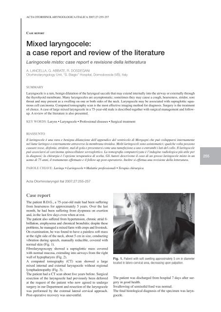
On examination, he was found to have a painless soft mass<br />
at the right side of the neck, about 5 cm in size, conducting<br />
vibration during speech, manually reducible, covered with<br />
normal skin (Fig. 1).<br />
Fibrolaryngoscopy showed a supraglottic mass covered<br />
with normal mucosa, extending into airways from the right<br />
wall of hypopharynx (Fig. 2).<br />
A computed tomography (CT) scan showed a large<br />
mixed internal and external laryngocele without regional<br />
lymphadenopathy (Fig. 3).<br />
The patient had a CT scan about five years before. Surgical<br />
resection of the laryngocele had previously been deferred<br />
at the request of the patient who now agreed to undergo<br />
surgery in our Department and resection of the laryngocele<br />
was performed by the external lateral cervical approach.<br />
Post-operative recovery was uneventful.<br />
Fig. 1. Patient with soft swelling approximately 5 cm in diameter<br />
located in latero-cervical area, decreasing upon palpation.<br />
The patient was discharged from hospital 7 days after surgery<br />
in good health.<br />
Swallowing of semisolid food was normal.<br />
The final histological diagnosis of the specimen was laryngocele.
A. Lancella et al.<br />
256<br />
Fig. 2. Flexible pre- and post-operative fi bro-endoscopic assessment.<br />
Fig. 3. Computed tomography image of patient’s neck: extended<br />
gaseous right laryngocele located on soft parts of neck.<br />
Fig. 4. Post-operative CT revealing disappearance of gaseous<br />
laryngeal mass.<br />
Post-operative CT 4 months after surgery, showed complete<br />
removal of the laryngocele (Fig. 4). The larynx and the other<br />
neck structures were normal.<br />
The patient remains free from disease.<br />
Review of the literature<br />
Laryngocele, an abnormal cystic dilatation of the laryngeal<br />
saccule 1-5 , is uncommon 2 4-6 , usually benign 7 and may occur<br />
in up to 5% of benign laryngeal lesions 4 .<br />
The aetiology is unknown and unclear 3 , but there is an interrelation<br />
between a congenital predisposition – represented<br />
by a large ventricular appendix 8 , for example, a congenital<br />
laryngocele which causes respiratory distress in a newborn 9 –<br />
and other post-natal acquired factors, for instance, laryngeal<br />
papillomatosis in a child 8 .<br />
An acquired laryngocele may develop when the laryngeal<br />
ventricle becomes functionally obstructed as a result of an increase<br />
in intra-glottic pressure, such as that caused by excessive<br />
coughing, playing a wind instrument, glass blowing 2-5 ,<br />
after performing Valsalva manoeuvre 10 or using ventricular<br />
phonation during speech 2 .<br />
Laryngoceles may extend internally into the airway or externally<br />
through the thyrohyoid membrane 2 , so they may<br />
present as internal, external or combined mixed internal and<br />
external laryngocele 11-19 , unilateral uncommon 2 3 6 11 20 , or<br />
bilateral rare 1 2 12 15 21 .<br />
Laryngocele may be asymptomatic and incidentally discovered<br />
through radiographic studies for unrelated symptoms 1 4 5 .<br />
The main symptoms, at presentation, are: airway obstruction<br />
5 7 9 13 16-18 , increasing stridor 4 9 14 , hoarseness 4 14-20 , sore<br />
throat, cough, pain, snoring, globus sensation 4 or a visible<br />
or palpable mass in the neck 9 10 15 16 19 20 .<br />
Serious forms of clinical emergency requiring tracheotomy<br />
may occur 5 7 12 16 .<br />
There is a rare, but well-documented, association of laryngocele<br />
with laryngeal carcinoma 1 11 15 17 20-23 . Therefore, if a<br />
laryngocele is detected clinically or radiologically, a carcinoma<br />
must be taken into consideration and appropriate tests<br />
be performed 12 22 23 .<br />
Supraglottic carcinoma is the most common laryngeal tumour<br />
17 .<br />
Fewer reports have appeared concerning the coexistence<br />
with other laryngeal diseases, for example, papillomatosis<br />
in children 8 , amyloidosis 6 , rheumatoid arthritis 3 , oncocytic<br />
cysts 21 .<br />
CT scan has proved to be the most accurate imaging method<br />
in defining the spatial relationship between the laryngocele<br />
and the laryngeal structures and extra-laryngeal<br />
soft tissues, in differentiating the laryngocele from other<br />
cystic formations and in identifying the coexistence of a<br />
laryngeal cancer 1 6 9 14 15 17 23 .<br />
Magnetic resonance imaging may be also useful 6 14 20 .<br />
Options in the management of laryngoceles include observation,<br />
endoscopic resection and resection via an external<br />
approach 24 .<br />
Surgery is the treatment of choice 15 .<br />
Endoscopic marsupialization with CO 2<br />
laser is frequently<br />
used to remove small internal laryngoceles 7 15-17 19 .<br />
According to some Authors, the external cervical approach,<br />
without tracheotomy, allows good exposure of the lesion<br />
with minimal functional disability 15 17 18 . It is recommended<br />
for the mixed and external laryngoceles 12 13 17 .<br />
Careful dissection of the neck, in the case of an external<br />
laryngocele sac, is important to prevent damage to the neurovascular<br />
bundle which penetrates the thyrohyoid membrane<br />
at the site of penetration of the external laryngocele 13 .<br />
Conclusion<br />
Laryngocele is a rare benign laryngeal disease which is often<br />
asymptomatic.<br />
The diagnosis may be incidentally discovered when the pa-
Mixed laryngocele<br />
tient undergoes a CT scan for a nagging cough or persistent<br />
hoarseness.<br />
In our opinion, the present case is of particular interest since<br />
the patient was affected by a large laryngocele unrelated to<br />
his profession.<br />
It is mandatory, in any patient presenting with a soft cervical<br />
mass, even if not a wind instrument player or a glass<br />
blower, to exclude the possibility of a laryngocele.<br />
In fact, the patient described had no predisposing factors for<br />
laryngocele although he presented increased intra-glottic<br />
pressure due to chronic bronchitis and emphysema.<br />
In the present case, laryngocele was not associated with laryngeal<br />
cancer, but it is most important to remember and to<br />
consider the possibility of this association.<br />
An external cervical approach to laryngocele gave adequate<br />
exposure of the lesion; post-operative recovery was free<br />
from complications.<br />
In our opinion, endoscopic laser treatment would not have<br />
permitted complete excision of this large and mixed (external<br />
and internal) lesion.<br />
References<br />
1<br />
Akbas Y, Unal M, Pata YS. Asymptomatic bilateral mixedtype<br />
laryngocele and laryngeal carcinoma. Eur Arch Otorhinolaryngol<br />
2004;261:307-9.<br />
2<br />
Dray TG, Waugh PF, Hillel AD. The association of laryngoceles<br />
with ventricular phonation. J Voice 2000;14:278-81.<br />
3<br />
Erdogmus B, Yazici B, Ozturk O, Ataoglu S, Yazici S. Laryngocele<br />
in association with ankylosing spondylitis. Wien Klin<br />
Wochenschr 2005;117:718-20.<br />
4<br />
Gallivan KH, Gallivan GJ. Bilateral mixed laryngoceles:<br />
simultaneous strobovideolaryngoscopy and external video<br />
examination. J Voice 2002;16:258-66.<br />
5<br />
Pennings RJ, van den Hoogen FJ, Marres HA. Giant laryngoceles:<br />
a cause of upper airway obstruction. Eur Arch<br />
Otorhinolaryngol 2001;258:137-40.<br />
6<br />
Aydin O, Ustundag E, Iseri M, Ozkarakas H, Oguz A.<br />
Laryngeal amyloidosis with laryngocele. J Laryngol Otol<br />
1999;113:361-3.<br />
7<br />
Detsouli M, Chelly H, Essaadi M, Mokrim B, Touhami M,<br />
Benchekroun Y. Laryngocele as an etiology of respiratory<br />
distress. Ann Otolaryngol Chir Cervicofac 1994;111:476-8.<br />
8<br />
Altamar-Rios J, Morales Rozo O. Laryngocele and pyolaryngocele.<br />
An Otorinolaringol Ibero Am 1992;19:393-9.<br />
9<br />
Zelman WH, Burke LI. External laryngocele: an unusual<br />
cause of respiratory distress in a newborn. Ear Nose Throat J<br />
1994;73:19-22.<br />
10<br />
Drozd M, Szuber K, Szuber D. The significance of the valve<br />
mechanism in pathology of laryngocele. Otolaryngol Pol<br />
1996;50:17-20.<br />
11<br />
Gierek T, Majzel K, Slaska-Kaspera A. Laryngocele.<br />
Otolaryngol Pol 1997;51:550-4.<br />
12<br />
Gil Tutor E. Laryngoceles. Clinical and therapeutic study. An<br />
Otorrinolaringol Ibero Am 1991;18:451-64.<br />
13<br />
Ingrams D, Hein D, Marks N. Laryngocele: an anatomical<br />
variant. J Laryngol Otol 1999;113:675-7.<br />
14<br />
Larsen JL, Lind O. Laryngocele. Tidsskr Nor Laegeforen<br />
1991;111:1488-9.<br />
15<br />
Luzzago F, Nicolai P, Tomenzoli D, Maroldi R, Antonelli AR.<br />
Laryngocele: analysis of 18 cases and review of the literature.<br />
<strong>Acta</strong> <strong>Otorhinolaryngologica</strong> <strong>Italica</strong> 1990;10:399-412.<br />
16<br />
Martinez Devesa P, Ghufoor K, Lloyd S, Howard D. Endoscopic<br />
CO 2<br />
laser management of laryngocele. Laryngoscope<br />
2002;112:1426-30.<br />
17<br />
Matino Soler E, Martinez Vecina V, Leon Vintro X, Quer<br />
Agusti M, Burgues Vila J, de Juan M. Laryngocele: clinical<br />
and therapeutic study of 60 cases. <strong>Acta</strong> Otorrinolaringol Esp<br />
1995;46:279-86.<br />
18<br />
Myssiorek D, Madnani D, Delacure MD. The external approach<br />
for submucosal lesions of the larynx. Otolaryngol<br />
Head Neck Surg 2001;125:370-3.<br />
19<br />
Szwarc BJ, Kashima HK. Endoscopic management of a combined<br />
laryngocele. Ann Otol Rhinol Laryngol 1997;106:556-9.<br />
20<br />
Brugel FJ, Grevers G, Vogl TJ. Coincidental appearance of<br />
laryngocele and laryngeal carcinoma. Laryngorhinootologie<br />
1991;70:511-4.<br />
21<br />
McDonald SE, Pinder DK, Sen C, Birchall MA. Oncocytic<br />
cyst presenting as laryngocele with surgical emphysema. Eur<br />
Arch Otorhinolaryngol 2006;263:237-40.<br />
22<br />
Harney M, Patil N, Walsh R, Brennan P, Walsh M. Laryngocele<br />
and squamous cell carcinoma of the larynx. J Laryngol<br />
Otol 2001;115:590-2.<br />
23<br />
Uguz MZ, Onal K, Karagoz S, Gokce AH, Firat U. Coexistence<br />
of laryngeal cancer and laryngocele: a radiologic and pathologic<br />
evaluation. Kulak Burun Bogaz Ihtis Derg 2002;9:46-52.<br />
24<br />
Ettema SL, Carothers DG, Hoffman HT. Laryngocele resection<br />
by combined external and endoscopic laser approach.<br />
Ann Otol Rhinol Laryngol 2003;112:361-4.<br />
257<br />
Received: October 10, 2006 - Accepted: March 22, 2007<br />
Address for correspondence: Dr. A. Lancella, Reparto ORL, Ospedale<br />
“San Biagio”, piazza Vittime dei Lager Nazifascisti 1, 28845 Domodossola<br />
(VB), Italy. Fax +39 0324/491301. E-mail: antolancy@tiscali.it