Read it in pdf format - Baystate Health
Read it in pdf format - Baystate Health
Read it in pdf format - Baystate Health

Create successful ePaper yourself
Turn your PDF publications into a flip-book with our unique Google optimized e-Paper software.
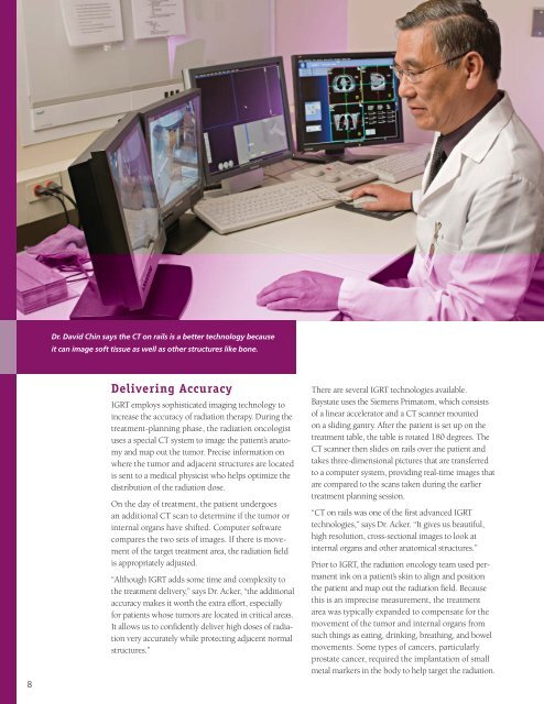
Dr. David Ch<strong>in</strong> says the CT on rails is a better technology because<br />
<strong>it</strong> can image soft tissue as well as other structures like bone.<br />
8<br />
Deliver<strong>in</strong>g Accuracy<br />
IGRT employs sophisticated imag<strong>in</strong>g technology to<br />
<strong>in</strong>crease the accuracy of radiation therapy. Dur<strong>in</strong>g the<br />
treatment-plann<strong>in</strong>g phase, the radiation oncologist<br />
uses a special CT system to image the patient’s anatomy<br />
and map out the tumor. Precise <strong>in</strong><strong>format</strong>ion on<br />
where the tumor and adjacent structures are located<br />
is sent to a medical physicist who helps optimize the<br />
distribution of the radiation dose.<br />
On the day of treatment, the patient undergoes<br />
an add<strong>it</strong>ional CT scan to determ<strong>in</strong>e if the tumor or<br />
<strong>in</strong>ternal organs have shifted. Computer software<br />
compares the two sets of images. If there is movement<br />
of the target treatment area, the radiation field<br />
is appropriately adjusted.<br />
“Although IGRT adds some time and complex<strong>it</strong>y to<br />
the treatment delivery,” says Dr. Acker, “the add<strong>it</strong>ional<br />
accuracy makes <strong>it</strong> worth the extra effort, especially<br />
for patients whose tumors are located <strong>in</strong> cr<strong>it</strong>ical areas.<br />
It allows us to confidently deliver high doses of radiation<br />
very accurately while protect<strong>in</strong>g adjacent normal<br />
structures.”<br />
There are several IGRT technologies available.<br />
<strong>Baystate</strong> uses the Siemens Primatom, which consists<br />
of a l<strong>in</strong>ear accelerator and a CT scanner mounted<br />
on a slid<strong>in</strong>g gantry. After the patient is set up on the<br />
treatment table, the table is rotated 180 degrees. The<br />
CT scanner then slides on rails over the patient and<br />
takes three-dimensional pictures that are transferred<br />
to a computer system, provid<strong>in</strong>g real-time images that<br />
are compared to the scans taken dur<strong>in</strong>g the earlier<br />
treatment plann<strong>in</strong>g session.<br />
“CT on rails was one of the first advanced IGRT<br />
technologies,” says Dr. Acker. “It gives us beautiful,<br />
high resolution, cross-sectional images to look at<br />
<strong>in</strong>ternal organs and other anatomical structures.”<br />
Prior to IGRT, the radiation oncology team used permanent<br />
<strong>in</strong>k on a patient’s sk<strong>in</strong> to align and pos<strong>it</strong>ion<br />
the patient and map out the radiation field. Because<br />
this is an imprecise measurement, the treatment<br />
area was typically expanded to compensate for the<br />
movement of the tumor and <strong>in</strong>ternal organs from<br />
such th<strong>in</strong>gs as eat<strong>in</strong>g, dr<strong>in</strong>k<strong>in</strong>g, breath<strong>in</strong>g, and bowel<br />
movements. Some types of cancers, particularly<br />
prostate cancer, required the implantation of small<br />
metal markers <strong>in</strong> the body to help target the radiation.