Tiefenbrunn - American Society for Mohs Surgery
Tiefenbrunn - American Society for Mohs Surgery
Tiefenbrunn - American Society for Mohs Surgery

You also want an ePaper? Increase the reach of your titles
YUMPU automatically turns print PDFs into web optimized ePapers that Google loves.
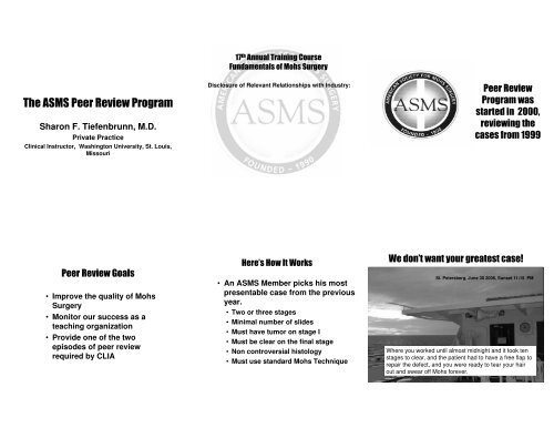
Sharon F. <strong>Tiefenbrunn</strong>, M.D.<br />
Private Practice<br />
Clinical Instructor, Washington University, St. Louis,<br />
Missouri<br />
<br />
<br />
Disclosure of Relevant Relationships with Industry:<br />
<br />
<br />
<br />
<br />
<br />
<br />
• Improve the quality of <strong>Mohs</strong><br />
<strong>Surgery</strong><br />
• Monitor our success as a<br />
teaching organization<br />
• Provide one of the two<br />
episodes of peer review<br />
required by CLIA<br />
<br />
• An ASMS Member picks his most<br />
presentable case from the previous<br />
year.<br />
• Two or three stages<br />
• Minimal number of slides<br />
• Must have tumor on stage I<br />
• Must be clear on the final stage<br />
• Non controversial histology<br />
• Must use standard <strong>Mohs</strong> Technique<br />
<br />
St. Petersberg, June 30 2006, Sunset 11:15 PM<br />
Where you worked until almost midnight and it took ten<br />
stages to clear, and the patient had to have a free flap to<br />
repair the defect, and you were ready to tear your hair<br />
out and swear off <strong>Mohs</strong> <strong>for</strong>ever.
St I<br />
St II<br />
1. Positive stage I<br />
2. Clear Stage II<br />
3. Stage II adequate<br />
to encompass<br />
tumor on stage I<br />
allowing <strong>for</strong> the<br />
inherent<br />
inaccuracy of<br />
<strong>Mohs</strong> surgery.<br />
This is a good case to send <strong>for</strong> peer review.<br />
4. Can be reviewed<br />
by looking a two<br />
selected sections.<br />
5. No controversial<br />
histology.<br />
6. No complicated<br />
maps to follow.<br />
Keep<br />
It<br />
Simple<br />
Stupid<br />
<br />
<br />
<br />
<br />
<br />
Atypical Fibroxanthoma Stage I,<br />
Inflammation stage II--? Residual<br />
tumor<br />
<br />
<br />
<br />
<br />
<br />
<br />
This was the tumor<br />
on stage I, called a<br />
microcystic adnexal<br />
carcinoma.<br />
The eStructure<br />
<br />
<br />
<br />
<br />
<br />
<br />
These structures remained on<br />
stage III that was called clear.<br />
Six dermatopathologists could<br />
not agree if they were <strong>for</strong>eign<br />
body giant cells, sweat duct<br />
tumors or regenerating sweat<br />
ducts in scar tissue.<br />
We felt these were<br />
fibrofollicuomas and<br />
there<strong>for</strong>e the case<br />
did meet the criteria<br />
<strong>for</strong> peer review.<br />
Similar structure
•Cases using nonstandard<br />
technique<br />
should not be selected.<br />
<br />
<br />
<br />
Don’t let it<br />
show that<br />
your tech is<br />
struggling to<br />
get a section<br />
of the slide<br />
at all.<br />
<br />
<br />
<br />
<br />
<br />
Tumor mapped but not taken<br />
<br />
<br />
<br />
As <strong>for</strong> the<br />
secret<br />
handshake–<br />
Nobody<br />
seems to<br />
know what<br />
it is. There<br />
are three<br />
or four<br />
basic<br />
styles out<br />
there.<br />
The selected case should con<strong>for</strong>m<br />
to
I<br />
• Complete skin edge (90%)<br />
• Complete Deep Margin<br />
• Intact tissue<br />
• Structural details are visible<br />
• Adequate stain<br />
• Visible ink<br />
• For orientation<br />
• To insure complete exam on<br />
sectioned wafers.<br />
<br />
What is meant by skin edge?<br />
Horny layer.
II<br />
• If skin edge is missing in stage I, it will be<br />
taken in stage II.<br />
• Stage II must include the tumor in stage I<br />
• The section as seen on the slide does not<br />
accurately reflect tumor left in the patient.<br />
• Overlap in all directions from edge of tumor<br />
• Connect to previous layer and confirm with<br />
inked margins.<br />
• Bevel all edges.
I<br />
Missing skin edge in<br />
stage I must be<br />
examined in stage II<br />
II<br />
<br />
<br />
Note map not marked<br />
<br />
• Traditionally <strong>Mohs</strong> surgeons have<br />
asssumed that the location of the tumor<br />
on the slide is exactly the same as it is in<br />
the patient.<br />
t<br />
• The ideas about to be presented<br />
contradict this theory and have<br />
implications <strong>for</strong> the planning of stage II.<br />
• It is in the planning and execution of stage<br />
II that many errors in <strong>Mohs</strong> <strong>Surgery</strong> occur.<br />
<br />
<br />
• A finite amount of tissue is sectioned<br />
away from the specimen be<strong>for</strong>e the first<br />
section is placed on a slide.<br />
• Assuming 20 turns of the cryostat be<strong>for</strong>e<br />
a section is obtained, at 6 microns with<br />
“sawdust effect” to equal 10 microns,<br />
200 microns are lost.<br />
• 200 microns = .2 mm, a space in which<br />
tumor can travel undetected.
Observed margin<br />
True margin/defect in patient<br />
<br />
• We take 20 turns of the cryostat be<strong>for</strong>e<br />
putting a section on the slide =.2mm<br />
• We want to examine 95% of possible angles<br />
of extension (180 degrees x.05=9 degrees)<br />
• (9/2 =4.5), tan 4.5=.078<br />
• .2/.078=2.564
• .<br />
<br />
<br />
<br />
<br />
<br />
<br />
<br />
<br />
<br />
Defect Size 1 cm 2cm 3cm 4cm<br />
St II overlap, each<br />
side 1.4mm 2mm 2.4mm 2.8mm<br />
Angle from center,<br />
each side 16 deg 11 deg 9 deg 8 deg
• 1mm of skin edge will give<br />
you 5 sections that<br />
contain skin edge if you<br />
select every 20 th section—<br />
if you save sections more<br />
frequently, it will give<br />
more.<br />
• A long narrow strip of<br />
skin edge attached to a<br />
wide area of dermis and<br />
fat bounded by ink is<br />
ideal.<br />
<br />
• Reviewers are your colleagues who are<br />
just as busy as you.<br />
• We want to whip through these cases and<br />
just circle satisfactory and go home.<br />
• So we need <strong>for</strong> your to fill out your<br />
presenter <strong>for</strong>m completely and accurately.<br />
• This is a job <strong>for</strong> the physician. It should<br />
not be assigned to anyone else.<br />
<br />
<br />
<br />
<br />
<br />
<br />
<br />
<br />
<br />
<br />
<br />
<br />
<br />
<br />
<br />
<br />
<br />
<br />
<br />
<br />
<br />
<br />
<br />
<br />
<br />
<br />
<br />
<br />
<br />
<br />
<br />
<br />
<br />
<br />
<br />
<br />
<br />
<br />
<br />
<br />
<br />
<br />
<br />
<br />
<br />
<br />
<br />
<br />
<br />
<br />
<br />
Submission date:<br />
Physican Name:<br />
Address:<br />
City State Zip:<br />
Phone:<br />
Fax:<br />
Two <strong>for</strong>ms will be sent.<br />
The Case Submission Form<br />
will have a number on it<br />
that is assigned by ASMS.<br />
ASMS Case Identification Form<br />
Physician Case Number: ____________________<br />
<strong>Surgery</strong> date:______________________________<br />
ASMS Case Number: __07001___(assigned by ASMS staff)<br />
The Case Identification Form<br />
will link the case with its<br />
presenter and the number it<br />
holds in his office. This Case<br />
ID <strong>for</strong>m will not be seen by the<br />
reviewers, only by the<br />
administrative staff. It will be<br />
destroyed after 6 mo.<br />
2/15/2007<br />
1<br />
2<br />
Don’t <strong>for</strong>get to<br />
label the sections,<br />
matching the<br />
labels on the<br />
slides.<br />
Don’t <strong>for</strong>get to<br />
map the tumor!!!!!!<br />
Date Submitted—<br />
Not the date<br />
done.<br />
Site (Location)<br />
Tumor Type<br />
Recurrent?<br />
Stain<br />
Diagram of site<br />
Number of stages<br />
Number of<br />
sections in stage<br />
I. The number of<br />
pieces into which<br />
you cut the<br />
tissue, not the<br />
number of pieces<br />
of tissue on the<br />
slides.<br />
Number of<br />
section in stage<br />
II.<br />
Ink legend -OK to<br />
use colored ink<br />
on <strong>for</strong>m.
I-A-R1<br />
I-1 I-2<br />
<br />
Map the location of the<br />
tumor.<br />
II<br />
III<br />
And there<br />
are many<br />
more like<br />
these in<br />
the files!!!<br />
<br />
<br />
<br />
<br />
<br />
Black<br />
dots, red<br />
dots, red<br />
circles,<br />
red x’s,<br />
black x’s.<br />
Just do it!
B2<br />
•Ideally B2 should be inked as a<br />
mirror image to B1, or even<br />
better as with a third color ink.<br />
Note the dotted line represents<br />
the defect in the patient and the<br />
solid line is stage II in<br />
relationship to that defect.<br />
B2<br />
This case is<br />
nontraditional<br />
in<br />
that is has a<br />
central<br />
vertical<br />
section taken<br />
from stage I.<br />
This results<br />
in tissue loss<br />
from stage I.<br />
No<br />
relationships<br />
are shown.<br />
<br />
<br />
<br />
<br />
<br />
A legend of dotted,<br />
solid, dashed and<br />
squiggly lines is<br />
acceptable, as is<br />
the use of colored<br />
pens or pencils.<br />
<br />
<br />
<br />
This case was inked on the skin edge. Even<br />
that ink is not very visible.<br />
Right!<br />
<br />
<br />
<br />
<br />
<br />
<br />
<br />
<br />
WRONG!!!!<br />
<br />
<br />
• This is probably the number one<br />
problem with the presentation<br />
of cases.<br />
The defect in the<br />
patent is represented<br />
with the dotted line.<br />
The second stage is<br />
shown by the solid<br />
line. Ink should be<br />
placed on the<br />
surface exposed at<br />
the end of the<br />
previous stage
Solid line was used to<br />
represent stage I and<br />
dotted to represent<br />
stage II; explanation<br />
that it was a deep only<br />
with no expected skin<br />
edge indicated which<br />
line was which.<br />
Solid line is<br />
the tissue<br />
taken on<br />
stage II.<br />
Straight lines drawn through defects imply<br />
straight down cuts with undermining brought<br />
up to it. More rounded stage II lines suggest<br />
beveled specimens were taken.<br />
<br />
<br />
<br />
<br />
<br />
<br />
<br />
<br />
<br />
<br />
<br />
<br />
<br />
<br />
<br />
<br />
<br />
<br />
<br />
<br />
<br />
<br />
We need to know if you put sections on slides from bottom to top or top<br />
to bottom. If you put two rows on a slide we need to know exactly how<br />
you do it. For example: 1 2<br />
Do you flip sections?<br />
3 4<br />
5 6<br />
7 8<br />
Missing skin edge should be<br />
indicated. It helps and you and us<br />
know if stage II took the skin edge<br />
that stage I missed.<br />
<br />
<br />
This case excludes itself<br />
from being satisfactory<br />
based on the map alone.<br />
If stage I is missing skin<br />
edge, stage II needs to<br />
examine the skin edge in<br />
that area.<br />
<br />
• Do you know what<br />
this means?
The section is<br />
then teased<br />
from underneath<br />
onto the knife<br />
and picked up<br />
with the slide.<br />
It is flipped<br />
vertically from<br />
its normal<br />
orientation.<br />
<br />
Stage I taken<br />
from file<br />
folder, lying<br />
just below<br />
defect,<br />
epidermis up.<br />
Section<br />
on chuck<br />
dermal<br />
side<br />
toward<br />
blade.<br />
Section<br />
on x-ray<br />
film<br />
transfer<br />
media,<br />
epidermis<br />
up<br />
Section<br />
on<br />
blade,<br />
dermal<br />
side<br />
up.<br />
<br />
<br />
<br />
Tissue picked<br />
up by slide now<br />
has epidermal<br />
side up, dermal<br />
side against<br />
slide.<br />
Map seen through<br />
microsope Slide rotated 180<br />
<br />
<br />
<br />
<br />
As seen through hth<br />
the<br />
microscope.<br />
<br />
<br />
<br />
<br />
<br />
Rotated to match<br />
the orientation in<br />
the patient.
• Follicular<br />
abnormalities<br />
• Areas of<br />
Inflammation<br />
• AKs<br />
• Nevi<br />
• SKs
• Circle the relevant<br />
sections--<br />
YOURSELF.<br />
• This makes it easy<br />
<strong>for</strong> the reviewers to<br />
quickly focus on<br />
the essentials.<br />
• Also, sometimes a<br />
a case with a flaw<br />
can get past.<br />
<br />
<br />
• Mark outside<br />
the skin edge<br />
• This is most<br />
appropriate <strong>for</strong><br />
superficial<br />
tumors<br />
• Do not mark<br />
over sections.<br />
<br />
<br />
<br />
Has the mounting<br />
medium<br />
deteriorated?<br />
<br />
• Draw what you see<br />
• Rotate<br />
• Don’t flip and turn<br />
microscopic images<br />
in your head.
•Label the case with the case<br />
number assigned by ASMS<br />
•Use Roman Numerals <strong>for</strong><br />
Stage I,II,III<br />
•Use Arabic numerals <strong>for</strong><br />
sections<br />
•Use R-Arabic numerals <strong>for</strong><br />
recuts - R1, R2, R3,<br />
I-1 2<br />
O6001 O6001 O6001 O6001 O6001 O6001<br />
I-1-R0 1-1-R1 I-2-R0 I-2-R1 II-R0 II-R1<br />
II<br />
<br />
<br />
<br />
<br />
<br />
<br />
<br />
<br />
<br />
<br />
<br />
<br />
<br />
<br />
<br />
<br />
<br />
<br />
<br />
<br />
<br />
• To the ASMS staff<br />
• To reviewers<br />
• To the presenter
It will be classified into one of the<br />
following categories:<br />
1. Satisfactory<br />
2. Comments only<br />
3. Unreviewable<br />
Case No.__________________<br />
Reviewer Form<br />
____Case Not Reviewable –please submit another case or correct the<br />
presentation of this one.<br />
Map issues-ink legend, Tumor not marked on map Relationship not shown<br />
Sections not labeled Confusing slide labeling Non-standard <strong>Mohs</strong> technique<br />
Bubbles Too complex a case – over 3 Stages. No Identifiable Tumor Stage I<br />
Other (Specify)<br />
____Case Reviewed _____Satisfactory _____Comments<br />
Mark “U” where unsatisfactory<br />
General gross appearance of slides<br />
Stage I-A<br />
Pertains to all Stages<br />
Skin Edge Adequate (90% with<br />
exceptions)<br />
Deep Margin (90% with exceptions)<br />
Structure Details (thick sections, etc.)<br />
Stain<br />
Ink <strong>for</strong> orientation<br />
Ink to ascertain completeness<br />
ess<br />
Tumor identified and accurately mapped<br />
Normal structures ID’d as tumor<br />
Pertains to Stage II<br />
Missing Skin Edge Stage I taken in<br />
Stage II<br />
Missing Deep Tissue I taken in Stage II<br />
Tumor from Stage I taken in stage II<br />
Adequate Overlap<br />
Adequate connection- Visible ink on<br />
non-epidermal edge and adequate ink<br />
visualized.<br />
Clear Final Stage<br />
Comments:<br />
<br />
• Satisfactory cases are<br />
beautiful cases with<br />
obvious tumor on stage<br />
I and absence of tumor<br />
on stage II
Comments<br />
Not enough overlap to determine clear margin.<br />
<br />
<br />
<br />
This case can be reviewed<br />
and gets comments:<br />
Stain is too dark<br />
Tumor is near epidermis in<br />
stage II and there<strong>for</strong>e<br />
stage III needs to have<br />
skin edge, which it doesn’t<br />
<br />
<br />
• The slides will be scanned, and<br />
possibly photographed.<br />
• The map will be scanned.<br />
• A critique of the case, including the<br />
first reviewers comments will be<br />
created and sent to the presenter.<br />
• The case may become a teaching<br />
case.<br />
<br />
• Satisfactory means pass<br />
• Comments means fail<br />
• Loss of ASMS Fellowship is no longer<br />
associated with lack of satisfactory<br />
completion of peer review.<br />
• Participation is not only required <strong>for</strong> the<br />
first four years.<br />
However, if you get Comments, hide the<br />
case and meet your CLIA requirements by<br />
cross-reading with another lab.<br />
<br />
• One cause is lack of in<strong>for</strong>mation.
• But until we<br />
get a map, we<br />
are not going<br />
to agonize<br />
over it.<br />
• It’s<br />
unreviewable.<br />
• Are these follicular<br />
structure or tumor?<br />
• The peer review<br />
committee split 6 to<br />
6.<br />
<br />
<br />
<br />
<br />
<br />
• Map issues-ink legend,<br />
• Tumor not marked on map<br />
• Relationship not shown<br />
• Sections not labeled<br />
• Confusing slide labeling<br />
• Non-standard <strong>Mohs</strong> technique<br />
• Bubbles<br />
• Too complex a case – over 3 Stages.<br />
• No Identifiable Tumor Stage I<br />
• Other (Specify)<br />
<br />
<br />
• Fix the case and send it back—map<br />
the tumor, draw the relationships.<br />
• Sometimes this can be done by fax.<br />
• If it is unreviewable because of an<br />
unsolvable controversy, a new case<br />
must be picked and submitted. The<br />
first case does not count toward the<br />
limit of two free tries per year.<br />
<br />
<br />
<br />
• A Satisfactory outcome can be used as<br />
one of the two peer review episodes<br />
required by CLIA.<br />
• Comments are meant to steer the<br />
presenter into improving his <strong>Mohs</strong>.<br />
• Unreviewable is used when there is no<br />
map, or the map is not marked, or when<br />
the panel cannot agree if a structure is<br />
tumor or not. A second case should be<br />
sent <strong>for</strong> review.<br />
<br />
• Select your case carefully<br />
y<br />
y<br />
• Present it well<br />
• OR THIS COULD HAPPEN TO YOU