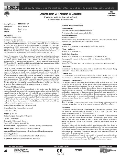
Desmoglein 3 + Napsin A Cocktail
Desmoglein 3 + Napsin A Cocktail
Desmoglein 3 + Napsin A Cocktail
You also want an ePaper? Increase the reach of your titles
YUMPU automatically turns print PDFs into web optimized ePapers that Google loves.
<strong>Desmoglein</strong> 3 + <strong>Napsin</strong> A <strong>Cocktail</strong><br />
Prediluted Multiplex <strong>Cocktail</strong> (4-Step)<br />
Control Number: 901-428DS-021611<br />
ISO<br />
9001:2000<br />
CERTIFIED<br />
Catalog Number:<br />
Description:<br />
Dilution:<br />
Diluent:<br />
PPM 428DS AA<br />
6.0 ml, prediluted<br />
Ready-to-use<br />
N/A<br />
Intended Use:<br />
For In Vitro Diagnostic Use<br />
Summary and Explanation:<br />
<strong>Desmoglein</strong> 3 (DSG3) is a calcium-binding transmembrane glycoprotein component of<br />
desmosomes in vertebrate epithelial cells. Studies have shown DSG3 to have 83-95%<br />
sensitivity and 100% specificity in detecting squamous cell carcinoma (SqCC) vs. lung<br />
adenocarcinoma. DSG3 is associated with shorter survival for all lung cancer patients<br />
regardless of the histologic subtype (5-year survival of 20.9% vs. 49.5%, P < .001).<br />
Patients with atypical carcinoid tumors, lacking <strong>Desmoglein</strong> 3 expression showed a 5-<br />
year survival of 0% compared with 36.8% for DSG3 positive cases (P < .001).<br />
<strong>Napsin</strong> A is expressed in type II pneumocytes of normal lung and in adenocarcinomas<br />
of the lung and kidney. Studies have shown that <strong>Napsin</strong> A is more sensitive (80-87%)<br />
and more specific marker than TTF-1. <strong>Napsin</strong> A is 100% specific for lung<br />
adenocarcinoma vs. 100% negative in lung SqCC. <strong>Napsin</strong> A used in combination with<br />
TTF-1 provides 93% sensitivity and 100% specificity for lung adenocarcinoma, if CK5<br />
and <strong>Desmoglein</strong> 3 are both negative in the same section.<br />
DSG3 is a cell membrane stain that marks lung SqCC (DAB). <strong>Napsin</strong> A is a<br />
cytoplasmic/granular stain that marks lung adenocarcinomas (Fast Red). In the vast<br />
majority of lung cancers tested, only a single antibody stain will be observed. Coexpression<br />
of both antibodies may be observed in adenosquamous cell carcinomas, or<br />
in some cases residual normal lung will stain with <strong>Napsin</strong> A. <strong>Desmoglein</strong> 3 + <strong>Napsin</strong><br />
A are very sensitive and specific markers for discriminating between lung SqCC and<br />
lung adenocarcinoma. This antibody cocktail is extremely accurate and is 100%<br />
specific. In grades 1-2, <strong>Desmoglein</strong> 3 + <strong>Napsin</strong> A provide staining sensitivity in the<br />
mid 90% range; thus the antibody cocktail of <strong>Desmoglein</strong> 3 + <strong>Napsin</strong> A is a first-line<br />
screener for discriminating between lung adenocarcinoma vs. lung SqCC.<br />
Principle of Multiplex Staining:<br />
A Multiplex IHC stain can be accomplished in four major steps. The initial step<br />
consists of an antibody cocktail with at least one mouse and one rabbit antibody. This<br />
cocktail is applied to the tissue and will bind with two or more target antigens. A<br />
multiplex detection cocktail of horseradish peroxidase (HRP) and alkaline phosphatase<br />
(AP) conjugated secondary antibodies is applied. The third step consists of the addition<br />
of DAB-Substrate that binds to the HRP and produces a brown chromogenic reaction<br />
product. The fourth step consists of a Fast Red-Substrate that binds to the AP and<br />
produces a red chromogenic reaction product.<br />
Source: Mouse monoclonal and Rabbit polyclonal<br />
Species Reactivity: Human, others not tested<br />
Clone: BC11 + N/A<br />
Isotype: IgG1 + N/A<br />
Epitope/Antigen: <strong>Desmoglein</strong> 3 + <strong>Napsin</strong> A<br />
Cellular Localization:<br />
<strong>Desmoglein</strong> 3 (Membrane): Brown<br />
<strong>Napsin</strong> A (Cytoplasmic - granular): Red<br />
Positive Control: Lung squamous cell carcinoma and lung adenocarcinoma<br />
Normal Tissue: Skin or tonsil (DSG3); lung (<strong>Napsin</strong> A)<br />
Abnormal Tissue: Lung squamous cell carcinoma and lung adenocarcinoma<br />
Known Applications:<br />
Immunohistochemistry (formalin-fixed paraffin-embedded tissues)<br />
Supplied As: Buffer with protein carrier and preservative.<br />
Protocol Recommendations<br />
Peroxide Block:<br />
Block for 5 minutes with Biocare's Peroxidazed 1.<br />
Pretreatment Solution (recommended): Diva<br />
Pretreatment Protocol:<br />
Heat Retrieval Method:<br />
Retrieve sections using Biocare’s Decloaking Chamber at 125°C for 30 seconds. Allow<br />
solution to cool for 10 minutes then wash in distilled water<br />
Protein Block:<br />
Incubate for 10 minutes at RT with Biocare's Background Punisher.<br />
Primary Antibody:<br />
Incubate for 30 minutes at RT.<br />
Double Stain Detection:<br />
Incubate for 30 minutes at RT using Biocare's MACH 2 Double Stain 2.<br />
Chromogen (1): Incubate for 5 minutes at RT with Biocare's Betazoid DAB.<br />
Chromogen (2):<br />
Incubate for 5-7 minutes at RT with Biocare's Warp Red. Rinse in deionized water.<br />
Counterstain:<br />
Counterstain with Hematoxylin. Rinse with deionized water. Apply Tacha's Bluing<br />
solution for 1 minute. Rinse with deionized water.<br />
Technical Note:<br />
This antibody has been standardized with Biocare's MACH 2 Double Stain 2. It can<br />
also be used on an automated staining system. Use TBS buffer for washing steps.<br />
Performance Characteristics:<br />
The optimum antibody dilution and protocols for a specific application can vary. These<br />
include, but are not limited to: fixation, heat-retrieval method, incubation times, tissue<br />
section thickness and detection kit used. Due to the superior sensitivity of these unique<br />
reagents, the recommended incubation times and titers listed are not applicable to other<br />
detection systems, as results may vary. The data sheet recommendations and protocols<br />
are based on exclusive use of Biocare products. Ultimately, it is the responsibility of<br />
the investigator to determine optimal conditions. These products are tools that can be<br />
used for interpretation of morphological findings in conjunction with other diagnostic<br />
tests and pertinent clinical data by a qualified pathologist.<br />
Quality Control:<br />
Refer to NCCLS Quality Assurance for Immunocytochemistry approved guidelines,<br />
December 1999 MM4-A Vol.19 No.26 for more information about tissue controls.<br />
Precautions:<br />
This antibody contains less than 0.1% sodium azide. Concentrations less than 0.1% are<br />
not reportable hazardous materials according to U.S. 29 CFR 1910.1200, OSHA<br />
Hazard communication and EC Directive 91/155/EC.<br />
Sodium azide (NaN3) used as a preservative is toxic if ingested. Sodium azide may<br />
react with lead and copper plumbing to form highly explosive metal azides. Upon<br />
disposal, flush with large volumes of water to prevent azide build-up in plumbing.<br />
(Center for Disease Control, 1976, National Institute of Occupational Safety and<br />
Health, 1976)<br />
Specimens, before and after fixation, and all materials exposed to them should be<br />
handled as if capable of transmitting infection and disposed of with proper precautions.<br />
Never pipette reagents by mouth and avoid contacting the skin and mucous membranes<br />
with reagents and specimens. If reagents or specimens come in contact with sensitive<br />
areas, wash with copious amounts of water.<br />
Microbial contamination of reagents may result in an increase in nonspecific staining.<br />
Incubation times or temperatures other than those specified may give erroneous results.<br />
The user must validate any such change. The MSDS is available upon request.<br />
Page 1 of 2
Storage and Stability:<br />
Store at 2ºC to 8ºC. Do not use after expiration date printed on vial. If reagents are<br />
stored under conditions other than those specified in the package insert, they must be<br />
verified by the user. Diluted reagents should be used promptly; any remaining reagent<br />
should be stored at 2ºC to 8ºC.<br />
Troubleshooting:<br />
Follow the antibody specific protocol recommendations according to data sheet<br />
provided. If atypical results occur, contact Biocare's Technical Support at<br />
1-800-542-2002.<br />
Limitations and Warranty:<br />
There are no warranties, expressed or implied, which extend beyond this description.<br />
Biocare is not liable for property damage, personal injury, or economic loss caused by<br />
this product.<br />
References:<br />
1. Tacha D, Yu C, Haas T. TTF-1, <strong>Napsin</strong> A, p63, TRIM29, <strong>Desmoglein</strong>-3 and CK5:<br />
An Evaluation of Sensitivity and Specificity and Correlation of Tumor Grade for Lung<br />
Squamous Cell Carcinoma vs. Lung Adenocarcinoma. Modern Pathology; Abstract,<br />
USCAP, 2011<br />
2. Tacha D, Zhou D, Henshall-Powell RL¹. Distinguishing Adenocarcinoma from<br />
Squamous Cell Carcinoma in Lung Using Double Stains p63+ CK5 and TTF-1 +<br />
<strong>Napsin</strong> A. Modern Pathology; Pathology Volume 23, Supplement 1, Feb 2010;<br />
Abstract 1852, page 222A.<br />
3. Terry J, et al. Optimal immunohistochemical markers for distinguishing lung<br />
adenocarcinomas from squamous cell carcinomas in small tumor samples. Am J Surg<br />
Pathol. 2010 Dec; 34(12):1805-11.<br />
4. Savci-Heijink CD, et al. The role of desmoglein-3 in the diagnosis of squamous cell<br />
carcinoma of the lung. Am J Pathol. 2009 May; 174(5):1629-37. Epub 2009 Mar 26.<br />
5. Center for Disease Control Manual. Guide: Safety Management, NO. CDC-22,<br />
Atlanta, GA. April 30, 1976 "Decontamination of Laboratory Sink Drains to Remove<br />
Azide Salts."<br />
6. National Committee for Clinical Laboratory Standards (NCCLS). Protection of<br />
laboratory workers from infectious diseases transmitted by blood and tissue; proposed<br />
guideline. Villanova, PA 1991;7(9). Order code M29-P.<br />
<strong>Desmoglein</strong> 3 + <strong>Napsin</strong> A <strong>Cocktail</strong><br />
Prediluted Multiplex <strong>Cocktail</strong> (4-Step)<br />
Control Number: 901-428DS-021611<br />
ISO<br />
9001:2000<br />
CERTIFIED<br />
Page 2 of 2
p63 + TRIM29 <strong>Cocktail</strong> (SqCC)<br />
Prediluted Multiplex <strong>Cocktail</strong> (4-Step)<br />
Control Number: 901-427DS-021611<br />
ISO<br />
9001:2000<br />
CERTIFIED<br />
Catalog Number:<br />
Description:<br />
Dilution:<br />
Diluent:<br />
PPM 427DS AA<br />
6.0 ml, prediluted<br />
Ready-to-use<br />
N/A<br />
Intended Use:<br />
For In Vitro Diagnostic Use<br />
Summary and Explanation:<br />
Tumor protein p63, also known as transformation-related protein 63 is a protein that in<br />
humans is encoded by the TP63 gene. Many studies have shown that p63 is a sensitive<br />
(90%) and fairly specific marker for squamous cell carcinoma and may be used in<br />
distinguishing poorly differentiated squamous cell carcinomas from adenocarcinomas.<br />
p63 has been shown to mark approximately 5 to 10% of lung adenocarcinomas.<br />
Tripartite motif-containing 29 (TRIM29) is a relatively new marker. A comprehensive<br />
study has shown that TRIM29 is a sensitive marker (93.7%) for lung squamous cell<br />
carcinoma (SqCC) and is a fairly specific marker staining only 6.1% of lung<br />
adenocarcinomas.<br />
p63 is a nuclear stain that marks lung SqCC (DAB), and TRIM29 is a<br />
cytoplasmic/membrane stain that also marks lung SqCC (Fast Red). In most cases, a<br />
co-expression of both antibodies will be observed in lung SqCC. Studies have also<br />
shown that when p63 and/or TRIM29 is expressed in lung SqCC, a 95.4% sensitivity<br />
and 100% specificity was achieved, if <strong>Napsin</strong> A and TTF-1 were both negative in the<br />
same case. Therefore, the antibody cocktail of p63 + TRIM29 is an excellent screener<br />
for discriminating lung SqCC vs. lung adenocarcinoma.<br />
Principle of Multiplex Staining:<br />
A Multiplex IHC stain can be accomplished in four major steps. The initial step<br />
consists of an antibody cocktail with at least one mouse and one rabbit antibody. This<br />
cocktail is applied to the tissue and will bind with two or more target antigens. A<br />
multiplex detection cocktail of horseradish peroxidase (HRP) and alkaline phosphatase<br />
(AP) conjugated secondary antibodies is applied. The third step consists of the addition<br />
of DAB-Substrate that binds to the HRP and produces a brown chromogenic reaction<br />
product. The fourth step consists of a Fast Red-Substrate that binds to the AP and<br />
produces a red chromogenic reaction product.<br />
Source: Mouse monoclonal and Rabbit polyclonal<br />
Species Reactivity: Human, others not tested<br />
Clone: BC4A4 + N/A<br />
Isotype: IgG2a/kappa + Rabbit IgG<br />
Epitope/Antigen: p63 + TRIM29<br />
Cellular Localization:<br />
p63 (Nuclear): Brown<br />
TRIM29 (Cytoplasmic & membrane): Red<br />
Positive Control: Lung squamous cell carcinoma<br />
Normal Tissue: Prostate, bladder (p63); prostate, placenta (TRIM29)<br />
Abnormal Tissue: Lung squamous cell carcinoma<br />
Known Applications:<br />
Immunohistochemistry (formalin-fixed paraffin-embedded tissues)<br />
Supplied As: Buffer with protein carrier and preservative.<br />
Storage and Stability:<br />
Store at 2ºC to 8ºC. Do not use after expiration date printed on vial. If reagents are<br />
stored under conditions other than those specified in the package insert, they must be<br />
verified by the user. Diluted reagents should be used promptly; any remaining reagent<br />
should be stored at 2ºC to 8ºC.<br />
Protocol Recommendations<br />
Peroxide Block:<br />
Block for 5 minutes with Biocare's Peroxidazed 1.<br />
Pretreatment Solution (recommended): Diva<br />
Pretreatment Protocol:<br />
Heat Retrieval Method:<br />
Retrieve sections using Biocare’s Decloaking Chamber at 125°C for 30 seconds. Allow<br />
solution to cool for 10 minutes then wash in distilled water.<br />
Protein Block:<br />
Incubate for 10 minutes at RT with Biocare's Background Punisher.<br />
Primary Antibody:<br />
Incubate for 30 minutes at RT.<br />
Double Stain Detection:<br />
Incubate for 30 minutes at RT using Biocare's MACH 2 Double Stain 2.<br />
Chromogen (1): Incubate for 5 minutes at RT with Biocare's Betazoid DAB.<br />
Chromogen (2):<br />
Incubate for 5-7 minutes at RT with Biocare's Warp Red. Rinse in deionized water.<br />
Counterstain:<br />
Counterstain with Hematoxylin. Rinse with deionized water. Apply Tacha's Bluing<br />
solution for 1 minute. Rinse with deionized water.<br />
Technical Note:<br />
This antibody has been standardized with Biocare's MACH 2 Double Stain 2. It can<br />
also be used on an automated staining system. Use TBS buffer for washing steps.<br />
Performance Characteristics:<br />
The optimum antibody dilution and protocols for a specific application can vary. These<br />
include, but are not limited to: fixation, heat-retrieval method, incubation times, tissue<br />
section thickness and detection kit used. Due to the superior sensitivity of these unique<br />
reagents, the recommended incubation times and titers listed are not applicable to other<br />
detection systems, as results may vary. The data sheet recommendations and protocols<br />
are based on exclusive use of Biocare products. Ultimately, it is the responsibility of<br />
the investigator to determine optimal conditions. These products are tools that can be<br />
used for interpretation of morphological findings in conjunction with other diagnostic<br />
tests and pertinent clinical data by a qualified pathologist.<br />
Quality Control:<br />
Refer to NCCLS Quality Assurance for Immunocytochemistry approved guidelines,<br />
December 1999 MM4-A Vol.19 No.26 for more information about tissue controls.<br />
Precautions:<br />
This antibody contains less than 0.1% sodium azide. Concentrations less than 0.1% are<br />
not reportable hazardous materials according to U.S. 29 CFR 1910.1200, OSHA<br />
Hazard communication and EC Directive 91/155/EC.<br />
Sodium azide (NaN3) used as a preservative is toxic if ingested. Sodium azide may<br />
react with lead and copper plumbing to form highly explosive metal azides. Upon<br />
disposal, flush with large volumes of water to prevent azide build-up in plumbing.<br />
(Center for Disease Control, 1976, National Institute of Occupational Safety and<br />
Health, 1976)<br />
Specimens, before and after fixation, and all materials exposed to them should be<br />
handled as if capable of transmitting infection and disposed of with proper precautions.<br />
Never pipette reagents by mouth and avoid contacting the skin and mucous membranes<br />
with reagents and specimens. If reagents or specimens come in contact with sensitive<br />
areas, wash with copious amounts of water.<br />
Microbial contamination of reagents may result in an increase in nonspecific staining.<br />
Incubation times or temperatures other than those specified may give erroneous results.<br />
The user must validate any such change. The MSDS is available upon request.<br />
Page 1 of 2
p63 + TRIM29 <strong>Cocktail</strong> (SqCC)<br />
Prediluted Multiplex <strong>Cocktail</strong> (4-Step)<br />
Control Number: 901-427DS-021611<br />
ISO<br />
9001:2000<br />
CERTIFIED<br />
Troubleshooting:<br />
Follow the antibody specific protocol recommendations according to data sheet<br />
provided. If atypical results occur, contact Biocare's Technical Support at 1-800-542<br />
-2002.<br />
Limitations and Warranty:<br />
There are no warranties, expressed or implied, which extend beyond this description.<br />
Biocare is not liable for property damage, personal injury, or economic loss caused by<br />
this product.<br />
References:<br />
1. Tacha D, Yu C, Haas T. TTF-1, <strong>Napsin</strong> A, p63, TRIM29, <strong>Desmoglein</strong>-3 and CK5:<br />
An Evaluation of Sensitivity and Specificity and Correlation of Tumor Grade for Lung<br />
Squamous Cell Carcinoma vs. Lung Adenocarcinoma. Modern Pathology; USCAP,<br />
2011 (Abstract Accepted)<br />
2. Tacha D, Zhou D, Henshall-Powell RL¹. Distinguishing Adenocarcinoma from<br />
Squamous Cell Carcinoma in Lung Using Double Stains p63+ CK5 and TTF-1 +<br />
<strong>Napsin</strong> A. Modern Pathology; Pathology Volume 23, Supplement 1, Feb 2010;<br />
Abstract 1852, page 222A.<br />
3. Terry J, et al. Optimal immunohistochemical markers for distinguishing lung<br />
adenocarcinomas from squamous cell carcinomas in small tumor samples. Am J Surg<br />
Pathol. 2010 Dec; 34(12):1805-11.<br />
4. Ring BZ, et al. A novel five-antibody immunohistochemical test for subclassification<br />
of lung carcinoma. Mod Pathol. 2009 Aug; 22(8):1032-43. Epub 2009 May 8.<br />
5. Center for Disease Control Manual. Guide: Safety Management, NO. CDC-22,<br />
Atlanta, GA. April 30, 1976 "Decontamination of Laboratory Sink Drains to Remove<br />
Azide Salts."<br />
6. National Committee for Clinical Laboratory Standards (NCCLS). Protection of<br />
laboratory workers from infectious diseases transmitted by blood and tissue; proposed<br />
guideline. Villanova, PA 1991;7(9). Order code M29-P.<br />
Page 2 of 2
TTF-1 + CK5 <strong>Cocktail</strong><br />
Prediluted Multiplex <strong>Cocktail</strong> (4-Step)<br />
Control Number: 901-425DS-021611<br />
ISO<br />
9001:2000<br />
CERTIFIED<br />
Catalog Number:<br />
Description:<br />
Dilution:<br />
Diluent:<br />
PM 425DS AA<br />
6.0 ml, prediluted<br />
Ready-to-use<br />
N/A<br />
Intended Use:<br />
For In Vitro Diagnostic Use<br />
Summary and Explanation:<br />
Thyroid transcription factor-1 (TTF-1) is a member of the NKX2 family of<br />
homeodomain transcription factors. It is expressed in epithelial cells of the thyroid<br />
gland and lung. TTF-1 has been shown to be a sensitive (65-81%) and specific marker<br />
(94%) in the majority of primary lung adenocarcinomas. Studies have shown that TTF<br />
-1, used in combination with <strong>Napsin</strong> A, provided 93% sensitivity and 100% specificity<br />
for lung adenocarcinoma, if CK5 and <strong>Desmoglein</strong> 3 were both negative in the same<br />
case.<br />
CK5 is a type II intermediate filament protein that is expressed in active basal layers of<br />
most stratified squamous epithelia. In a published study, rabbit monoclonal CK5<br />
antibody was compared to mouse monoclonal CK5/6. CK5 was 84% sensitive and<br />
100% specific for lung squamous cell carcinoma (SqCC) when compared to CK5/6<br />
(80% sensitivity and 97% specificity). CK6 mRNA has been detected in lung<br />
adenocarcinomas; thus CK5 alone, may be a more specific marker than CK5/6. Studies<br />
have shown that CK5, used in combination with <strong>Desmoglein</strong> 3, provided 93.7%<br />
sensitivity with 100% specificity for lung SqCC.<br />
TTF-1 (lung adenocarcinoma) is stained with DAB chromogen, and CK5 rabbit<br />
monoclonal (lung SqCC) is stained with a Fast Red chromogen. In most lung cancers<br />
tested, only a single antibody stain will be observed. Co-expressions of both antibodies<br />
may be an indication of adenosquamous cell carcinomas. When used in combination<br />
with <strong>Desmoglein</strong> 3 and <strong>Napsin</strong> A, a 93% staining sensitivity and 100% specificity was<br />
achieved for lung adenocarcinoma, and a 93.7% sensitivity and 100% specificity was<br />
achieved for lung SqCC; therefore, the antibody cocktail of TTF-1 + CK5 is a first<br />
class screener for discriminating between lung adenocarcinoma (TTF-1) vs. lung SqCC<br />
(CK5).<br />
Principle of Multiplex Staining:<br />
A Multiplex IHC stain can be accomplished in four major steps. The initial step<br />
consists of an antibody cocktail with at least one mouse and one rabbit antibody. This<br />
cocktail is applied to the tissue and will bind with two or more target antigens. A<br />
multiplex detection cocktail of horseradish peroxidase (HRP) and alkaline phosphatase<br />
(AP) conjugated secondary antibodies is applied. The third step consists of the addition<br />
of DAB-Substrate that binds to the HRP and produces a brown chromogenic reaction<br />
product. The fourth step consists of a Fast Red-Substrate that binds to the AP and<br />
produces a red chromogenic reaction product.<br />
Source: Mouse monoclonal and Rabbit monoclonal<br />
Species Reactivity: Human, others not tested<br />
Clone: 8G7G3/1 + EP1601Y<br />
Isotype: IgG1 + Rabbit IgG<br />
Epitope/Antigen: TTF-1 + CK5<br />
Cellular Localization:<br />
TTF-1 (Nuclear): Brown<br />
CK5 (Cytoplasmic): Red<br />
Positive Control: Lung adenocarcinoma (TTF-1) and lung SqCC (CK5)<br />
Normal Tissue: Lung (TTF-1); normal prostate (CK5)<br />
Abnormal Tissue: Lung adenocarcinoma (TTF-1); lung SqCC (CK5)<br />
Known Applications:<br />
Immunohistochemistry (formalin-fixed paraffin-embedded tissues)<br />
Supplied As: Buffer with protein carrier and preservative.<br />
Protocol Recommendations<br />
Peroxide Block:<br />
Block for 5 minutes with Biocare's Peroxidazed 1.<br />
Pretreatment Solution (recommended): Diva<br />
Pretreatment Protocol:<br />
Heat Retrieval Method:<br />
Retrieve sections using Biocare’s Decloaking Chamber at 125°C for 30 seconds. Allow<br />
solution to cool for 10 minutes then wash in distilled water.<br />
Protein Block:<br />
Incubate for 10 minutes at RT with Biocare's Background Punisher.<br />
Primary Antibody:<br />
Incubate for 30 minutes at RT.<br />
Double Stain Detection:<br />
Incubate for 30 minutes at RT using Biocare's MACH 2 Double Stain 2.<br />
Chromogen (1): Incubate for 5 minutes at RT with Biocare's Betazoid DAB.<br />
Chromogen (2):<br />
Incubate for 5-7 minutes at RT with Biocare's Warp Red. Rinse in deionized water.<br />
Counterstain:<br />
Counterstain with Hematoxylin. Rinse with deionized water. Apply Tacha's Bluing<br />
solution for 1 minute. Rinse with deionized water.<br />
Technical Note:<br />
This antibody has been standardized with Biocare's MACH 2 Double Stain 2. It can<br />
also be used on an automated staining system. Use TBS buffer for washing steps.<br />
Performance Characteristics:<br />
The optimum antibody dilution and protocols for a specific application can vary. These<br />
include, but are not limited to: fixation, heat-retrieval method, incubation times, tissue<br />
section thickness and detection kit used. Due to the superior sensitivity of these unique<br />
reagents, the recommended incubation times and titers listed are not applicable to other<br />
detection systems, as results may vary. The data sheet recommendations and protocols<br />
are based on exclusive use of Biocare products. Ultimately, it is the responsibility of<br />
the investigator to determine optimal conditions. These products are tools that can be<br />
used for interpretation of morphological findings in conjunction with other diagnostic<br />
tests and pertinent clinical data by a qualified pathologist.<br />
Quality Control:<br />
Refer to NCCLS Quality Assurance for Immunocytochemistry approved guidelines,<br />
December 1999 MM4-A Vol.19 No.26 for more information about tissue controls.<br />
Precautions:<br />
This antibody contains less than 0.1% sodium azide. Concentrations less than 0.1% are<br />
not reportable hazardous materials according to U.S. 29 CFR 1910.1200, OSHA<br />
Hazard communication and EC Directive 91/155/EC.<br />
Sodium azide (NaN3) used as a preservative is toxic if ingested. Sodium azide may<br />
react with lead and copper plumbing to form highly explosive metal azides. Upon<br />
disposal, flush with large volumes of water to prevent azide build-up in plumbing.<br />
(Center for Disease Control, 1976, National Institute of Occupational Safety and<br />
Health, 1976)<br />
Specimens, before and after fixation, and all materials exposed to them should be<br />
handled as if capable of transmitting infection and disposed of with proper precautions.<br />
Never pipette reagents by mouth and avoid contacting the skin and mucous membranes<br />
with reagents and specimens. If reagents or specimens come in contact with sensitive<br />
areas, wash with copious amounts of water.<br />
Microbial contamination of reagents may result in an increase in nonspecific staining.<br />
Incubation times or temperatures other than those specified may give erroneous results.<br />
The user must validate any such change. The MSDS is available upon request.<br />
Page 1 of 2
TTF-1 + CK5 <strong>Cocktail</strong><br />
Prediluted Multiplex <strong>Cocktail</strong> (4-Step)<br />
Control Number: 901-425DS-021611<br />
ISO<br />
9001:2000<br />
CERTIFIED<br />
Storage and Stability:<br />
Store at 2ºC to 8ºC. Do not use after expiration date printed on vial. If reagents are<br />
stored under conditions other than those specified in the package insert, they must be<br />
verified by the user. Diluted reagents should be used promptly; any remaining reagent<br />
should be stored at 2ºC to 8ºC.<br />
Troubleshooting:<br />
Follow the antibody specific protocol recommendations according to data sheet<br />
provided. If atypical results occur, contact Biocare's Technical Support at<br />
1-800-542-2002.<br />
Limitations and Warranty:<br />
There are no warranties, expressed or implied, which extend beyond this description.<br />
Biocare is not liable for property damage, personal injury, or economic loss caused by<br />
this product.<br />
References:<br />
1. Mukhopadhyay S, Katzenstein AL. Subclassification of non-small cell lung<br />
carcinomas lacking morphologic differentiation on biopsy specimens: Utility of an<br />
immunohistochemical panel containing TTF-1, napsin A, p63, and CK5/6. Am J Surg<br />
Pathol. 2011 Jan; 35(1):15-25.<br />
2. Tacha D, Yu C, Haas T. TTF-1, <strong>Napsin</strong> A, p63, TRIM29, <strong>Desmoglein</strong>-3 and CK5:<br />
An Evaluation of Sensitivity and Specificity and Correlation of Tumor Grade for Lung<br />
Squamous Cell Carcinoma vs. Lung Adenocarcinoma. Modern Pathology; USCAP,<br />
2011 (Abstract Accepted)<br />
3. Tacha D, Zhou D, Henshall-Powell RL¹. Distinguishing Adenocarcinoma from<br />
Squamous Cell Carcinoma in Lung Using Double Stains p63+ CK5 and TTF-1 +<br />
<strong>Napsin</strong> A. Modern Pathology; Pathology Volume 23, Supplement 1, Feb 2010;<br />
Abstract 1852, page 222A.<br />
4. Terry J, et al. Optimal immunohistochemical markers for distinguishing lung<br />
adenocarcinomas from squamous cell carcinomas in small tumor samples. Am J Surg<br />
Pathol. 2010 Dec; 34(12):1805-11.<br />
5. Kargi A, Gurel D, Tuna B. The diagnostic value of TTF-1, CK 5/6, and p63<br />
immunostaining in classification of lung carcinomas. Appl Immunohistochem Mol<br />
Morphol. 2007 Dec; 15(4):415-20.<br />
6. Downey P, et al. If it's not CK5/6 positive, TTF-1 negative it's not a squamous cell<br />
carcinoma of lung. APMIS 2008 Jun; 116(6):526-9.<br />
7. Center for Disease Control Manual. Guide: Safety Management, NO. CDC-22,<br />
Atlanta, GA. April 30, 1976 "Decontamination of Laboratory Sink Drains to Remove<br />
Azide Salts."<br />
8. National Committee for Clinical Laboratory Standards (NCCLS). Protection of<br />
laboratory workers from infectious diseases transmitted by blood and tissue; proposed<br />
guideline. Villanova, PA 1991;7(9). Order code M29-P.<br />
Page 2 of 2


