EDM Product Brochure - Deltex Medical
EDM Product Brochure - Deltex Medical
EDM Product Brochure - Deltex Medical

Create successful ePaper yourself
Turn your PDF publications into a flip-book with our unique Google optimized e-Paper software.
Where there’s flow,<br />
there’s life
When fluid management really matters,<br />
Not all cardiac output<br />
devices are the same<br />
Widely proven and suitable for use across<br />
the surgical population, esophageal Doppler<br />
monitoring (<strong>EDM</strong>) using the CardioQ-<strong>EDM</strong>, is<br />
the only minimally invasive therapy to measure<br />
blood flow directly in the central circulation.<br />
The clinical benefits of the CardioQ-<strong>EDM</strong> stem directly from<br />
the use of a low-frequency ultrasound signal to measure blood<br />
flow directly in the central circulation.<br />
Doppler works<br />
CardioQ-<strong>EDM</strong> has the precision necessary to successfully guide a<br />
10% Stroke Volume Optimization (SVO) protocol. Its considerable<br />
evidence base is testimony to the unique ability of the CardioQ-<br />
<strong>EDM</strong> to recognize and monitor 10% changes in Stroke Volume.<br />
Other cardiac output devices do not have the required<br />
precision. Technologies using pressure as a surrogate for flow<br />
are confounded by changes in arterial compliance or impedance,<br />
regularly reporting changes in the wrong direction. As such, they<br />
are not appropriate to guide Stroke Volume Optimization (SVO)<br />
without frequent, expensive, and time-consuming recalibration<br />
by a more precise technology. To date, more than 500,000<br />
patients have benefited from the use of the CardioQ-<strong>EDM</strong>.<br />
Flow versus Pressure<br />
• During surgery, hemodynamics change<br />
frequently.<br />
• Only direct flow measurement can detect<br />
such change precisely; surrogates cannot.<br />
• Pulse Pressure Wave Analysis (PPWA)<br />
devices measure pressure not flow and are<br />
confounded by changes in resistance.<br />
VELOCITY<br />
The CardioQ-<strong>EDM</strong> waveform<br />
Stroke Distance<br />
(cm)<br />
Mean Acceleration<br />
(cm/sec 2 )<br />
Peak Velocity<br />
(cm/sec)<br />
Flow Time (msec)<br />
Enhanced Recovery<br />
Protocols involve numerous<br />
pre and postoperative elements,<br />
with perioperative stroke volume –<br />
targeted volume expansion,<br />
being a key intervention.<br />
Tony Roche, Associate Professor, Anesthesiology and Pain Medicine,<br />
UW Medicine, Harborview <strong>Medical</strong> Center.<br />
FIGURE 1<br />
TIME<br />
The green line indicates the velocity/time envelope that the monitor uses<br />
to make calculations. The white arrows indicate time and velocity values<br />
used for CardioQ-<strong>EDM</strong> calculations.<br />
The Stroke Distance (SD) is the area under the waveform and is the basic<br />
measured parameter upon which calculations of Stroke Volume (SV) and all<br />
other Cardiac Output (CO) and indexed measurements are made.<br />
Stroke Volume is the parameter of choice for fluid management protocols,<br />
however changes in Stroke Distance (SD) or Stroke Volume Index (SVI) can<br />
also be utilised.
think Doppler.<br />
Perioperative studies show that<br />
Doppler-guided optimization of fluid<br />
loading – a simple and cheap intervention<br />
– will significantly inpact on<br />
outcome and hospital stay.<br />
Prof. Mervyn Singer, University College London, UK.<br />
<strong>EDM</strong> allows us to measure<br />
and understand blood flow and<br />
its components of preload, contractility,<br />
and afterload impedance. This helps<br />
us improve outcomes by optimizing<br />
hemodynamics across wide patient<br />
populations, not just the ‘sickest’.<br />
Paul W. Corey MD, Director Cardiac Anesthesiology,<br />
Sharp Memorial Hospital, San Diego, CA.<br />
Systemic Vascular Resistance dyn·s·cm −5<br />
2500 10<br />
2000<br />
1500<br />
1000<br />
500<br />
0<br />
3<br />
10:51:36 10:52:19 10:53:02 10:53:46 10:54:29 10:55:12<br />
FIGURE 2<br />
Effect of a vasoconstrictor on flow-based and<br />
pressure-based cardiac output monitors<br />
Vasoconstrictor<br />
Calibration<br />
Time<br />
A<br />
B<br />
C<br />
9<br />
8<br />
7<br />
6<br />
5<br />
4<br />
Cardiac Output l/min<br />
Unique insight<br />
The graph to the left (FIGURE 2)<br />
illustrates the effect of a<br />
vaso-active drug on Systemic<br />
Vascular Resistance (SVR).<br />
It demonstrates dramatically<br />
the superiority of a<br />
flow-based technology<br />
(CardioQ-<strong>EDM</strong>) over a<br />
pressure-based (PPWA)<br />
approach.<br />
This is a real patient event in which a vaso-active drug was<br />
administered. Almost immediately after the drug is administered,<br />
the pressure-based system (B) records the increased SVR (A)<br />
as an increase in flow. However, the unique and direct flow<br />
measurement of the CardioQ-<strong>EDM</strong> (C) shows the true - and<br />
opposite – result. The increased SVR causes a small fall in flow<br />
as the heart pumps against the increased vascular resistance.<br />
PPWA devices using pressure as a surrogate for flow<br />
measurement lack the precision necessary to guide the SVO<br />
protocol and often indicate that flow has increased when in fact,<br />
the opposite has occurred. The unreliability of the PPWA approach<br />
is due to the frequent changes in arterial compliance during the<br />
operative period.<br />
A<br />
B<br />
C<br />
Systemic Vascular Resistance (SVR)<br />
Pressure-based cardiac output monitor<br />
CardioQ-<strong>EDM</strong><br />
(Flow-based cardiac output monitor)
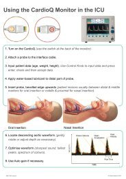
Direct flow measurement<br />
Placing a single-use probe in the esophagus, the<br />
CardioQ-<strong>EDM</strong> monitor uses Doppler ultrasound<br />
technology to determine directly a patient’s central<br />
vascular blood flow and fluid status during the<br />
intraoperative period.<br />
Easy to use and quick to focus, the device<br />
generates a low-frequency ultrasound signal, which<br />
is highly sensitive to changes in flow and measures<br />
them immediately.<br />
A<br />
Transmit crystal<br />
B<br />
Receive crystal<br />
B<br />
A<br />
FIGURE 3<br />
A An esophageal Doppler probe is inserted into the patient’s<br />
esophagus, either nasally or orally.<br />
B The transmit and receive piezo electric crystals at the tip of the<br />
probe measure velocity of blood flow in the descending aorta.
think Doppler.<br />
CMS has determined that there is<br />
sufficient evidence to conclude<br />
that esophageal Doppler monitoring of<br />
cardiac output for ventilated patients in the<br />
ICU and operative patients with a need for<br />
intra-operative fluid optimization<br />
is reasonable and necessary.<br />
CMS Final Coverage Decision Memorandum for Ultrasound<br />
Diagnostic Procedures. May 22, 2007 (CAG-00309R).<br />
Doppler is recommended<br />
The Centers for Medicare and Medicaid Services (CMS) issued<br />
a National Coverage Determination for esophageal Doppler<br />
monitoring (<strong>EDM</strong>) in 2007 covering the procedure “for ventilated<br />
patients in the ICU and operative patients with a need for intraoperative<br />
fluid optimization.” As a result of the coverage policy,<br />
CMS also established reimbursement for physicians using <strong>EDM</strong>.<br />
AHRQ reported:<br />
• Clinically significant reduction in major<br />
complications during surgery<br />
• Clinically significant reduction in the total<br />
number of complications during surgery, and<br />
• Reduction in hospital length of stay (LOS)<br />
Randomized, controlled trials of individually guided fluid<br />
management using the CardioQ-<strong>EDM</strong> have demonstrated that<br />
early fluid management intervention during surgery, will reduce<br />
post-operative complications, reduce intensive care admissions,<br />
and reduce the length of hospital stay. The device has established<br />
an incomparable evidence base that is acknowledged and<br />
endorsed by The Agency for Healthcare Research and Quality<br />
(AHRQ), who conducted a technology assessment of Doppler<br />
guided fluid management in 2007.<br />
The UK National Institute for Health and Clinical Excellence<br />
(NICE), issued medical technology guidance in 2011 on the<br />
CardioQ-ODM (MTG3). NICE asserts that the technology should<br />
be considered for use in patients undergoing major or high-risk<br />
surgery or other surgical patients in whom a clinician would<br />
consider using invasive cardiovascular monitoring. In addition to<br />
reducing complications and length of stay, NICE estimated savings<br />
of £1,100 per patient.<br />
The National Health Service (NHS) National Technology<br />
Adoption Centre (NTAC) audit reported the benefits of <strong>EDM</strong><br />
implementation in three hospitals in over 1,300 patients.<br />
NTAC reported:<br />
• The post-operative stay was reduced<br />
by 3.5 days<br />
• CVC use was reduced by 23%.<br />
• The results also indicate a trend towards<br />
a reduction in readmission rates,<br />
re-operations and mortality.
References<br />
Mythen MG, Webb AR<br />
Perioperative plasma volume expansion reduces the incidence<br />
of gastric mucosal hypoperfusion during cardiac surgery. Arch Surg.<br />
1995; 130: 423-429.<br />
Sinclair S, James S, Singer M<br />
Intraoperative intravascular volume optimisation and length of stay<br />
after repair of proximal femoral fracture: randomised control trial.<br />
BMJ. 1997; 315: 909-912.<br />
Gan Tong J, Soppitt A, Maroof M, El-Moalem H, Robertson KM,<br />
Moretti E, Dwane P, Glass PS<br />
Goal-directed intraoperative fluid administration reduces length of hospital<br />
stay after major surgery. Anaesthesiology 2002; 97(4): 820-826.<br />
Venn R, Steele A, Richardson P, Poloniecki J, Grounds M,<br />
Newman P<br />
Randomized controlled trial to investigate influence of the fluid<br />
challenge on duration of hospital stay and perioperative morbidity in<br />
patients with hip fractures. Br J Anaesth. 2002; 88: 65-71.<br />
Wakeling HG, McFall MR, Jenkins CS, Woods WGA, Miles WFA,<br />
Barclay GR, Fleming SC<br />
Intraoperative oesophageal Doppler guided fluid management<br />
shortens postoperative hospital stay after major bowel surgery.<br />
Br J Anaesth. 2005; 95(5):634-642.<br />
Noblett SE, Snowden CP, Shenton BK, Horgan AF<br />
Randomized clinical trial assessing the effect of Doppler-optimized<br />
fluid management on outcome after elective colorectal resection.<br />
Br J Surg 2006; 93:1069-1076.<br />
Senagore AJ, Emery T, Luchtefeld M, Kim D, Dujovny N, Hoedema R<br />
Fluid management for laparoscopic colectomy: a prospective,<br />
randomized assessment of goal-directed administration of balanced<br />
salt solution or hetastarch coupled with an enhanced recovery<br />
program. Dis Colon Rectum. 2009 Dec; 52(12):1935-40.<br />
Pillai P, McEleavy I, Gaughan M, Snowden C, Nesbitt I, Durkan G,<br />
Johnson M, Cosgrove J, Thorpe A.<br />
A double-blind randomized controlled clinical trial to assess the effect<br />
of Doppler optimized intraoperative fluid management on outcome<br />
following radical cystectomy. J Urol. 2011 Dec; 186(6):2201-6.<br />
Figus A, Wade RG, Oakey S, Ramakrishnan VV<br />
Intraoperative esophageal Doppler hemodynamic monitoring in<br />
free perforator flap surgery. Ann Plast Surg. 2011 Dec 9.<br />
Agency For Healthcare Research and Quality (AHRQ) January<br />
16, 2007 Esophageal Doppler Ultrasound-Based Cardiac Output<br />
Monitoring for Real-Time Therapeutic Management of Hospitalized<br />
Patients – A Review. http://www.cms.hhs.gov/mcd/viewtechassess.<br />
asp?where=index&tid=45<br />
Centers for Medicare and Medicaid Services (CMS), Decision<br />
Memo for Ultrasound Diagnostic Procedures (CAG-00309R) May 22, 2007<br />
http://www.cms.gov/medicare-coverage-database/details/ncadecision-memo.aspx?<br />
NHS: National Institute for Health and Clinical Excellence.<br />
CardioQ-ODM Oesophageal Doppler Monitor.<br />
http://guidance.nice.org.uk/MTG3, 2011.<br />
NHS National Technology Adoption Centre (NTAC)<br />
http://www.ntac.nhs.uk/HowToWhyToGuides/<br />
DopplerGuidedIntraoperative/Doppler-Executive-Summary.aspx.<br />
<strong>Product</strong> Description<br />
CardioQ-<strong>EDM</strong> Monitor (<strong>Product</strong> Code: 9051-7057)<br />
For adult use in operating room and critical care.<br />
Surgical Probes<br />
I 2 S Doppler Probe (<strong>Product</strong> Code: 9090-7015)<br />
6-hour oral/nasal Doppler probe for anesthetized,<br />
sedated and awake patients.<br />
I 2 P Doppler Probe (<strong>Product</strong> Code: 9090-7016)<br />
24-hour oral/nasal Doppler probe for anesthetized,<br />
sedated and awake patients.<br />
Critical Care Probes<br />
I 2 C Doppler Probe (<strong>Product</strong> Code: 9090-7017)<br />
72-hour oral/nasal Doppler probe for anesthetized,<br />
sedated and awake patients.<br />
EDP240 Doppler Probe (<strong>Product</strong> Code: 9070-7006)<br />
10-day oral/nasal Doppler probe for patients under<br />
anesthesia or full sedation.<br />
When fluid management really matters, think Doppler<br />
<strong>Deltex</strong> <strong>Medical</strong>, SC, Inc.<br />
330 E. Coffee Street, Greenville, SC 29601<br />
Telephone: 864 527 5913<br />
Fax: 864 527 5914<br />
Email: ussales@deltexmedical.com<br />
www.deltexmedical.com<br />
AKA 9051-5521 Issue 2