Using the CardioQ Monitor in the ICU - Deltex Medical
Using the CardioQ Monitor in the ICU - Deltex Medical
Using the CardioQ Monitor in the ICU - Deltex Medical

Create successful ePaper yourself
Turn your PDF publications into a flip-book with our unique Google optimized e-Paper software.
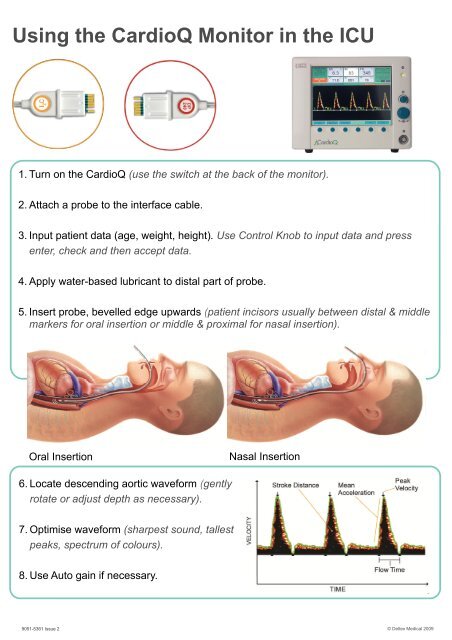
<strong>Us<strong>in</strong>g</strong> <strong>the</strong> <strong>CardioQ</strong> <strong>Monitor</strong> <strong>in</strong> <strong>the</strong> <strong>ICU</strong><br />
1. Turn on <strong>the</strong> <strong>CardioQ</strong> (use <strong>the</strong> switch at <strong>the</strong> back of <strong>the</strong> monitor).<br />
2. Attach a probe to <strong>the</strong> <strong>in</strong>terface cable.<br />
3. Input patient data (age, weight, height). Use Control Knob to <strong>in</strong>put data and press<br />
enter, check and <strong>the</strong>n accept data.<br />
4. Apply water-based lubricant to distal part of probe.<br />
5. Insert probe, bevelled edge upwards (patient <strong>in</strong>cisors usually between distal & middle<br />
markers for oral <strong>in</strong>sertion or middle & proximal for nasal <strong>in</strong>sertion).<br />
Oral Insertion<br />
Nasal Insertion<br />
6. Locate descend<strong>in</strong>g aortic waveform (gently<br />
rotate or adjust depth as necessary).<br />
7. Optimise waveform (sharpest sound, tallest<br />
peaks, spectrum of colours).<br />
8. Use Auto ga<strong>in</strong> if necessary.<br />
9051-5361 Issue 2<br />
© <strong>Deltex</strong> <strong>Medical</strong> 2009
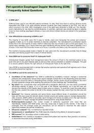
The <strong>CardioQ</strong> Waveform<br />
The green l<strong>in</strong>e <strong>in</strong>dicates <strong>the</strong> velocity/time envelope which <strong>the</strong><br />
monitor uses to make calculations. The white arrows<br />
<strong>in</strong>dicate time and velocity values used for <strong>CardioQ</strong> TM calculations.<br />
The Stroke Distance (SD) is <strong>the</strong> area under <strong>the</strong> waveform<br />
and is <strong>the</strong> basic measured parameter upon which calculations<br />
of Stroke Volume (SV) and all o<strong>the</strong>r Cardiac Output<br />
(CO) and <strong>in</strong>dexed measurements are made. Stroke Volume<br />
is <strong>the</strong> parameter of choice for fluid management protocols,<br />
however changes <strong>in</strong> Stroke Distance (SD) or Stroke Volume<br />
Index (SVI) can also be utilised.<br />
The waveform base, (flow time) depends on heart rate, left<br />
ventricular fill<strong>in</strong>g and afterload. The flow time corrected to a<br />
heart rate of 60bpm (FTc) is <strong>in</strong>versely correlated with <strong>the</strong><br />
systemic vascular resistance (SVR).<br />
The most common cause of a short FTc (10%<br />
Yes<br />
No<br />
Treatment Algorithm<br />
Organ Hypoperfusion<br />
Hypotension<br />
Circulatory Optimisation<br />
<strong>Monitor</strong> SV/SD & FTc<br />
200ml Colloid Challenge<br />
over 10 m<strong>in</strong>utes<br />
Yes<br />
Patient los<strong>in</strong>g fluid at<br />
rate exceed<strong>in</strong>g <strong>in</strong>put<br />
No<br />
Still compromised<br />
(eg Low BP, Oliguria)<br />
<strong>Monitor</strong> SV/SD & FTc<br />
No<br />
Treatment Algorithm suggested by Prof. M. S<strong>in</strong>ger, University College London<br />
<strong>Deltex</strong> <strong>Medical</strong><br />
Tel: +44(0)1243 774837<br />
www.deltexmedical.com<br />
O<strong>the</strong>r <strong>the</strong>rapies as appropriate eg:<br />
Dilators (+ more fluid) if low FTc, low PV and BP acceptable.<br />
Inotropes if low PV and low BP.<br />
Vasopressors if high FTc, high SV and low BP.