Chapter 9 Part 1 Lecture Notes.pdf
Chapter 9 Part 1 Lecture Notes.pdf
Chapter 9 Part 1 Lecture Notes.pdf

Create successful ePaper yourself
Turn your PDF publications into a flip-book with our unique Google optimized e-Paper software.
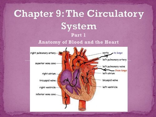
<strong>Part</strong> 1<br />
Anatomy of Blood and the Heart
• What’s in your blood?<br />
• Functions of Blood Cells<br />
• Anatomy of the Heart
• The heart pumps<br />
blood throughout<br />
your body<br />
• Blood picks up and<br />
drops off different<br />
substances to<br />
ensure that cells<br />
have oxygen and<br />
carbon dioxide as<br />
well as other waste<br />
products are<br />
properly disposed<br />
of
• Considered a connective<br />
tissue<br />
• When your blood is<br />
centrifuged (spun really<br />
fast) it separates<br />
materials by density<br />
• Your blood consists of a<br />
liquid component called<br />
plasma<br />
• You blood also consists<br />
of 3 formed components<br />
• Red Blood Cells<br />
• White Blood Cells<br />
• Platelets
• 92% is water<br />
• 8% is made of plasma<br />
proteins, salt, nutrients,<br />
urea, hormones and<br />
vitamins<br />
• 3 Types of Plasma<br />
Proteins<br />
• Albumin – maintains<br />
proper osmotic<br />
pressure<br />
• Fibrinogen – helps<br />
with clotting<br />
• Immunoglobulin –<br />
AKA antibodies
• AKA Erythrocytes<br />
• RBCs contain a protein<br />
called hemoglobin<br />
which carries oxygen<br />
• Oxyhemoglobin is<br />
bright red (makes your<br />
arteries look red)<br />
• Deoxyhemoglobin is<br />
purplish-blue (makes<br />
your veins look blue<br />
• Live for about 4 months
• AKA leukocytes<br />
• Not as many WBCs as<br />
RBCs<br />
• Fight off invading<br />
microbes, bacteria,<br />
viruses<br />
• Two main types of<br />
WBCs<br />
• Granular Leukocytes<br />
• Agranular Leukocytes
White Blood Cell Functions<br />
Type of White Blood Cell<br />
Specific Functions<br />
Granular Leukocytes<br />
Eosinophils<br />
Neutrophils<br />
Basophils<br />
Numbers increase during allergic reactions<br />
and parasitic infections<br />
First to respond to infections - phagocytize<br />
Seep out of vessels at site of injury and<br />
release histamine to dilate vessels<br />
Agranular Leukocytes - Lymphocytes<br />
B lymphocytes<br />
T lymphocytes<br />
Monocytes<br />
Form antibodies to fight infection<br />
Destroy cells that contain foreign material<br />
Mature into macrophages, engulf diseasecausing<br />
microbes, stimulate other WBCs into<br />
action
• AKA thrombocyte<br />
• Tiny fragments of cells<br />
• Large cells in the bone<br />
marrow called<br />
megakaryocytes break<br />
into fragments which are<br />
platelets<br />
• Help the clotting process<br />
by plugging up the<br />
injured blood vessels
• Main organ of the<br />
circulatory system<br />
• The heart is the driving<br />
force behind the<br />
movement of the blood<br />
• The pressure is<br />
generates by the<br />
pumping action, forces<br />
the blood through the<br />
vessels<br />
• The heart lies between<br />
the lungs and behind<br />
and slightly to the left<br />
of the sternum
• Pericardium/Pericardial<br />
Tissue: thick layer of<br />
muscle tissue and a<br />
protective membrane that<br />
folds into two layers<br />
surrounding the heart<br />
• Endothelial Tissue:<br />
endothelial tissue that<br />
lines the inside of the heart<br />
and is continuous with all<br />
your blood vessels
• Pericardial Cavity:<br />
Coronary vessels –<br />
blood vessels that<br />
supply the tissues<br />
of the heart with<br />
nutrients and<br />
oxygen<br />
• Myocardium:<br />
muscular layer of<br />
the heart
• Epicardium: inner layer of<br />
the pericardium, covers the<br />
myocardium and secretes<br />
perocardial fluid to help<br />
lubricate so tissues don’t<br />
rub together during<br />
contraction<br />
• Parietal Pericardium:<br />
outermost layer of the heart,<br />
thin, white, fibrous<br />
connective tissue that joins<br />
the major blood vessels
• Right Atrium<br />
• Left Atrium<br />
• Right Ventricle<br />
• Left Ventricle<br />
• Interatrial Septum –<br />
wall dividing the two<br />
atria<br />
• Interventricular<br />
Septum – wall<br />
dividing the two<br />
ventricles
• The heart contains several<br />
valves<br />
• Valves keep blood flowing in<br />
the right direction on the<br />
pathway and allows the right<br />
amount of blood into each<br />
chamber<br />
• The names of the valves can<br />
tell you their location or<br />
certain characteristics<br />
• Semilunar Valves - half<br />
moons<br />
• Atrioventricular Valves<br />
(AV) – between the atria and<br />
ventricles<br />
• Bicuspid Valve – 2 flaps<br />
• Tricuspid Valve – 3 flaps
• What are the 3 formed components of blood?<br />
• List some of the types of white blood cells<br />
• What is the name of the thin fibrous tissue that<br />
covers the heart?<br />
• What are the four chambers of the heart?<br />
• Under what conditions does blood look purplish<br />
blue?<br />
• What types of substances are carried in plasma?<br />
• What is the purpose of the circulatory system?