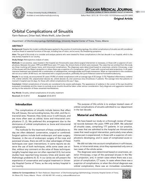
Orbital Complications of Sinusitis - Balkan Medical Journal
Orbital Complications of Sinusitis - Balkan Medical Journal
Orbital Complications of Sinusitis - Balkan Medical Journal
Create successful ePaper yourself
Turn your PDF publications into a flip-book with our unique Google optimized e-Paper software.
© Trakya University Faculty <strong>of</strong> Medicine<br />
Available at www.balkanmedicaljournal.org<br />
BALKAN MEDICAL JOURNAL<br />
THE OFFICIAL JOURNAL OF TRAKYA UNIVERSITY FACULTY OF MEDICINE<br />
<strong>Balkan</strong> Med J 2013; 30: 151-4 • DOI: 10.5152/balkanmedj.2013.8005<br />
Original Article<br />
151<br />
<strong>Orbital</strong> <strong>Complications</strong> <strong>of</strong> <strong>Sinusitis</strong><br />
Pjerin Radovani, Dritan Vasili, Mirela Xhelili, Julian Dervishi<br />
Department <strong>of</strong> Otorhinolaryngology-Ophthalmology, University Hospital Center <strong>of</strong> Tirana, Tirana, Albania<br />
ABSTRACT<br />
Background: Despite the modern antibiotherapies applied in the practice <strong>of</strong> otorhinolaryngology, the orbital complications <strong>of</strong> sinusitis are still considered<br />
a serious threat to essential functions <strong>of</strong> the eye, including loss <strong>of</strong> vision, and at worst, life threatening symptoms.<br />
Aims: The goal <strong>of</strong> this study is to consider and analyse patients who were treated for these complications in the last decade in our hospital, which is the<br />
only tertiary hospital in our country.<br />
Study Design: Retrospective analysis <strong>of</strong> cases.<br />
Methods: In our practice, cases treated in the hospital are rhinosinusitis cases where surgical intervention is necessary, or those with a suspicion <strong>of</strong> complications.<br />
Between the years 1999 and 2009 there were 177 cases, the clinical charts <strong>of</strong> which were reviewed. The cases that are omitted from this study<br />
are those involving s<strong>of</strong>t tissues, bone, and intracranial complications. The diagnoses were determined based on anamnesis, anterior rhinoscopy, x-rays<br />
<strong>of</strong> the sinuses with the Water’s projection or where there was a suspicion <strong>of</strong> a complication, and CT scans with coronal and axial projections. In all cases,<br />
intensive treatment was initiated with a combination <strong>of</strong> cefalosporines, aminoglycosides and Proetz manoeuvre. When an improvement in the conditions<br />
did not occur within 24-48 hours, we intervened with a surgical procedure, preferably the Lynch-Patterson external frontoethmoidectomy.<br />
Results: In our study, we encountered 35 cases (19.8%) <strong>of</strong> orbital complications with an average age <strong>of</strong> 25 (range: 3-75); Palpebral inflammatory oedema<br />
(15), orbital cellulitis (10), subperiosteal abscess (6), orbital abscess (3), and cavernous sinus thrombosis (1 patient). The average time that patients remained<br />
in hospital was 4.6 days; for those with orbital complications this was 7 days.<br />
Conclusion: <strong>Orbital</strong> complications <strong>of</strong> sinusitis are considered to be severe pathologies. The appearance <strong>of</strong> oedema in the corner <strong>of</strong> the eye should be<br />
evaluated immediately and the means to exclude acute sinusitis should be taken under serious consideration. Early diagnosis and aggressive treatment<br />
are key to the reduction <strong>of</strong> these unwanted manifestations.<br />
Key Words: <strong>Sinusitis</strong>, orbital complications <strong>of</strong> sinusitis, treatment<br />
Received: 25.10.2012 Accepted: 20.02.2013<br />
Introduction<br />
The complications <strong>of</strong> sinusitis include lesions that affect<br />
the s<strong>of</strong>t tissues, the surrounding bones, the orbit, and the intracranial<br />
area. However, they rarely occur in s<strong>of</strong>t tissues, and<br />
are more <strong>of</strong>ten seen as orbital, bone and intracranial complications<br />
(1, 2). We preferred this arrangement due to the<br />
dominance <strong>of</strong> orbital complications vs. bone and intracranial<br />
complications.<br />
The methods for the treatment <strong>of</strong> these complications today<br />
are <strong>of</strong>ten debated: conservative, surgical or combined.<br />
Surgical options include both endoscopic and open surgery.<br />
However, in the era <strong>of</strong> endoscopic surgery <strong>of</strong> the nose and<br />
paranasal sinuses, there are still no clear advantages <strong>of</strong> these<br />
techniques over open interventions. In some reports we observe<br />
the use <strong>of</strong> both techniques, while some cases use either,<br />
without any comment on the value <strong>of</strong> these techniques.<br />
Lengthy hospital stays are not <strong>of</strong>ten reported (3-5), but some<br />
studies have reported long recovery times and severe ophthalmologic<br />
problems (4, 5). In our series we will present the<br />
experience <strong>of</strong> conservative and surgical treatment in University<br />
Hospital Centre <strong>of</strong> Tirana, as the only tertiary hospital in<br />
our country.<br />
The purpose <strong>of</strong> this article is to analyse treated cases <strong>of</strong><br />
orbital complications <strong>of</strong> sinusitis admitted to our department<br />
in the last decade.<br />
Material and Methods<br />
We have based our study on a thorough review <strong>of</strong> hospital<br />
records between the years 1999 and 2009. We selected<br />
all sinusitis cases, comprising 177 patients. In our practice,<br />
the cases that are admitted to the hospital are rhinosinusitis<br />
cases that need surgical intervention, particularly ones where<br />
intense pain is experienced, in those with a suspicion <strong>of</strong> complications<br />
developing, and in cases <strong>of</strong> complicated acute sinusitis.<br />
We did not take into account complications in s<strong>of</strong>t tissues,<br />
bone, or intracranial areas, since they represented only one or<br />
two cases <strong>of</strong> the whole series.<br />
The diagnoses were determined based on anamnesis, anterior<br />
rhinoscopy, x-rays <strong>of</strong> the paranasal sinuses in the Water’s<br />
projection, and, in certain cases where there was a suspicion<br />
<strong>of</strong> complication, CT scans. In cases where there was a<br />
suspicion <strong>of</strong> complications, or when the clinical diagnosis was<br />
orbital complication, there was an immediate consultation<br />
Address for Correspondence: Dr. Pjerin Radovani, Department <strong>of</strong> Otorhinolaryngology-Ophthalmology, University Hospital Center <strong>of</strong> Tirana,<br />
Rr. Dibres 372, Tirana, Albania Phone: +35 542 455 195 e-mail: p_radovani@hotmail.com
152<br />
Radovani et al.<br />
<strong>Orbital</strong> <strong>Complications</strong> <strong>of</strong> <strong>Sinusitis</strong><br />
<strong>Balkan</strong> Med J<br />
2013; 30: 151-4<br />
with the ophthalmologist and a collaborative treatment approach<br />
was chosen. In fact, a lot <strong>of</strong> these cases were referred<br />
to us by the Department <strong>of</strong> Ophthalmology or private clinics.<br />
In all cases, intensive treatment was initiated with a combination<br />
<strong>of</strong> cefalosporines, aminoglycosides, and a modified<br />
Proetz procedure in accordance with recent guidelines. When<br />
an improvement in the patient’s condition did not occur within<br />
24-48 hours, we intervened with surgical procedures, preferably<br />
the external frontoethmoidectomy in combination with<br />
the Lynch-Patterson technique, because this allowed a wide<br />
opening, good drainage and clear entry to the nasal cavity. In<br />
two cases with one-sided pansinusitis, the Caldwell-Luc intervention<br />
was used, approaching the sinuses externally. When<br />
the imaging produced a clear view <strong>of</strong> the intraorbital complication,<br />
or if the patient experienced problems with vision,<br />
the intervention was applied within the first 24 hours. In this<br />
analysis <strong>of</strong> clinical material, we took into account age, gender,<br />
conservative treatment vs. surgical intervention and the average<br />
time <strong>of</strong> hospital stay.<br />
Results<br />
From this study <strong>of</strong> 177 cases there were 35 cases with orbital<br />
complications, which were further analysed. The youngest<br />
case encountered in the series was a 3 year old child, while<br />
the oldest was a 75 year old. Cases affecting younger patients<br />
dominated the series. The average age was 25.3, which was<br />
an average similar to that reported by many other series. The<br />
average time that patients stayed in hospital was 4.6 days,<br />
while for those with orbital complications it was 7 days.<br />
<strong>Orbital</strong> complications <strong>of</strong> sinusitis were classified primarily<br />
by Hubert in 1937, but it was Chandler who categorised them<br />
in 1970, followed by later modifications by others (6, 7, 8). The<br />
classifications <strong>of</strong> Chandler and Moloney remain the most commonly<br />
used to date (8, 9). Modifications were made in relation<br />
to CT-scan and MRI findings. We have referred to Chandler’s<br />
classification; however, in Table 1 both classifications were included.<br />
The difference between the two systems only appears<br />
in the palpebral region.<br />
In our study, we encountered 35 cases (19.8%) with orbital<br />
complications. The cases and treatments based on Chandler’s<br />
classification are found in Table 2.<br />
The case <strong>of</strong> cavernous sinus thrombosis presented as<br />
pansinusitis <strong>of</strong> the left side and received ambulatory medical<br />
Table 1. The most commonly accepted orbital classifications<br />
<strong>of</strong> sinuses<br />
Group Chandler Moloney<br />
First Inflammatory oedema Preseptal cellulitis<br />
Second <strong>Orbital</strong> cellulitis Subperiosteal abscess<br />
Third Subperiosteal abscess <strong>Orbital</strong> cellulitis<br />
Fourth <strong>Orbital</strong> abscess <strong>Orbital</strong> abscess<br />
Fifth Cavernous sinus Cavernous sinus<br />
thrombosis<br />
thrombosis<br />
treatment for 10 days. The patient was admitted to the ophthalmology<br />
department, but was transferred to the hospital<br />
after two days. The patient had lost vision due to an orbital<br />
abscess, so we performed a complete opening <strong>of</strong> the left side<br />
<strong>of</strong> the sinuses. All other cases recovered without any functional<br />
problems.<br />
Discussion<br />
Even though the occurrences are significantly lower than<br />
decades ago, complications <strong>of</strong> sinusitis, whether acute or<br />
chronic, continue to appear despite progresses in therapy that<br />
have been achieved in recent years. The topographic anatomy<br />
<strong>of</strong> the orbit, which shows intimate relations with paranasal<br />
sinuses, provides a convincing explanation to these events.<br />
The Lamina Papyracea, which divides the orbit from the nasal<br />
space, is a thin layer <strong>of</strong> bone. Inside it are multiple thin blood<br />
vessels, which allow the spread <strong>of</strong> aggressive infection in the<br />
orbit. Inside the orbital vessels is where the palpebral vessels<br />
and those <strong>of</strong> the centre <strong>of</strong> the face drain and travel parallel<br />
with the lamina papyracea toward the sinus cavernosus. None<br />
<strong>of</strong> these vessels contain valves; therefore, there is an accelerated<br />
spread <strong>of</strong> infection. The periorbita is a strong barrier<br />
to infection and provides clinicians with time before they are<br />
faced with serious complications.<br />
In our series, just as in many others, male patients dominated<br />
female patients with a ratio <strong>of</strong> 2 to 1. The question <strong>of</strong><br />
why this happens has been repeatedly asked through the years,<br />
but there is still no definite explanation for this observation. It<br />
is believed that the female immune system is more pr<strong>of</strong>icient<br />
than that <strong>of</strong> males (8). Also, the higher number <strong>of</strong> occurrences<br />
<strong>of</strong> these complications in young patients is another observation<br />
noted not only here, but by many other researchers (7-10). This<br />
study also found a dominance <strong>of</strong> orbital complications involving<br />
bone (1 case) and intracranial complications (2 cases). This<br />
observation is imperative to a better understanding <strong>of</strong> orbital<br />
complications from both clinical and imaging points <strong>of</strong> view.<br />
Due to the acceptance <strong>of</strong> several classifications, the nomencla-<br />
Table 2. Classifications <strong>of</strong> our cases and the corresponding<br />
treatment<br />
Complication Cases Treatment<br />
according Chandler<br />
Palpebral inflammatory 15 12 conservative<br />
oedema<br />
3 conservative + surgical<br />
<strong>Orbital</strong> cellulitis 10 Surgery with intensive<br />
antibiotherapy<br />
Subperiosteal abscess 6 Surgery with intensive<br />
antibiotherapy<br />
<strong>Orbital</strong> abscess 3 Surgery with intensive<br />
antibiotherapy<br />
Cavernous sinus 1 Surgery with intensive<br />
thrombosis<br />
antibiotherapy
<strong>Balkan</strong> Med J<br />
2013; 30: 151-4<br />
Radovani et al.<br />
<strong>Orbital</strong> <strong>Complications</strong> <strong>of</strong> <strong>Sinusitis</strong><br />
153<br />
ture becomes more difficult, blurring clinical aspects. As stated<br />
above, this study used Chandler’s classification <strong>of</strong> orbital complications<br />
<strong>of</strong> sinusitis due to its simplicity, even though the classification<br />
was designed prior to computer imaging.<br />
The most commonly occurring complication is palpebral<br />
inflammatory oedema, which Moloney referred to as preseptal<br />
cellulitis and reported it as being encountered more <strong>of</strong>ten<br />
in children; this was noted in our study too. The upper eyelid<br />
becomes swollen and hyperaemic due to a blockage <strong>of</strong> vein<br />
drainage from an ethmoidal sinusitis. Chemosis and proptosis<br />
are absent, both <strong>of</strong> which usually indicate postseptal infection.<br />
Vision remains unaffected and the eyeball moves in all directions.<br />
Usually, the oedema is most notable in the morning and<br />
persists throughout the day, with a slight reduction. When the<br />
oedema does not improve, it shows a postseptal inflammation<br />
inside the orbital cave. The appearance <strong>of</strong> proptosis indicates<br />
an advance <strong>of</strong> the orbital infection toward cellulitis. Proptosis,<br />
as well as chemosis, indicates the spread <strong>of</strong> an inflammatory<br />
process in the anterior <strong>of</strong> the orbit. The movements <strong>of</strong> the<br />
eyeball begin to become limited, and vision starts to weaken<br />
from the pressure on the optic nerve. Conservative treatment<br />
does not produce a fast reduction <strong>of</strong> the symptoms; therefore,<br />
surgical intervention is obligatory in these cases (Figure 1).<br />
Another complication is subperiosteal abscess <strong>of</strong> the orbit,<br />
where an accumulation <strong>of</strong> pus forms between the bones<br />
Figure 1. A patient with orbital cellulitis and the corresponding<br />
CT-Scan<br />
<strong>of</strong> the orbit and the periorbita, usually between the lamina<br />
papyracea and the periorbita. Proptosis, chemosis and limited<br />
movement <strong>of</strong> the eyeball are present, but the eyeball makes<br />
a lateral downward switch. Pain is experienced when the superior-medial<br />
corner <strong>of</strong> the orbit is touched. All cases in this<br />
series were in patients over the age <strong>of</strong> 20, all <strong>of</strong> whom underwent<br />
surgical intervention under the protection <strong>of</strong> combined<br />
antibiotherapy.<br />
When cases <strong>of</strong> orbital cellulitis do not receive appropriate<br />
treatment, the inflammatory oedema can progress towards a<br />
purulent inflammation. An accumulation <strong>of</strong> pus begins in the<br />
retroorbital adipose tissue. This is a more severe complication<br />
because it makes up the precedents for the septic thrombosis<br />
<strong>of</strong> the cavernous sinus, or the passing <strong>of</strong> the infection through<br />
nervous routs into the intracranial space. Besides the symptoms<br />
that were mentioned, exophthalmos, ophthalmoplegia<br />
and a weakening or loss <strong>of</strong> vision can also occur. The patient<br />
experiences constant pain and the affected eyeball is sensitive<br />
to touch. There were three such cases in our series, one <strong>of</strong><br />
which was under the age <strong>of</strong> 20. Only one patient lost vision: he<br />
was treated in an ophthalmology outpatient clinic, and after<br />
a few days was admitted to the Otorhinolaryngology Department.<br />
When admitted to our care, the vision had already been<br />
lost. The starting point was an acute frontoethmoiditis.<br />
Lastly, when bacterial infection is not stopped, or when<br />
treatment is delayed, the infection will follow the orbital veins<br />
toward the cavernous sinus. There was only one case with orbital<br />
complications involving bilateral oedemas <strong>of</strong> the eyelids,<br />
and there was a suspicion <strong>of</strong> septic thrombosis <strong>of</strong> the cavernous<br />
sinus. All <strong>of</strong> the sinuses <strong>of</strong> one side and the orbital abscess were<br />
opened up.<br />
Despite the fact that we mainly relied on clinical and surgical<br />
exploration to determine these diagnoses, CT scans<br />
played an undisputable role in the determination <strong>of</strong> a topical<br />
diagnosis and the correct differentiation <strong>of</strong> such pathologies.<br />
Therefore, today, follow-ups are expected to be performed<br />
every hour via computer imaging in order to track the development<br />
<strong>of</strong> the pathology and to determine the appropriate<br />
time for intervention (11-13).<br />
Today, there are discussions regarding endoscopic surgery<br />
<strong>of</strong> the sinuses as treatment for these complications (3-5,<br />
13, 14), but this does not seem the most appropriate in the<br />
presence <strong>of</strong> acute infection and haemorrhage that <strong>of</strong>ten accompany<br />
an intervention <strong>of</strong> this kind; however, it can always<br />
be kept under consideration. In our country, endoscopic surgery<br />
<strong>of</strong> the nose and paranasal sinuses is a new development,<br />
since 2005 in private clinics, but is never used in cases<br />
<strong>of</strong> orbital complication. Two cases in our series came from<br />
private clinics. However, this argument does not diminish the<br />
value <strong>of</strong> endoscopic interventions as the future <strong>of</strong> surgery <strong>of</strong><br />
the nose and sinuses.<br />
Young patients, especially children, respond better to conservative<br />
treatment. This is an observation in accordance with<br />
other observations (15), but our cases mainly presented with<br />
inflammatory palpebral oedema. In other cases <strong>of</strong> orbital complications<br />
where conservative treatments did not show major<br />
improvements to the symptoms in the first 24 hours, we moved<br />
forward with surgery. Other authors insist on conservative
154<br />
Radovani et al.<br />
<strong>Orbital</strong> <strong>Complications</strong> <strong>of</strong> <strong>Sinusitis</strong><br />
<strong>Balkan</strong> Med J<br />
2013; 30: 151-4<br />
treatment in children, basing their point <strong>of</strong> reference for surgical<br />
intervention on contact with the optic nerve (16-19). These<br />
authors may be being reserved regarding surgery due to the<br />
classical idea <strong>of</strong> osteomyelitis intervening on an acute sinusitis<br />
(20-21). We believe that this idea is outdated and belongs to<br />
an era prior to the antibiotics; also, our interventions occurred<br />
under the protection <strong>of</strong> antibiotics. Also, reports from other<br />
authors have mentioned average stays in hospital, which were<br />
higher than the average reported in this series.<br />
In conclusion, we concur that, despite their classification,<br />
orbital complications <strong>of</strong> sinusitis should be considered severe<br />
pathologies. They are life-threatening and also threaten the<br />
patient’s quality <strong>of</strong> life. The appearance <strong>of</strong> oedema in the corner<br />
<strong>of</strong> the eye in a case with acute sinusitis should be recognised<br />
immediately and should be taken under serious consideration<br />
(22).<br />
<strong>Complications</strong> <strong>of</strong> sinusitis, whether acute or chronic, appear<br />
to be severe morbidity, which may sometimes have a<br />
fatal outcome. Early diagnosis and aggressive treatment are<br />
key to the reduction <strong>of</strong> unwanted manifestations. Compared<br />
to previous decades, these conditions have been declining<br />
thanks to proper and timely communication, diagnostic methods,<br />
and the intervention <strong>of</strong> a series <strong>of</strong> updated antibiotics.<br />
The otorhinolaryngologists, ophthalmologists and general<br />
physicians should always be aware <strong>of</strong> symptoms that could<br />
indicate complications <strong>of</strong> sinusitis.<br />
Ethics Committee Approval: N/A.<br />
Informed Consent: Informed consent was obtained from patients who<br />
participated in this study.<br />
Peer-review: Externally peer-reviewed.<br />
Author contributions: Concept – P.R.; Design – P.R., D.V.; Supervision<br />
- D.V., J.D. Resource - P.R., J.D.; Materials – Department <strong>of</strong> ORL-Ophthalmology<br />
; Data Collection&/or Processing – M.X., J.D.; Analysis&/<br />
or Interpretation – P.R., D.V.; Literature Search – P.R.; Writing – P.R.;<br />
Critical Reviews - P.R.<br />
Conflict <strong>of</strong> Interest: No conflict <strong>of</strong> interest was declared by the authors.<br />
Financial Disclosure: No financial disclosure was declared by the authors.<br />
References<br />
1. Stankiewicz JA, Nevell DJ, Park AH. <strong>Complications</strong> <strong>of</strong> inflamatory<br />
disease <strong>of</strong> the sinuses. Otolaryngol Clin North Am 1993;26:<br />
639-55.<br />
2. Gardiner LJ. Complicated frontal sinusitis:evaluation and management.<br />
Otolaryngol Head Neck Surg 1986;95:333-43.<br />
3. Ali A, Kurien M, Matheus SS, Mathew J. <strong>Complications</strong> <strong>of</strong> acute<br />
infective rhinosinusitis:experience from a developing country.<br />
Singapore Med J 2005;46:540-4.<br />
4. Suhaili DN, Goh BS, Gendeh BS. A ten year retrospective review<br />
<strong>of</strong> orbital complications secondary to acute sinusitis in children.<br />
Med J Malaysia 2010;65:49-52.<br />
5. Siedek V, Kremer A, Betz CS, Tschiesner U, Berghaus A, Leunig A.<br />
Management <strong>of</strong> orbital complications due to rhinosinusitis. Eur<br />
Arch Otorhinolaryngol 2010;267:1881-6. [CrossRef]<br />
6. Chandler JR, Langenbrunner DJ, Stevens ER. The pathogenesis<br />
<strong>of</strong> <strong>of</strong> orbital complications in acute sinusitis. Laryngoscope<br />
1970;80:1414-28. [CrossRef]<br />
7. Moloney JR, Badham NJ, McRae A. The acute orbit preseptal<br />
(periorbital) cellulitis, subperiosteal abscess and orbital cellulitis<br />
due to sinusitis. J Laryngol Otol 1987;101(Suppl.12):1-14.<br />
8. Mortimore S, Wormald PJ. The Groote Schuur hospital classification<br />
<strong>of</strong> the orbital complications <strong>of</strong> sinusitis. J Laryngol Otol<br />
1997;111:719-23. [CrossRef]<br />
9. Spires JR, Smith RJ. Bacterial infections <strong>of</strong> the orbital and periorbital<br />
s<strong>of</strong>t-tissues in children. Laryngoscope 1986;96:763-8. [CrossRef]<br />
10. Fearon B, Edmonds B, Bird R. <strong>Orbital</strong>-facial complications <strong>of</strong> sinusitis<br />
in children. Laryngoscope 1979;89:947-53. [CrossRef]<br />
11. Bolinaga U, Perez N, Lobato R, Martinez-Seijas P, Sumitier<br />
Perez E. Abscess <strong>of</strong> the orbit as a complication <strong>of</strong> acute<br />
sinusitis:double surgical approach. Eur Arch <strong>of</strong> Otorhinolaryngol<br />
2009;266(A368):1037-8.<br />
12. Stankiewicz JA, Park AA, Newell DJ. <strong>Complications</strong> <strong>of</strong> sinusitis.<br />
Curr Opin Otolaryngol 1996;4:17-20. [CrossRef]<br />
13. El-Silimi O. The place <strong>of</strong> endonasal endoscopy in the treatment<br />
<strong>of</strong> orbital cellulitis. Rhinology 1995;33:93-6.<br />
14. Manning SC. Endoscopic management <strong>of</strong> medial subperiostal orbital<br />
abscess. Arch Otolaryngol Head Neck Surg 1993;119:789-91. [CrossRef]<br />
15. Brown CL, Graham SM, Griffin MC, Smith RJ, Carter KD, Nerad<br />
JA, et al. Pediatric medial subperiosteal orbital abscess:medical<br />
management where possible. Amer J Rhinol 2004;18:321-7.<br />
16. Durand ML. Intravenous antibiotics in sinusitis. Curr Opin in Otolaryngol<br />
1999;7:7-10. [CrossRef]<br />
17. Poole MD. Antimicrobial therapy for sinusitis. Otolaryngol Clin<br />
North Am 1997;30:331-9.<br />
18. Singh B. The management <strong>of</strong> sinogenic orbital complications. J<br />
Laryngol Otol 1995;109:300-3. [CrossRef]<br />
19. Nageswaran S, Woods CR, Benjamin DK Jr, Givner LB, Shetty AK. <strong>Orbital</strong><br />
cellulitis in children. Pediatr Infect Dis J 2006;25:695-9. [CrossRef]<br />
20. Ryan JT, Preciado DA, Bauman N, Pena M, Bose S, Zalzal GH,<br />
et al. Management <strong>of</strong> pediatric orbital cellulitis in patients with<br />
radiografic findings <strong>of</strong> subperiosteal abscess. Otolaryngol Head<br />
Neck Surg 2009;140:907-11. [CrossRef]<br />
21. Oxford LE, McClay J. <strong>Medical</strong> and surgical management <strong>of</strong> subperiosteal<br />
orbital abscess secondary to acute sinusitis in children.<br />
Int J Pediatr Otorhinolaryngol 2006;70:1853-61. [CrossRef]<br />
22. Radovani P, Xhumbi K, Golemi H. Ënjtjet e përsëritura rreth orbitës.<br />
Revista Mjeksore 1989;6:49-52.


