X-ray Structures and Analysis of 11 Cyclosporin Derivatives ...
X-ray Structures and Analysis of 11 Cyclosporin Derivatives ...
X-ray Structures and Analysis of 11 Cyclosporin Derivatives ...

Create successful ePaper yourself
Turn your PDF publications into a flip-book with our unique Google optimized e-Paper software.
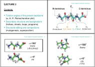
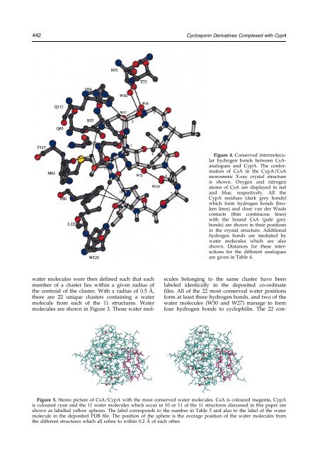
442 <strong>Cyclosporin</strong> <strong>Derivatives</strong> Complexed with CypA<br />
Figure 4. Conserved intermolecular<br />
hydrogen bonds between CsAanalogues<br />
<strong>and</strong> CypA. The conformation<br />
<strong>of</strong> CsA in the CypA/CsA<br />
monomeric X-<strong>ray</strong> crystal structure<br />
is shown. Oxygen <strong>and</strong> nitrogen<br />
atoms <strong>of</strong> CsA are displayed in red<br />
<strong>and</strong> blue, respectively. All the<br />
CypA residues (dark grey bonds)<br />
which form hydrogen bonds (broken<br />
lines) <strong>and</strong> close van der Waals<br />
contacts (thin continuous lines)<br />
with the bound CsA (pale grey<br />
bonds) are shown in their positions<br />
in the crystal structure. Additional<br />
hydrogen bonds are mediated by<br />
water molecules which are also<br />
shown. Distances for these interactions<br />
for the different analogues<br />
are given in Table 4.<br />
water molecules were then de®ned such that each<br />
member <strong>of</strong> a cluster lies within a given radius <strong>of</strong><br />
the centroid <strong>of</strong> the cluster. With a radius <strong>of</strong> 0.5 AÊ ,<br />
there are 22 unique clusters containing a water<br />
molecule from each <strong>of</strong> the <strong>11</strong> structures. Water<br />
molecules are shown in Figure 3. Those water molecules<br />
belonging to the same cluster have been<br />
labeled identically in the deposited co-ordinate<br />
®les. All <strong>of</strong> the 22 most conserved water positions<br />
form at least three hydrogen bonds. <strong>and</strong> two <strong>of</strong> the<br />
water molecules (W30 <strong>and</strong> W27) manage to form<br />
four hydrogen bonds to cyclophilin. The 22 con-<br />
Figure 5. Stereo picture <strong>of</strong> CsA/CypA with the most conserved water molecules. CsA is coloured magenta, CypA<br />
is coloured cyan <strong>and</strong> the <strong>11</strong> water molecules which occur in 10 or <strong>11</strong> <strong>of</strong> the <strong>11</strong> structures discussed in this paper are<br />
shown as labelled yellow spheres. The label corresponds to the number in Table 5 <strong>and</strong> also to the label <strong>of</strong> the water<br />
molecule in the deposited PDB ®le. The position <strong>of</strong> the sphere is the average position <strong>of</strong> the water molecules from<br />
the different structures which all re®ne to within 0.2 AÊ <strong>of</strong> each other.