PDF - Orion Group
PDF - Orion Group
PDF - Orion Group

Create successful ePaper yourself
Turn your PDF publications into a flip-book with our unique Google optimized e-Paper software.
Review Article<br />
The length of the preitoneal catheter in VP Shunt Surgery : A review<br />
Barkatullah AM, Mahmood E, Barua KK, Alam MM<br />
The ORION Medical Journal 2004 Sep;20:248-249<br />
Abstract<br />
Complications of distal ventriculoperitoneal<br />
shunt tubing is quite common either due to a<br />
short length of the catheter or a very long length<br />
of catheter in the peritoneal cavity. This review<br />
article is based on two sample cases of<br />
hydrocephalous, in which one had a short and<br />
the other had a long peritoneal catheter<br />
placement in the peritoneal cavity during VP<br />
Shunt surgery.<br />
Keywords<br />
Cerebrospinal fluid shunts, complication<br />
Case no. one<br />
A thirteen years old right hander young boy, an<br />
amateur cricket player came to us in November<br />
1996 with the complaints of headache, vomiting<br />
and blurring vision. Fundoscopic examination<br />
revealed gross papilloedema. A CT Scan<br />
revealed gross hydrocephalous with dilatation of<br />
all four ventricles without any other CT<br />
findings.<br />
A medium pressure ventriculoperitoneal shunt<br />
system was inserted by canulating the right<br />
lateral ventricle through a right parietal in the<br />
right lower abdomen burr hole and the<br />
peritoneal catheter inserted through a right<br />
lower abdominal incision. After surgery the<br />
1. Dr. Asif Moazzam Barkatullah, MBBS,<br />
MS (Neurosurgery) Assistant Professor of<br />
Neurosurgery, Sir Salimullah Medical College<br />
and Mitford Hospital (SSMCH)<br />
2. Dr. Ehsan Mahmood, MBBS, Ph.D<br />
Associate Professor of Neurosurgery, SSMCH<br />
3. Dr. Kanak Kanti Barua, MBBS, FCPS,<br />
FICS, Ph.D Associate Professor of Neurosurgery,<br />
BSMMU, Dhaka.<br />
4. Dr. Md. Mahbub-ul-Alam, MBBS,<br />
FCPS, FICS Associate Professor of Paediatric<br />
surgery, SSMCH<br />
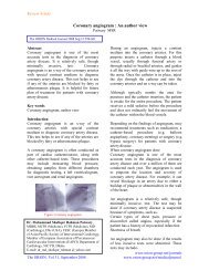
Figure 1: A swelling beneath the scar mark in the right<br />
lower abdomen<br />
patient had an excellent recovery. He started<br />
back his usual activities as a cricketer. He was<br />
very good in bowling. After one year of surgery<br />
he noticed a small swelling in the right lower<br />
abdomen.<br />
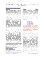
Figure 2: Pre-operative photograph of the cystic cavity<br />
He was advised for revision shunt surgery but<br />
he did not comply. About six and a half years<br />
later, in mid 2004 he developed headache and<br />
vomiting with gradually enlarging abdominal<br />
swelling, to the size of a cricket ball. An<br />
ultrasonogram of the abdomen revealed a cyst<br />
containing the peritoneal catheter of the VP<br />
Shunt in the anterior abdominal wall and a CT<br />
Scan of Brain revealed moderately dilated<br />
ventricular system suggesting improperly<br />
functioning VP Shunt system. He was<br />
hospitalized and revision of the peritoneal end<br />
of the ventricular catheter was planned. Upon<br />
surgery it was confirmed that the peritoneal end<br />
of the catheter had been pulled out of the<br />
peritoneal cavity and was lying between the<br />
external and internal oblique muscle layer of the<br />
anterior abdominal wall with the formation of a<br />
cyst. Excision of the cyst, repair of the anterior<br />
abdominal wall at this site and reintroduction of<br />
The ORION Vol. 20 January 2005<br />
www.orion-group.net/journals
Review Article<br />
the ventricular catheter in the peritoneal cavity<br />
with added length was undertaken.<br />
Case no. two<br />
A two months old female child was brought to<br />
the neurosurgery unit of Mitford Hospital<br />
Dhaka, Bangladesh on March 16, 2003 with an<br />
irregular mass in the lower back. On clinical<br />
examination it was found to be a lumber<br />
meningocele with no motor deficit of lower<br />
limbs and no urinary incontinence. The head<br />
circumference was moderately enlarged with<br />
tense and bulging anterior fontanel (OFC 45<br />
cm). The VP shunt was removed by opening up<br />
sub cutaneous tract below the valve of the shunt<br />
system in the neck under local anesthesia and<br />
then bisecting the peritoneal catheter there. The<br />
extruding catheter per anus was gently pulled<br />
out. Then the ventricular catheter along with the<br />
valve was also pulled nut by the neck opening<br />
of the catheter tract by gentle pull.<br />
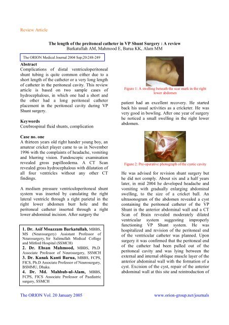
Figure 3: Per anal extrusion of the peritoneal catheter of<br />
VP Shunt<br />
The baby was kept on a third generation<br />
cephalosporin and metronidazole for five days<br />
and nothing by mouth and IV infusion for next<br />
twenty four hours. Oral feeds were subsequently<br />
started after confirming no bowel complications.<br />
A subsequent fresh VP shunt was inserted and<br />
the baby was doing quite fine after surgery.<br />
Discussion<br />
Shunt complications are numerous and can be<br />
classified under three main headings<br />
I. Infection<br />
ii. Functional failure<br />
iii. Mechanical failure<br />
Late anal extrusion of the peritoneal catheter is a<br />
very rare complication with a very few cases<br />
reported so far 1,2 . Factors relating to shunt<br />
failure have three potential origins: the surgeon<br />
the patient and the shunt. Shunt complications<br />
are in fact more often related to a combination<br />
of the factors 3 . Experience with the primary<br />
insertion of an extended length open-ended<br />
peritoneal tubing (120 cm) undertaken expressly<br />
to avoid the need for a lengthening procedure<br />
because of growth of the patient had been<br />
reported very successful 4 .<br />
In a review of new insertions of VP shunts using<br />
the extended length tubing over a 14-year period<br />
at Children’s Hospital of Los Angeles, a total<br />
998 shunts were placed in 952 patients, with a<br />
mean follow-up period of 6.7 years. The<br />
patients experienced a total of 52 distal shunt<br />
revisions for a variety of malfunction etiologies.<br />
In patients ranging in age from premature<br />
neonate to 20 years, there was no increase in the<br />
distal complication rate, and specifically no<br />
complications were experienced that were<br />
directly related to the use of the extended length<br />
tubing 4 . An extrd-abdominal cyst filled with<br />
cerebrospinal fluid was found postpartum in a<br />
patient with a ventriculoperitoneal (VP) shunt.<br />
No similar complication of VP shunting has<br />
been reported before except for our case no-1 5 .<br />
Conclusion<br />
Extra long peritoneal catheter and the rigidity of<br />
the tube along with the poor nutritional status of<br />
the patient appears to be the cause of the slow<br />
penetration of the catheter tip inside the lumen<br />
of the large gut. Pulsatile CSF out flow at the<br />
catheter tip and the peristaltic bowel movement<br />
added to the ultimate extrusion of the shunt per<br />
anus. This rare complication can be prevented<br />
by keeping the peritoneal end of the catheter to<br />
a minimum size, not more than 10 cm in the<br />
peritoneal cavity of the neonates and 25 -30 cm<br />
in infants 6 . Our experience reveals that keeping<br />
the peritoneal catheter in infants not more than<br />
20 cm. gives better results. As regards the<br />
The ORION Vol. 20 January 2005<br />
www.orion-group.net/journals
Review Article<br />
patient factor the nutritional status of the patient<br />
must be taken into good consideration in<br />
preventing such complications. So the answer<br />
for the question of what should be the length of<br />
the peritoneal catheter inside the peritoneal<br />
cavity be considered on the merit of each<br />
individual case.<br />
References<br />
1. O’Donoghue GT. Kumar R, Taleb F,<br />
Phillips J.: Per-anal extrusion of a disconnected<br />
ventriculoperitoneal catheter-an unusual<br />
complication; Ir Med J. 2002 Mar; 95(3):88-9.<br />
2. Simon W. Strudee, lake Timothy, Atul<br />
Tyagi; Total extrusion of a cranial peritoneal<br />
shunt per rectum; Journal of Clinical<br />
Neuroscience; 2002 Mar; 9(2):199-200.<br />
3. Saeedy-Boroujeni H R ; Photo Clinic;<br />
Archives of Iranian Medicine; 2002 Jan;5(1);61-<br />
62.<br />
4. Couldwell WI. LeMay DR. McComb JG,<br />
:Experience with use of extended length<br />
peritoneal shunt catheters. J Neurosurg. 1996<br />
Sep;85(3):425-7.<br />
5. Nugent P, Hoshek S.: Large extraabdominal<br />
cyst as a postpartum complication of<br />
peritoneal shunt. Case report: J Neurosurg. 1986<br />
Jan;64(1):151-2.<br />
6. Simon Linndsay, Thomas David GT, Clark<br />
K; Operative Surgery (neurosurgery); 4th<br />
edition; London; Butterworth;1989;127.<br />
The ORION Vol. 20 January 2005<br />
www.orion-group.net/journals