Role of radiology and imaging in the daignosis of ... - Orion Group
Role of radiology and imaging in the daignosis of ... - Orion Group
Role of radiology and imaging in the daignosis of ... - Orion Group

Create successful ePaper yourself
Turn your PDF publications into a flip-book with our unique Google optimized e-Paper software.
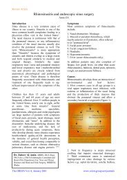
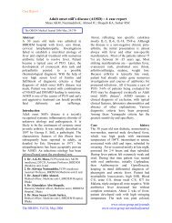
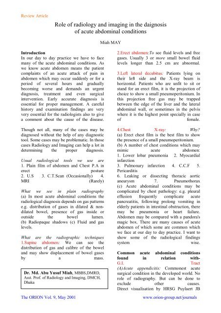
Review Article<strong>Role</strong> <strong>of</strong> <strong>radiology</strong> <strong>and</strong> <strong>imag<strong>in</strong>g</strong> <strong>in</strong> <strong>the</strong> <strong>daignosis</strong><strong>of</strong> acute abdom<strong>in</strong>al conditionsMiah MAYIntroductionIn our day to day practice we have to facemany <strong>of</strong> <strong>the</strong> acute abdom<strong>in</strong>al conditions. Aswe know acute abdomen means <strong>the</strong> patientcompla<strong>in</strong>ts <strong>of</strong> an acute attack <strong>of</strong> pa<strong>in</strong> <strong>in</strong>abdomen which may occur suddenly or for aperiod <strong>of</strong> several hours <strong>and</strong> graduallybecom<strong>in</strong>g worse <strong>and</strong> dem<strong>and</strong>s an urgentdiagnosis, treatment <strong>and</strong> even surgical<strong>in</strong>tervention. Early accurate diagnosis isessential for proper management. A carefulhistory <strong>and</strong> exam<strong>in</strong>ation f<strong>in</strong>d<strong>in</strong>gs are veryvery essential for <strong>the</strong> radiologists also to givea comment about <strong>the</strong> cause <strong>of</strong> <strong>the</strong> disease.Though not all, many <strong>of</strong> <strong>the</strong> cases may bediagnosed without <strong>the</strong> help <strong>of</strong> any diagnostictool. Some cases may be problematic. In thosecases Radiology <strong>and</strong> Imag<strong>in</strong>g can help a lot <strong>in</strong>determ<strong>in</strong><strong>in</strong>g <strong>the</strong> proper diagnosis.Usual radiological tools we use are1. Pla<strong>in</strong> film <strong>of</strong> abdomen <strong>and</strong> Chest P.A <strong>in</strong>erectposture2. U.S 3. C.T.Scan (Occasionally) 4.MRI(Rarely)What we see <strong>in</strong> pla<strong>in</strong> radiography(a) In most acute abdom<strong>in</strong>al conditions <strong>the</strong>radiological diagnosis depends on gas patternse.g. distribution <strong>of</strong> gases <strong>in</strong> dilated & nondilatedbowel, presence <strong>of</strong> gas <strong>in</strong>side oroutside <strong>the</strong> bowel lumen.(b) Radiopaque shadows (c) Fluid <strong>and</strong> gaslevels.What are <strong>the</strong> radiographic techniques1.Sup<strong>in</strong>e abdomen: We can see <strong>the</strong>distribution <strong>of</strong> gas <strong>and</strong> calibre <strong>of</strong> <strong>the</strong> bowel<strong>and</strong> may show displacement <strong>of</strong> bowel gasesby a mass.Dr. Md. Abu Yusuf Miah, MBBS,DMRD,Asst. Pr<strong>of</strong>. <strong>of</strong> Radiology <strong>and</strong> Imag<strong>in</strong>g, DMCH,DhakaThe ORION Vol. 9, May 20012.Erect abdomen:To see fluid levels <strong>and</strong> freegases. Usually 3 or more small bowel fluidlevels longer than 2.5 cm are abnormal.3.Left lateral decubitus: Patients ly<strong>in</strong>g on<strong>the</strong>ir left side <strong>and</strong> <strong>the</strong> X-ray beam ishorizontal. Patients who are unfit to sit orst<strong>and</strong> for an erect film, it is <strong>the</strong> projection <strong>of</strong>choice to show a small pneumoperitonium. Inthis projection free gas may be trappedbetween <strong>the</strong> edge <strong>of</strong> <strong>the</strong> liver <strong>and</strong> <strong>the</strong> lateralabdom<strong>in</strong>al wall, or sometimes <strong>in</strong> <strong>the</strong> pelviswhere it is <strong>the</strong> highest po<strong>in</strong>t specially <strong>in</strong> case<strong>of</strong>female.4.Chest X-ray: Why?(a) Erect chest film is <strong>the</strong> best film to show<strong>the</strong> presence <strong>of</strong> a small pneumoperitonium.(b) A number <strong>of</strong> chest conditions which maymimic acute abdomen:1. Lower lobar pneumonia 2. Myocardial<strong>in</strong>farction3. Pulmonary <strong>in</strong>farction 4. C.C.F 5.Pericarditis6. Leak<strong>in</strong>g or dissect<strong>in</strong>g thoracic aorticaneurysm 7. Pneumothorax(c) Acute abdom<strong>in</strong>al conditions may becomplicated by chest pathology: e.g. pleuraleffusion frequently complicate acutepancreatitis, follow<strong>in</strong>g prolong vomit<strong>in</strong>g <strong>in</strong>elderly patients <strong>in</strong> <strong>in</strong>test<strong>in</strong>al obstruction, <strong>the</strong>remay be pneumonia or heart failure.Abdomen may be compared with a p<strong>and</strong>ora'smagic box. There are many causes <strong>of</strong> acuteabdomen <strong>of</strong> which some are common whichwe face at our day to day practice. I want toshow some <strong>of</strong> <strong>the</strong> radiological f<strong>in</strong>d<strong>in</strong>gssystemwise.Common acute abdom<strong>in</strong>al conditionsfound <strong>in</strong> relation with-G.I.Tract(A)Acute appendicitis: Commonest acutesurgical condition <strong>in</strong> <strong>the</strong> developed world. Norole <strong>of</strong> radiography. But can be done toexclude o<strong>the</strong>r causes.Direct visualisation by HRSG Puylaert JBwww.orion-group.net/journals
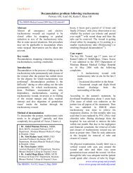
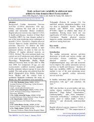
Review Articlegraded compression technique.The fat <strong>and</strong> <strong>the</strong> bowel loops are displaced bycompression. Inflamed appendix appears tobe a thick walled <strong>in</strong>compressible sausage liketube with concentric layers <strong>in</strong> transverseplane . Perifocal oedema is also present. Fluidis also seen <strong>in</strong> <strong>the</strong> lumen <strong>of</strong> appendix. It iselongated tube with one end bl<strong>in</strong>d. When wallthickness exceeds 6 mm it is suggestive <strong>of</strong><strong>in</strong>flammation.Fig. no.4- -Abdomensup<strong>in</strong>e -A triangularcollection <strong>of</strong> free gas <strong>in</strong><strong>the</strong> sub -hepatic regionFig. no.5--A b<strong>and</strong> <strong>of</strong>curvil<strong>in</strong>ear pulmonarycollapse with acrescent <strong>of</strong> normal lungbeneath it- simulat<strong>in</strong>gPseudo -pneumoperitoniumFig. no.l-Shows <strong>in</strong>flamedappendix <strong>in</strong> transverse plane.The ORION Vol. 9, May 2001Fig. no.2-Shows<strong>in</strong>flamed appendix <strong>in</strong>long axis.(B)Pneumoperitonium: Demonstration <strong>of</strong>small pneumoperitonium is very essential.Most <strong>of</strong> <strong>the</strong> cases <strong>the</strong> cause <strong>of</strong> <strong>the</strong>pneumoperitonium will require emergencysurgery.Fig. no.3-(a) Erect chest film- Free gasseen under both domes <strong>of</strong>diaphragm.(b) Abdomen erect ,free gas under tIledomes 6(diaphragm.(C) Pseudopneumoperitonium: A number <strong>of</strong>conditions which simulate pneumoperitoneume.g.-1. Chilaiditis syndrome -Distended bowelbetween liver <strong>and</strong> right. dome <strong>of</strong> diaphragm2. Sub diaphragmatic fat-omental fat3. Curvil<strong>in</strong>ear pulmonary collapse(D)lntest<strong>in</strong>al obstruction: Dilation <strong>of</strong> boweloccurs<strong>in</strong>1. Mechanical <strong>in</strong>test<strong>in</strong>al obstruction 2.Pseudoobstruction 3. Paralytic ileus 4. Airswallow<strong>in</strong>g5. Several o<strong>the</strong>r conditions.Radiological differentiation depends ma<strong>in</strong>lyon <strong>the</strong> size, mucosal appearance <strong>and</strong> <strong>the</strong>distribution <strong>of</strong> loops <strong>of</strong> bowel. The diagnosis<strong>of</strong> <strong>in</strong>test<strong>in</strong>al obstruction depends on <strong>the</strong>demonstration <strong>of</strong> dilated loops <strong>of</strong> bowelproximally with non-dilated or collapsedbowel distal to <strong>the</strong> presumed po<strong>in</strong>t <strong>of</strong>obstruction.(A)Acute gastric dilatation:Dilatation <strong>of</strong> <strong>the</strong> stomach can be caused by 4ma<strong>in</strong> groups <strong>of</strong> conditions--(I)Mechanical gastric outlet obstruction maybe a sequel <strong>of</strong> peptic ulcer or a carc<strong>in</strong>oma <strong>of</strong><strong>the</strong> antrum.(II)Paralytic ileus- This group <strong>of</strong> conditions isfrequently referred to as acute gastricdilatation.(III)Volvulus <strong>and</strong> air swallow<strong>in</strong>g are usuallyuncommonconditions.(B)Small bowel obstruction:Usual causes are <strong>the</strong> b<strong>and</strong>s,adhesions <strong>and</strong>strangulations. Complete obstruction<strong>of</strong> <strong>the</strong> small bowel usually causes small boweldilatation with accumulation <strong>of</strong> both gas <strong>and</strong>fluid <strong>and</strong> a reduction <strong>in</strong> calibre <strong>of</strong> <strong>the</strong> largebowel. Pla<strong>in</strong> film changes <strong>in</strong> small bowelobstruction may appear after 3-5 hours ifwww.orion-group.net/journals
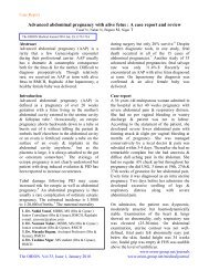
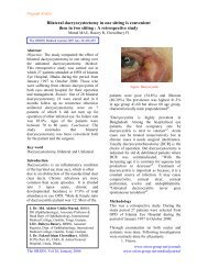
Review Article<strong>the</strong>re is complete small bowel obstruction <strong>and</strong>such changes are usually marked after 12hours. In <strong>in</strong>complete obstruction early filmsmay be normal.This slide shows <strong>the</strong> feature <strong>of</strong> gall stone ileus.Here you see multiple dilated loops <strong>of</strong> smallbowel with a b<strong>and</strong> <strong>of</strong> gas with<strong>in</strong> <strong>the</strong> CBD.Gallstone ileus is a mechanical obstruction causedby <strong>the</strong> impaction <strong>of</strong> one or more gall stones <strong>in</strong><strong>the</strong> <strong>in</strong>test<strong>in</strong>e, usually <strong>in</strong> <strong>the</strong> term<strong>in</strong>al ileum <strong>and</strong>rarely <strong>in</strong> <strong>the</strong> duodenum.Fig, No.9-Gall stoneileus.Slide no.6-Shows acutegastric dilatationThe ORION Vol. 9, May 2001Slide no. 7-ShowsSmall bowelobstruction: Multipledilated loops <strong>and</strong> airfluid level. A verysmall amount <strong>of</strong> gasesare noted <strong>in</strong> <strong>the</strong> largebowel.Differentiat<strong>in</strong>g po<strong>in</strong>ts between a small <strong>and</strong>large bowel obstruction:Str<strong>in</strong>g <strong>of</strong> beads sign:What is that: In dilated small bowel which isalmost completely filled with fluid. Smallbubbles <strong>of</strong> gas may be trapped <strong>in</strong> rowsbetween <strong>the</strong> Volvulae connivanties onhorizontal ray films- this is known as Str<strong>in</strong>g<strong>of</strong> beads sign. This sign if present is virtuallydiagnostic <strong>of</strong> small bowel obstruction.Fig. No.8- -Ano<strong>the</strong>rslide <strong>of</strong> small bowelobstruction show<strong>in</strong>gStr<strong>in</strong>g <strong>of</strong> beads sign.Signs <strong>of</strong> gallstone ileusa) Gas with<strong>in</strong> <strong>the</strong> bile duct b)Features <strong>of</strong> smallbowel obstruction c)Abnormallocation <strong>of</strong>gallstone d)O<strong>the</strong>r previous f<strong>in</strong>d<strong>in</strong>gs whichco<strong>in</strong>cides biliary stone.What are <strong>the</strong> o<strong>the</strong>r causes <strong>of</strong> gas <strong>in</strong> <strong>the</strong> biliarytree:a)Physiological if <strong>the</strong> sph<strong>in</strong>cter is laxed.b)Follow<strong>in</strong>g biliary surgery c)Malignantfistula d)Perforated peptic ulcer <strong>in</strong>to <strong>the</strong> bileduct.Its a case <strong>of</strong> <strong>in</strong>tussusception is seen<strong>in</strong> children under 2 years <strong>of</strong> age. In children itusually commences <strong>in</strong> <strong>the</strong> ileum as <strong>the</strong> result<strong>of</strong> <strong>in</strong>flammation <strong>of</strong> <strong>the</strong> lymphoid tissue <strong>and</strong>tends to be associated with mesentericadenitis. The enlarged lymphatic patches areforced <strong>in</strong>to <strong>the</strong> ileum by peristaltic movement<strong>and</strong> act<strong>in</strong>g as a tumour. So one part <strong>of</strong> <strong>the</strong>ileum is pushed <strong>in</strong>to <strong>the</strong> o<strong>the</strong>r <strong>and</strong> caus<strong>in</strong>g<strong>in</strong>tussusception. In adults <strong>in</strong>tussusception is<strong>in</strong>variably caused by a tumour <strong>of</strong> bowel. Abarium enema exam<strong>in</strong>ation is frequentlyrequired to establish a def<strong>in</strong>ite diagnosis <strong>and</strong>sometimes also relieves <strong>the</strong> patient.Fig. no. 10(a)--Multiple gas filledloops <strong>of</strong> dilated smallbowel <strong>the</strong>re is a s<strong>of</strong>ttissue mass <strong>in</strong> <strong>the</strong> rightiliac fossa,Contrast enema exam<strong>in</strong>ation <strong>of</strong> large gut <strong>of</strong> ababy shows <strong>the</strong> <strong>in</strong>tussusception produc<strong>in</strong>g awww.orion-group.net/journals
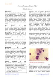
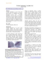
Review Articleconcave defect <strong>in</strong> <strong>the</strong> head <strong>of</strong> <strong>the</strong> contrastcolumn. Sometimes contrast medium maypass beyond this, caus<strong>in</strong>g <strong>the</strong> well knowncoiled spr<strong>in</strong>g appearance.obstruction, <strong>the</strong> radiographic appearance maybe identical to those <strong>of</strong> a paralytic ileus.Paralytic ileus causes when <strong>in</strong>test<strong>in</strong>alperistalsis ceases <strong>and</strong> as a result fluid <strong>and</strong> gasaccumulates <strong>in</strong> <strong>the</strong> dilated bowel. It isfrequently found <strong>in</strong> cases <strong>of</strong> peritonitis <strong>and</strong> <strong>in</strong><strong>the</strong> post operative cases. Along with <strong>the</strong>radiological f<strong>in</strong>d<strong>in</strong>gs, history <strong>and</strong> cl<strong>in</strong>icalsigns are helpful <strong>in</strong> differentiat<strong>in</strong>g betweenlarge bowel obstruction & paralytic ileus.Fig. no.10 (b)(iii) Large bowel obstruction:Usual causes are <strong>the</strong>a) Carc<strong>in</strong>oma -60% is situated <strong>in</strong> <strong>the</strong> sigmoidcolonb) Diverticular disease c)Volvulus <strong>of</strong> <strong>the</strong>colon.Key to <strong>the</strong> radiological appearance <strong>of</strong> <strong>the</strong>large bowel obstructiondepends on <strong>the</strong> state <strong>of</strong> <strong>the</strong> competence <strong>of</strong> <strong>the</strong>Ileocaecal valve. The obstructed colon<strong>in</strong>variably conta<strong>in</strong>s large amounts <strong>of</strong> air <strong>and</strong>can usually be identified by its location <strong>and</strong>by <strong>the</strong> presenc <strong>of</strong> haustral mark<strong>in</strong>gs.When both small <strong>and</strong> large bowel dilatationare present <strong>in</strong> a case <strong>of</strong> large bowelThe ORION Vol. 9, May 2001Fig. no.10 (c)Fig. no.11. Shows hugelydilated large gut. Its a case<strong>of</strong> sigmoid volvulus. Ahaustral loop <strong>of</strong> sigmoid isseen ris<strong>in</strong>g out <strong>the</strong> pelvis <strong>in</strong><strong>the</strong> shape <strong>of</strong> an <strong>in</strong>verted 'U'Dilated haustral colons arepresent on <strong>the</strong> two sides <strong>of</strong><strong>the</strong> volvulus.Fig.no.12--Th/s filmshowsgeneralised dilatatlon <strong>of</strong> both small <strong>and</strong>large bowel <strong>and</strong> its acase <strong>of</strong> paralytic ileus.Acute pacreatitis: Cl<strong>in</strong>ical diagnosis may bedifficult <strong>in</strong> <strong>the</strong> <strong>in</strong>itial stage .O<strong>the</strong>r acuteabdom<strong>in</strong>al conditions such as perforation,acute cholecystitis, acute peptic ulcer have tobe <strong>in</strong>cluded <strong>in</strong> <strong>the</strong> differential diagnosis. Alarge number <strong>of</strong> radiological signs have beendescribed, most <strong>of</strong> <strong>the</strong>m are non-specific.USG<strong>and</strong> CT are <strong>the</strong> diagnostic modality.Fig. no. 13- -Shows <strong>the</strong> transverse scan <strong>of</strong>normal pancreas <strong>and</strong> its duct.The normal pancreas has about <strong>the</strong> sameechogenecity as that <strong>of</strong> liver but itsechogenecity <strong>in</strong>crease with age. When wescan <strong>the</strong> pancreas we usually identify <strong>the</strong>follow<strong>in</strong>g anatomical l<strong>and</strong>marks:1.Aorta2. Superior mesenteric artery3. Superior mesenteric ve<strong>in</strong>4. Liver etc.Average diameter <strong>of</strong> <strong>the</strong> head <strong>of</strong> <strong>the</strong> pancreasis 2.8 cm. body 2 cm <strong>and</strong> tail 2.5cm, ductdiameter less than 2mm.In acute pancreatitis pancreas is diffuselyenlarged <strong>and</strong> oedematous which ishypoechogenic <strong>in</strong> comparison with <strong>the</strong> liverparenchyma. Diffusely enlarged irregularlyhypoechogenic pancreas may also seen <strong>in</strong>www.orion-group.net/journals
Review Articleacute pancreatitis when it is superimposed onchronic pancreatitis. C. T. is <strong>the</strong> <strong>in</strong>vestigation<strong>of</strong> choice for such cases.Fig. no. 14- -Showsdiffusely enlarged<strong>and</strong> oedematousPancreasThe ORION Vol. 9, May 2001www.orion-group.net/journals
Review ArticleThe ORION Vol. 9, May 2001www.orion-group.net/journals