PhD Arthur Decae 2010 - Ghent Ecology - Universiteit Gent
PhD Arthur Decae 2010 - Ghent Ecology - Universiteit Gent
PhD Arthur Decae 2010 - Ghent Ecology - Universiteit Gent

You also want an ePaper? Increase the reach of your titles
YUMPU automatically turns print PDFs into web optimized ePapers that Google loves.
Diversity and distribution of<br />
mygalomorph spiders in the<br />
Mediterranean region.<br />
<strong>Arthur</strong> Emile <strong>Decae</strong>
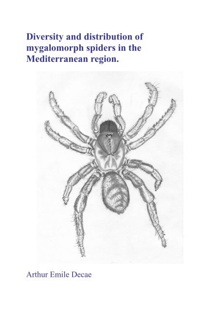
Cover drawing:<br />
Nemesia randa <strong>Decae</strong> 2005 Female, dorsal view. Drawing by the author.
Voor<br />
Jean-Pierre MAELFAIT<br />
Ik heb je veel te kort gekend.<br />
“Almost the whole summer was lost with this agonizing labor. At last on a quite trifling<br />
occasion I came nearer to the truth.”<br />
Johannes Kepler in Mysterium Cosmographicum 1596.<br />
English translation from D.J. Boorstin 1983<br />
The Discoverers Vol. 1
UNIVERSITEIT GENT<br />
Faculteit Wetenschappen<br />
Onderzoeksgroep Terrestrische Ecologie (TEREC)<br />
Diversity and distribution of mygalomorph spiders in the Mediterranean<br />
region.<br />
Diversiteit en verspreiding van mygalomorfe spinnen in het Middellandse Zeegebied.<br />
<strong>Arthur</strong> Emile <strong>Decae</strong><br />
Thesis submitted to the University of <strong>Ghent</strong> in order to acquire the <strong>PhD</strong>-degree in Sciences.<br />
Proefschrift ter verwerving van het doctoraat in de wetenschappen aan de <strong>Universiteit</strong> van<br />
<strong>Gent</strong><br />
Promotor: Prof. Dr. D.Bonte<br />
examencommissie: Dr. R. Jocqué & Prof. Dr. F. Hendrickx<br />
leescommissie: Prof. Dr. J. Mertens, Dr. R. Bosmans, Dr. J. Bosselaers
Contents<br />
(page numbers and running heads present in the hard cover are absent in this<br />
PDF File)<br />
Acknowledgements. 1<br />
Chapter 1 General introduction. 2 - 15<br />
Chapter 2 Trapdoor spiders of the genus Nemesia Audouin, 1826<br />
on Majorca and Ibiza: taxonomy, distribution and behaviour<br />
(Araneae, Mygalomorphae, Nemesiidae). 16 -46<br />
Chapter 3<br />
Chapter 4<br />
Chapter 5<br />
Chapter 6<br />
Chapter 7<br />
Taxonomic Review of the Portuguese Nemesiidae<br />
(Araneae, Mygalomorphae). 47 -68<br />
Iberesia, a new genus of trapdoor spiders (Araneae, Nemesiidae)<br />
from Portugal & Spain. 69 -79<br />
Sub-generic diversity and distribution in the Mediterranean<br />
trapdoor spider genus Nemesia Audouin 1826<br />
(Araneae, Mygalomorphae, Nemesiidae). 80 - 103<br />
The genus Ummidia Thorell, 1875 in the western Mediterranean,<br />
a review (Araneae, Mygalomorphae, Ctenizidae). 104 -119<br />
Systematics of the trapdoor spider genus Cyrtocarenum<br />
Ausserer, 1871 (Araneae, Ctenizidae). 120 - 134<br />
Chapter 8 General conclusions. 135 - 149<br />
Summary/Samenvatting. 150 - 153<br />
Literature. 154 -162<br />
Epilogue. 163
Acknowledgements<br />
Because this thesis consists of a series of papers that have already been published, or that are<br />
submitted for publication, acknowledgements are provided separately with each paper or in<br />
each chapter. The thesis could not have been written however without the help of others not<br />
directly involved in the published material.<br />
My special thanks go to Dr. Rudy Jocqué and the late Prof. Dr. Jean–Pierre Maelfait for their<br />
warm enthusiasm, active help and provision of the conditions in which this project could<br />
succeed. I furthermore thank Jean-Pierre for taking-up the position as my promotor and for<br />
the valuable advice he gave me in the earlier stages of the work. I am also very grateful to<br />
Prof. Dr. Dries Bonte for taking over the difficult task of being my promotor after the tragic<br />
and much too early death of Jean-Pierre and to Prof. Dr. Luc Lens for his friendly help and<br />
willingness to give me a place to work at the TEREC laboratories. I thank the members of the<br />
promotion committees Prof. Dr. D. Bonte, Dr. R. Jocqué, Prof. Dr. F. Hendrickx, Prof. Dr. J.<br />
Mertens, Dr. R. Bosmans and Dr. Jan Bosselaers for their enthusiasm to contribute to my<br />
studies and I thank all my colleague <strong>PhD</strong> students for the friendly reception they provided me<br />
at the TEREC laboratories. I thank the University of <strong>Gent</strong> for accepting me as a student and<br />
for financing the expenses made in order to successfully carry out this thesis work. I am also<br />
greatly indebted to many personal friends and family members for their mental support and<br />
the constant interest they showed in the progression of my spider work. Finally I thank Nollie<br />
Hallensleben, my wife and life’s companion for over forty years, for her help in preparing this<br />
thesis.<br />
A thesis usually marks the beginning of a scientific career and is therefore accompanied by a<br />
curriculum vitae and a list of publications aimed to help the young doctor at a prospective<br />
professional start. That is not the case here. This thesis is written at the end of a professional<br />
career in which science did not play a central role, but was constantly present as a matter of<br />
special interest and useful background to achieve all sorts of practical goals. The motivation<br />
to nevertheless produce this thesis is that I hope to encourage interest in a branch of science<br />
that, after a providential start in the last quarter of the nineteenth century, got bogged down in<br />
its own confusion and the changing interests in the biological sciences. The study of<br />
mygalomorph spiders started off in the Mediterranean where early savants of Natural History<br />
such as J.T. Moggridge, E. Simon, A. Ausserer, O. Pickard-Cambridge, T. Thorell and others<br />
carried out their pioneering research. Their work, building a solid taxonomic basis for<br />
understanding mygalomorph spider diversity in the Mediterranean Region, unfortunately has<br />
never been satisfactorily finished. When in the course of the twentieth century the focus of<br />
biological science shifted from classical Systematics and Taxonomy to more predictive and<br />
process orientated studies, attention for biological description faded and no further progress<br />
was made in the knowledge of the Mediterranean mygalomorph spider fauna. In the<br />
awakening of modern biodiversity studies however, there is a renewed interest in questions of<br />
species identity and identification. Knowledge about how many species there are and which<br />
species occurs where is important again. Due to the unfinished taxonomical work these<br />
questions, when asked in relation to Mediterranean mygalomorph spiders, are very difficult to<br />
answer. Nevertheless, these questions are now asked by a number of young researchers that<br />
live and work in countries bordering the Mediterranean or that are located in the<br />
Mediterranean climate zone. As an older arachnologist I hope to be able to act as a mediator<br />
between the generations providing helpful background information for new studies in<br />
Phylogenetics and Phylogeography. This thesis is therefore dedicated to all these young<br />
researchers whose names feature in the acknowledgements of the here collected papers.<br />
1
Chapter 1<br />
General introduction.<br />
<strong>Arthur</strong> <strong>Decae</strong><br />
August <strong>2010</strong><br />
Front-page illustration. Cladograms of higher classifications of spiders (Order Araneae) Top:<br />
conventional following Platnick & Gertsch 1976. Bottom: here proposed alternative cladogram<br />
based on functional apomorphies. Lateral views of spider bodies, appendages omitted.
Motivation for this thesis<br />
This thesis is written as an effort to contribute basic knowledge to the study of Arachnology,<br />
hoping this will help future research in a branch of biology that I regard as a potentially rich<br />
source of important scientific insight. All papers in this thesis contain original information on<br />
Mediterranean mygalomorph spiders provided in order to facilitate and stimulate more<br />
detailed future research and understanding of these fascinating, yet little known, animals.<br />
Aim<br />
The central goal of this thesis is to gain insight in the historical build-up of regional<br />
mygalomorph diversity in the expectation that this will aid a more general understanding of<br />
growing faunal complexity as it has occurred on earth.<br />
Personal Note<br />
When in 1983 I took my exams for a Master Degree in Biology at the University of<br />
Groningen one of the members in the jury asked me if I did not think that my studies had been<br />
too narrowly focused at just one animal group. I had worked on subjects in taxonomy,<br />
biogeography, ecology, ethology and comparative morphology of sensory organs all in one<br />
genus of mygalomorph spiders; the genus Cyrtocarenum Ausserer 1871. To my relief, and<br />
before I could answer, another member on the committee countered that there would have<br />
been no critical questions on this point if the one animal group had been fruit flies<br />
(Drosophila melanogaster), rats (Rattus norvegicus) or zebra fish (Brachydanio rerio). The<br />
point of course is that fruit flies, rats, zebra fish and a hand full of other animals are widely<br />
considered ideal model organisms for a range of interests in zoological research and that it<br />
remains to be seen if we could learn anything important from mygalomorph spiders. In fact, I<br />
believe we can learn something new and important from indiscriminately which animal we<br />
choose to study, but that the study of mygalomorph spiders offers more.<br />
The study of spiders in general offers more than the study of other animal groups because<br />
spiders are unique in producing a wide range of intricate and seemingly intelligent and/or<br />
geometric constructions made of a combination of material found in nature and self produced<br />
excretions: such as saliva and silk. These constructions, nests, webs, drag lines, cocoons,<br />
trapdoors, wrappings, ties and a number of other functional devices, may be seen as material<br />
expressions of complex behavior that no other animal, save the human species, leaves<br />
behind 1 . To produce their complex constructs spiders of course possess a range of<br />
morphological, physiological and neurological qualities not found in any other animal group<br />
and it is only because spiders are not of any obvious economical or medical interest that<br />
araneology (the study of spiders) is a relatively remote branch of the biological sciences 2 .<br />
This is to be regretted, because spiders not only possess all these unique qualities of direct<br />
biological interest, they also preserve, within their diversity, the reflection of an evolutionary<br />
history that dates back to the beginning of terrestrial animal life (<strong>Decae</strong> 1984). Although the<br />
fossil record of spiders is relatively poor, spiders do not fossilize easily (Selden & Penney<br />
<strong>2010</strong>), much of the history of spiders can be read from extant species. Apparently spiders<br />
have been a marked biological success throughout evolutionary time, surviving in great<br />
1 Beavers, birds, termites, bees and other animals produce amazing constructions in size and<br />
complexity, but rarely as versatile, intricate, geometrical and multifunctional as spiders and<br />
humans do (see e.g. Von Frisch 1974, Hansell 2007).<br />
2 Although properties of spider silk and venom in particular might offer prospects of medical,<br />
agricultural and other applications (e.g. Bailey & Chada 1968, Sterling et. al 1992, Patrick &<br />
Canard 1997, Novak 2001, Scheller et. al. 2001, Escoubas & Rash 2004) it seems to be very<br />
difficult to economically exploit spider products.
diversity (Penney et. al. 2003) all cataclysmic mass extinctions that mark the divisions of the<br />
geological timescale and that have punctuated the histories of many animal groups. There is<br />
no clear evidence of major evolutionary bottle necks 3 in the contemporary spider fauna. Three<br />
major lineages all dating far back in geological time have living representatives (see frontpage<br />
illustration). The primordial Mesothelae have 5 known genera and 87 species in eastern<br />
Asia, the primitive Mygalomorphae have 320 genera and 2643 known species distributed on<br />
all continents, being absent only from circum polar regions, and the Araneomorphae finally<br />
with 3777 genera and 38523 species 4 have an even wider cosmopolitan distribution.<br />
Moreover, in their degree of evolutionary development the Araneomorphae contain both<br />
rather generalized forms such as the lampshade web spiders 5 (Hypochilidae), the tube web<br />
spiders (Segestriidae) and the daddy-long-legs spiders (Pholcidae) and superbly specialized<br />
lineages such as the orb weavers (Araneidae), the wolf spiders (Lycosidae) and the jumping<br />
spiders (Salticidae). Paleontology has shown that many extant spider families can be traced<br />
back in time deep into the Mesozoic era and some Mesothelae even into Paleozoic times<br />
(Selden & Penney <strong>2010</strong>). This means that a well developed phylogenetic tree of spiders would<br />
provide a rare telescopic view into the evolutionary history of a major animal group. Over the<br />
passed decades arachnologists have greatly advanced in the reconstruction of this tree but<br />
advances have been biased towards the more advanced Araneomorphae whereas knowledge<br />
of the primitive Orthognatha (Mesothelae + Mygalomorphae) is relatively underdeveloped<br />
(Coddington 2005). This is to be regretted again because precisely the living existence of<br />
these primitive spiders in great abundance and diversity alongside advanced forms give<br />
spiders their unmatched evolutionary scope. Moreover, the very effective mechanism for<br />
aerial dispersal (ballooning), characteristic for most araneomorph spiders, is lacking in most<br />
orthognathe lineages (Main 1976, Coyle 1985b). This means that whereas araneomorph<br />
distributions (at lower taxonomic levels) are generally to be understood in terms of ecological<br />
opportunity and capacity for dispersal, orthognathe spiders tend to be ‘locked-up’ in their<br />
centers of origin as distinct local endemics. This special character of orthognathe spiders<br />
means that our telescopic view of spider evolution can be focused in space as well as in time<br />
and that primitive spiders have outstanding qualities for studies in historical biogeography and<br />
other aspects of evolutionary biology.<br />
To conclude this motivation for working on mygalomorph spiders I’ll turn back to my earlier<br />
studies at the University of Groningen, where after graduation, I successfully applied for a<br />
three year grant to study the ecology and behavior of Neotropical mygalomorph spiders at the<br />
Smithsonian Tropical Research Institute (STRI) in Panama. My supervisor at STRI, the late<br />
Dr. Mike Robinson, remarked on my arrival that he was sure mygalomorphs were present in<br />
the area but that, except for the large bird-spiders (Theraphosidae), virtually nothing was<br />
known about their whereabouts and their identities. A study of ecology and behavior<br />
necessarily had to be preceded by a study in mygalomorph taxonomy if future results were to<br />
be comprehensively communicated to the scientific community. In a nut shell this is the<br />
situation that students of mygalomorphs still encounter today. In most parts of the world little<br />
can be studied on mygalomorphs until their taxonomy is reasonably resolved. The problem is<br />
that studies of mygalomorph taxonomy have a reputation of being difficult and frustrating<br />
3 Although the Permian-Triassic extinction event might have seriously affected spider<br />
diversity and modern cladograms (e.g. Penney et. al 2003 Fig. 1) suggest that most spider<br />
radiation occurred after the P-T event.<br />
4 As a result of new discoveries of species and genera and changing insights following<br />
taxonomical revisions these numbers constantly change. The numbers given here follow the<br />
World Spider Catalog version 10.5 (Platnick <strong>2010</strong>).<br />
5 Vernacular names follow Jocqué & Dippenaar-Schoeman 2006
(Coddington 2005) and therefore lags behind other developments in biological knowledge.<br />
The rewards of studying mygalomorphs however will be in keeping with the efforts and a<br />
much improved vista on the evolutionary history of a major animal group can thus be<br />
obtained.<br />
Outline and objectives<br />
The Mediterranean mygalomorph fauna, as it is presently known, contains representatives of<br />
seven different spider families, eleven genera and approximately 120 species (see Chapter 8<br />
general conclusions). Current knowledge of Mediterranean Mygalomorphae largely rests on<br />
old and often incoherent information that has accumulated over the past 200 years. However<br />
all families and genera need thorough taxonomical revision before deeper biological insight<br />
can be developed. This elementary though necessary work is beyond the scope of this thesis.<br />
However two genera − Ummidia Thorell 1875 and Cyrtocarenum Ausserer 1871 − have been<br />
fully revised on all material available (Chapters 6 & 7) and the complex and diverse genus<br />
Nemesia Audouin 1826 has been revised for local geographic regions (the Balearic Islands in<br />
Chapter 2 and Portugal in Chapter 3). To structure future research on the Mediterranean<br />
Nemesiidae a genus level revision was carried out (Chapter 4) resulting in the splitting-off of<br />
a new genus (Iberesia <strong>Decae</strong> & Cardoso 2005) from traditional Nemesia stock, and the<br />
establishment of two subgenera (Pronemesia and Holonemesia) within Nemesia (Chapter 5).<br />
Within both Pronemesia and Holonemesia several distinct species-groups are recognized and<br />
discussed (Chapter 5). Work on the remaining mygalomorph genera occurring in the<br />
Mediterranean Region −Atypus, Brachythele, Chaetopelma, Cteniza, Cyrtauchenius,<br />
Ischnocolus, Macrothele and Idops− is restricted to descriptions of their species diversity and<br />
Mediterranean distributions. The probable regions of origin for all these genera and their<br />
affinities to related genera outside the Mediterranean are discussed in Chapter 8.<br />
The central object of study is the Mediterranean mygalomorph spider fauna. To provide an<br />
informative backdrop for understanding the arachnological information contained in this work<br />
the history and biological importance of the Mediterranean Region as an area of high<br />
biodiversity will be described below separate from an exposé on mygalomorph spiders. The<br />
general background information on Mygalomorphae and their presumed sister-group the<br />
Liphistiomorphae is intended to shed a new and original light on spider phylogeny and spider<br />
evolution that departs from conventional arachnological views.<br />
The Mediterranean Region<br />
Why study the Mediterranean<br />
Obviously there are many very good answers to this question depending on whatever your<br />
line of interest is. One very good reason is that, for people, the Mediterranean is a very<br />
pleasant place to be. This is not only evidenced by the huge success of modern mass tourism<br />
in the region, it is also evidenced by early Homo erectus visitors coming straight from Africa.<br />
H. erectus settled in the Mediterranean at least half a million years ago (Dennel & Roebroeks<br />
1994, Turner 1999) and since that early period mankind has never left the beautiful places that<br />
surround the bluest of all seas. Within the Mediterranean Region humans developed their<br />
incredible success as a biological species. They started off as small tribes equipped with<br />
stones and sticks looking for whatever nature would provide, but in the benign environment of<br />
the Mediterranean they became farmers, citizens, soldiers, empire builders, discoverers of the<br />
world and of the universe. For the rise of human civilization the Mediterranean certainly is a<br />
‘hotspot’.
Recently the Mediterranean has acquired another qualification for being a hotspot. Within the<br />
field of biological conservation the Mediterranean is proclaimed one of the world’s hotspots<br />
of biodiversity (Meyers et.al. 2000). This qualification is used by the International Union for<br />
the Conservation of Nature (IUCN) in order to focus human attention on the impoverishing<br />
effects on nature of unbound exploitation of the environment. The Mediterranean, as a source<br />
of natural wealth and beauty, is of special significance because of its unique evolutionary<br />
history that makes it one of the richest biological regions outside the tropics (Naveh 2007).<br />
Moreover the Mediterranean flora and fauna contains a very high proportion of endemic<br />
species, species that only occur in the Mediterranean, which gives the region the status of<br />
being of critical importance for nature conservation 6 . Present knowledge of Mediterranean<br />
biodiversity however is based on only a very thin slice of the local flora and fauna. It is<br />
mainly focused on vascular plants, vertebrates and very few invertebrate groups 7 . Very little<br />
information is available for the bulk of smaller animals and plants that actually build the<br />
world’s biodiversity. This means that, from a biodiversity point of view, there is much to<br />
discover in and around the Mediterranean, providing numerous outstandingly good reasons to<br />
study the region.<br />
A first question coming-up from the biodiversity perspective would be; where does it come<br />
from Why is the Mediterranean so rich in endemic and other species of plants and animals<br />
To answer that question we will have to go far back in time, long before the first human<br />
settlements in the region, to the birth of the Mediterranean itself and its subsequent history. To<br />
do that, we will have to turn to geology and geography and we may start with a map of the<br />
world. The map of the world, as we all know it (Fig. 1), is only a reflection of the current<br />
coastlines and the distribution of land and water over the surface of the planet. Since a number<br />
of scientific discoveries and realizations, settling in human conscience over the past 250<br />
years 8 , we now know that the map of the world is changing continuously and has changed<br />
dramatically over millions of years of geological time. The main features of geography, such<br />
as oceans, seas, continents and mountain ranges have not always been where we see them<br />
today, but they have come and gone in different configurations. This shifting of continents<br />
and oceans has had major effects on the evolution of plant- and animal species. Evolutionary<br />
processes have broadly followed the dynamics in the earth’s crust to produce the regional<br />
florae and faunae of the different continents, including that of the Mediterranean.<br />
In connection to the here presented studies of Mediterranean mygalomorph spiders this idea<br />
of related shifts in geography and faunal composition led to asking three fundamental<br />
questions: (1) what is the origin of the Mediterranean, (2) why does it have such a high<br />
species diversity, and (3) how did it get its mygalomorph spider fauna<br />
Fig. 1 Familiar map of the World<br />
showing existing coastlines and the<br />
distribution of land and water over<br />
the earth’s surface. Source: Google<br />
maps of the world.<br />
6 For actual numbers see www.biodiversity/hotspots.org/...<br />
7 See IUCN, Red List on: www.iucn.org/redlist/<br />
8 Discoveries of (1) ‘deep time’ (Hutton 1788, Lyell 1830-1833), (2) evolution by natural<br />
selection (Darwin 1859), (3) continental drift (Wegener 1916), (4) seafloor spreading (Hess<br />
1954, 1960).
Origin of the Mediterranean<br />
Questions of origin are always difficult to approach because we have to find a starting point.<br />
In the case of the Mediterranean this is particularly difficult, but we might start from the idea<br />
that the forces that broke-up the ancient super continent Pangaea, some 170 million years ago,<br />
led to the formation of the separate ‘continental plates’ that much later in time, were to form<br />
the Mediterranean. This formation of the Mediterranean finally took place in a gigantic<br />
‘tombola’ of collisions and segregations of fractions of the earths crust (see footnote 7 for<br />
discoveries and discoverers of continental drift and seafloor spreading). The main processes to<br />
be considered here are: (1) the opening of the North Atlantic Ocean and the vanishing of the<br />
ancient Tethys Ocean, (2) the movements of the African and Arabian continental plates, (3)<br />
birth of the Mediterranean Sea. For information on these phenomena I lean heavily on the<br />
following sources: D. Ager’s (1980) celebrated book The Geology of Europe; Rögl &<br />
Steiniger’s (1984) Chapter 10 in Fossils & Climate (Wiley Books) and Rögl’s (1999) short<br />
discussion of the Mediterranean Palaeogeography in Geologica Carpatica.<br />
1. The Atlantic and Tethys Oceans<br />
The formation of the Atlantic Ocean started<br />
near the equator with cracking open the<br />
solid continental block of Pangaea, roughly<br />
at a place where we now find the Caribbean.<br />
A subsequent rapture moved from there to<br />
the southwest to separate North and South<br />
America. A second giant rapture moved<br />
northeast to separate North America from<br />
Africa and Europe. In the process southern<br />
Europe was broken-up in a series of microplates<br />
that formed an archipelago within the<br />
northwestern Tethys Ocean (Fig. 2). It all<br />
must have happened early in Jurassic times,<br />
approximately 170 million years ago<br />
(Pannekoek 1973), when giant dinosaurs<br />
roamed the earth. The rapture of Pangaea<br />
separated the northern continents from one<br />
large southern continent that is here denoted<br />
as Neogondwana 9 . To the east the young<br />
Atlantic Ocean had a seaway connection with<br />
the western parts of the ancient Tethys Ocean.<br />
This inter-oceanic seaway is the location where<br />
the Mediterranean was going to be formed. In<br />
time the Tethys Ocean would be replaced by the<br />
Indian Ocean (late Mesozoic to mid Cenozoic)<br />
Fig. 2 Showing the young Atlantic Ocean (approx.<br />
150myBP) separating the northern from the still<br />
united southern continents and the seaway<br />
connection with the western Tethys. Source:<br />
R.Blakey.ucc.nau.edu/<br />
through the northward movement of the Indo-Australian plate and the rotation of the Arabian<br />
plate away from Africa to close the Atlantic-Tethys connection so shaping the Mediterranean.<br />
9 An earlier separation between northern and southern continental blocks broadly denoted as<br />
Laurasia and Gondwana had existed in the Palaeozoicum (400myBP-350myBP) before the<br />
formation of Pangaea, hence the term Neogondwana for the southern continental block that<br />
originated from the breakdown of Pangaea.
2. The African-Arabic Continent<br />
Around 120myBP the African-Arabian<br />
plate broke away from the great southern<br />
continent of Neogondwana and drifted<br />
towards the northeast ever closer to the<br />
European archipelago. These movements<br />
of the African-Arabian Plate in the late<br />
Eocene and early Oligocene made the<br />
old Tethys Ocean finally disappear<br />
between 35myBP and 25myBP. In the<br />
western parts of the largely shallow<br />
seaway that remained, the Mediterranean<br />
was gradually taking shape. The<br />
Neoeuropean 10 archipelago was pushedup<br />
against the Mesoeuropean mainland<br />
and the formation of the modern<br />
European continent slowly took shape.<br />
To the south, the continuous stable and<br />
passive continental front of Africa<br />
contrasts sharply with the fragmented<br />
archipelago of Neoeurope along the northern<br />
shores of the Proto-Mediterranean (Fig.3). A<br />
following anti-clockwise rotation of Arabia away<br />
from Africa was going to terminate the last<br />
remnants of the Tethys Ocean. To the north of the<br />
Proto-Mediterranean the Paratethys became a<br />
more and more isolated epicontinental sea in<br />
Central and eastern Europe and in western Asia.<br />
Fig. 3 Proto-Mediterranean and Paratethys<br />
developing during late Oligocene- early Miocene<br />
times (25myBP-15myBP). Source:<br />
R.Blakey.ucc.nau.edu/<br />
3. Birth of the Mediterranean Sea<br />
The final formation of the Mediterranean Sea took shape in the Miocene between<br />
approximately 20myBP and 10myBP. The rotation of the Arabian plate more and more closed<br />
off the connection of the eastern Mediterranean with the former Tethys Ocean, and Africa<br />
pushing-up in the West formed a separate western Mediterranean Basin surrounded by rising<br />
Alpine mountain ranges. Within this western zone the former islands of the Maghreb, the<br />
Betic micro-plate, Iberia and the Tyrrhenian micro-plate (including most of what is now the<br />
Italian mainland) formed a separate geographical unit. In the Northeast continental microplates<br />
that had existed as islands in the eastern Neoeuropean Archipellago (Taurus, Pindos,<br />
Adriatica, Pannonica) were pushed against southwestern Asia reducing the Paratethys to a few<br />
isolated basins that were to form the Black Sea, Caspian Sea and other smaller basins (Fig. 4).<br />
Connections between the Paratethys and the Mediterranean and between the Mediterranean<br />
and the Atlantic Ocean were closed. In the course of the Miocene this would change. The<br />
disappearing shallow seaway to the East would finally shut off the Mediterranean from the<br />
10 Terminology from Ager 1980, the Geology of Europe, who distinguishes between<br />
Eoeurope in the far north, Palaeoeurope in Scandinavia and the British Isles, Mesoeuropes for<br />
the Varischian effected parts of Central Europe and Neoeurope for the largely southern Alpine<br />
regions.
Indian Ocean and the Strait of Gibraltar would open (probably periodically) to connect the<br />
western Mediterranean with the Atlantic again. The western Mediterranean in this period was<br />
a distinct sea from the eastern Mediterranean. Opening of the Strait of Sicily in time would<br />
renew the connection between the eastern and western parts of the Mediterranean Basin. The<br />
familiar geography of the region is now becoming visible although the major peninsulas and<br />
archipelagos as well as the connection between the Black Sea (Bosporus – Dardanelle<br />
connection) still had to develop (Fig. 4).<br />
Fig. 4 Mediterranean in the early Miocene (approx. 20myBP). The western Mediterranean is separated<br />
from the eastern Mediterranean, The Paratethyis is reduced to isolated basins and the European<br />
Continent is taking its final shape. Source: R.Blakey.ucc.nau.edu/<br />
Mediterranean Biodiversity<br />
What is the connection between the above exposé on plate tectonics and the high biodiversity<br />
in the Mediterranean The geological history sketched-out here shows that the Mediterranean<br />
was formed at the crossroads of the African, Asian and European continents (Fig. 3). So, parts<br />
of the native florae and faunae of these three continents are expected to have met and mixed<br />
within the Mediterranean leading to an increase of species richness. Moreover one of the three<br />
contributing continents, Europe, had existed as an extensive archipelago of larger and smaller<br />
islands for many millions of years. In this archipelago the evolution of island endemics must<br />
have been high and the contribution from Europe to the later Mediterranean flora and fauna<br />
must have been particularly rich. This reasoning however only relates to terrestrial<br />
biodiversity. The repeated opening and closing of seaways and the constant formation of<br />
separate aquatic basins both in the Mediterranean and the Paratethys must have had further<br />
effects of the enrichment on the marine flora and fauna of the Mediterranean. These basic<br />
conditions for the development of a particularly rich biodiversity in the Mediterranean have<br />
prevailed throughout the geological formation of the region to our present time. And there is<br />
even more than the historical high diversity of terrestrial and marine biomes in the
Mediterranean. Recently a very diverse deep-sea fauna was discovered that is believed to<br />
have originated from special conditions of fragmentation and isolation of local deep water<br />
holes that pertained oceanic conditions during the Messinian salinity crisis. The Messinian<br />
salinity crisis has been a major event (6myBP) in the history of the Mediterranean in which<br />
the Atlantic connection was blocked and much of the sea bottom fell dry (Danovaro et.al<br />
<strong>2010</strong>). Another instant of severe fragmentation and isolation of local elements of flora and<br />
fauna has occurred repeatedly in the course of numerous Pleistocene glaciations (Médial &<br />
Diadema 2009). During the height of these glaciations, when the permafrost covered most of<br />
Europe and the permafrost front reached the Alps, the particularly heterogenic habitat of the<br />
Mediterranean produced local refuge zones in which species could survive. Again the<br />
repeated fragmentation and isolation of populations is expected to have produced increasing<br />
biodiversity. Finally the present flora and fauna of the Mediterranean still survives in, and is<br />
adapted to the small scale heterogeneity that is typical for the Mediterranean environment,<br />
where soil depth, micro-relief, availability of moisture, nutrients, rocky outcrops, tree cover,<br />
litter cover and shade are all patchily distributed (Rundel 1998). All this has attributed to the<br />
very high species diversity in the Mediterranean that needs to be studied before too severe<br />
destruction overcomes the natural resilience of the local flora and fauna. In conclusion, the<br />
rich species diversity of the Mediterranean can not be fully understood in terms of prevalent<br />
ecological conditions, it must be understood in the context of complex historical and<br />
environmental factors that go back deep into geological time. The history of Mediterranean<br />
biodiversity is expected to be reflected in the diversity and distributions of smaller species<br />
with strong qualities for survival and a limited capacity for dispersal. Such species are<br />
particularly common among mygalomorph spiders.<br />
Arachnology<br />
Arachnology is the scientific study of spiders and spider-like animals. Spiders are descendents<br />
of an ancient animal group, the Chelicerata, traces of which are found as fossils in rocks<br />
dating back to the Cambrian Period (>500myBP). This is the period when recognizable<br />
animals appear for the first time in nature (Meglitsch 1972). At that time all animals were<br />
aquatic in habit and no complex forms of life occurred on the land at all. Still fully aquatic,<br />
during the Silurian period (430-413myBP), the Chelicerata had diversified in several animal<br />
groups, one of which was the Order Arachnida. These early arachnids might well have been<br />
among the very first animals that invaded the land (<strong>Decae</strong> 1984). Such an invasion of animal<br />
groups into a new and pristine environment promotes adaptive radiation and the arachnids<br />
showed strong diversification probably leading to the origin of all fifteen taxa we presently<br />
recognize (Table 1). Although we have no Paleozoic records for the small and fragile bodied<br />
Schizomida and Palpigradi the major radiation of arachnids is likely to have occurred in the<br />
Devonian and Carboniferous periods (Dunlop & Selden 2009). In the course of evolutionary<br />
history only four arachnid Orders became extinct and eleven Orders are still alive today<br />
(Table 1).<br />
Because the Arachnida were early colonizers of the terrestrial environment it is only to be<br />
expected that initially they were adapted to sheltered habitat situations where risks of<br />
desiccation were minimized. In fact, nearly all arachnids that have survived the eons of<br />
geological time are still adapted to very sheltered habitat situations. They survive in cracks<br />
and crevices only to venture out at under conditions of high humidity or darkness. Many<br />
occur in caves, under litter, moss, stones, logs or rubbish, and only very few have adapted to<br />
exposed living conditions. True spiders, such as the araneomorph orb-web weavers<br />
(Araneidae), jumping spiders (Salticidae), wolf spiders (Lycosidae) and a few other groups<br />
are aberrant arachnids in a sense that they have adapted to living in exposed conditions, often<br />
in bright sun shine and hot climates. Also among spiders, these exposed living groups are the
most seen and studied groups, but actually they represent the exception rather than the rule. It<br />
is insufficiently realized by students of spiders (arachnologists) that the most conspicuous<br />
spiders naturally draw most of the attention and that much hypothesizing and theorizing in<br />
Arachnology is biased to explaining the ways of these ‘aberrant’ spiders. Most spider species,<br />
as virtually all their arachnid relatives, are nocturnal and secretive creatures that live in hidden<br />
and sheltered positions and that, because of this, have attracted little attention from zoologists.<br />
Following conventional taxonomical wisdom the Order Araneae (spiders) contains two<br />
Suborders: Mesothelae and Opisthothelae. The Mesothelae contain only one family of living<br />
spiders: Liphistiidae. The Opisthothelae are further subdivided into two Infraorders (see frontpage<br />
illustration): Mygalomorphae and Araneomorphae (Platnick & Gertsch 1976). Only the<br />
Mygalomorphae are of further concern here.<br />
Mygalomorphae<br />
Mygalomorph spiders are a prime example of a group of understudied secretive living<br />
arachnids. Nevertheless, the mygalomorph spiders are a major group of animals in themselves<br />
(320 genera, 2643 species known). They occur in most terrestrial and some semi-aquatic<br />
environments on all continents except Antarctica. In many habitats they are among the<br />
dominant predators of ground living arthropods and some species at least incidentally prey on<br />
other animal groups including small vertebrates (Raven <strong>2010</strong>). Mygalomorphae are primitive<br />
spiders that have retained many ancestral traits including their way of living. Just as in the<br />
true spiders of the Infraorder Araneomorphae however, some successful diversification has<br />
taken place within the Mygalomorphae. The large bird-spiders of the family Theraphosidae<br />
are a fine example. Some species in this family that have adopted a more actively hunting and<br />
wandering way of life, have been very successful. Their biological success is reflected in the<br />
highest known species diversity among mygalomorph spiders and an almost worldwide<br />
distribution in the warmer climate zones.<br />
Table1. List of Arachnida taxa, with their first appearance in the fossil record and their status of being,<br />
living or extinct (after Dunlop & Selden 2009). Note the Paleozoic origin of all taxa except<br />
Schizomida and Palpigradi. Fossil traces of these two taxa of small and fragile bodied animals have<br />
not as yet turned up in Paleozoic rocks.<br />
TAXON OLDEST RECORD GEOLOGICAL PERIOD STATUS<br />
myBP<br />
1 Scorpiones 428 Silurian Living<br />
2 Trichonotarbida 419 Silurian Extinct<br />
3 Phalangiotarbida 411 Devonian Extinct<br />
4 Opiliones 410 Devonian Living<br />
5 Acari 410 Devonian Living<br />
6 Pseudoscorpiones 392 Devonian Living<br />
7 Uraraneida 392 Devonian Extinct<br />
8 Ricinulei 319 Carboniferous Living<br />
9 Thelyphonida 319 Carboniferous Living<br />
10 Haptopoda 312 Carboniferous Extinct<br />
11 Araneae 312 Carboniferous Living<br />
12 Amblypygi 312 Carboniferous Living<br />
13 Solifigae 308 Carboniferous Living<br />
14 Schizomida 34 Oligocene Living<br />
15 Palpigradi 5 Pliocene Living
The funnel-web spiders of the families Dipluridae and Hexathelidae may also be regarded as<br />
advanced mygalomorph spiders in their adopted life strategies 11 . They build elaborated webs<br />
for capturing prey and in this respect closely resemble some of the ‘true spiders’ (e.g.<br />
members of the family Agelenidae).<br />
Most mygalomorph spiders however have retained an ancestral lifestyle that they share with<br />
the truly primitive Liphistiomorphae (the segmented trapdoor spiders of SE Asia). The central<br />
aspect of this ancestral lifestyle is the construction of tunnels in which the spider spends most,<br />
if not all of its life. Because individual mygalomorph spiders may live for many years their<br />
burrows need to be solid and stable structures. Usually the burrow tunnels are dug in the<br />
ground (occasionally in rotting logs), in few species they exist as tunnel shaped extensions,<br />
socks or cells on rock faces or tree trunks. Because we cannot travel back in geological time<br />
we can never be sure that tunneling actually is the ancestral lifestyle of all spiders and the best<br />
evidence to prove it is when we can collect sufficient circumstantial evidence. A good piece<br />
of circumstantial evidence would be if tunneling was found to be the habit among the majority<br />
of primitive Orthognatha (Mygalomorphae + Mesothelae). And actually it is.<br />
Currently 16 families of orthognate spiders are recognized; one family in the Mesothelae and<br />
fifteen families in the Mygalomorphae (Table 2). At least thirteen of these families (>80%)<br />
have species that dig holes in the ground. Three of these families (Table 2) are not specially<br />
noted for the burrows they dig. In eleven families of orthognate spiders however, burrow<br />
construction is the dominant lifestyle. Actually the great majority of the species included in<br />
these families are obligate burrowers. In other words, excavating burrows in the ground is by<br />
far the majority lifestyle of primitive spiders living today and this alone should serve as strong<br />
circumstantial evidence to defend the proposition that burrowing is the ancestral lifestyle for<br />
all spiders. The fact that this burrowing lifestyle is ancestral for all spiders is insufficiently<br />
realized in Arachnology. This is the basis of much misconception in attempts to understand<br />
spider evolution. The prime misconception originates from interpretations of the function of<br />
the orthognate chelicerae, the abdominal position of the spinnerets, the compact build with<br />
short strong legs of Orthognatha (particularly the rear two pairs of legs) and the function of<br />
the remarkable narrow and flexible pedicel that is present in all spiders. Interpretations of<br />
these structures in arachnological literature are usually strict morphological and not<br />
functional. Nevertheless understanding the adaptive function of this typical orthognate spider<br />
morphology has major consequences for our view on the phylogeny of spiders in general. The<br />
questions of why do primitive spiders have orthognate chelicerae, why are spinnerets located<br />
on the abdomen, why do Orthognatha have stubby legs and why do all spiders have this<br />
vulnerable narrow stalk, the pedicel, to connect their two bulky body parts, are rarely asked.<br />
The reason is I think psychological; it is because our conception of nature is shaped by our<br />
experience, perception and interest, and since we are predominantly visual animals our<br />
experience, perception and interests are primarily fed by the things we readily see. Orthognate<br />
spiders do not belong to those objects we readily see alive, we only find them dead in<br />
museum collections and therefore very few arachnologists have ever looked at primitive<br />
spiders beyond the things visible in dead specimens; static morphology.<br />
If a civil engineer is asked to develop a machine for tunneling in the earth he/she will comeup<br />
with a machine as shown in Fig. 1. The machine shown is called a TBM (tunnel boring<br />
machine). The essence of its construction is that it has a strong cutting and scraping device up<br />
front, a compact body, a mechanism for grip on the tunnel wall that allows it to be driven<br />
11 Mecicobothriidae and/or Microstigmatidae may also fall in this category, although little is<br />
known of their ways of life.
forward with great force, and a facility for stabilizing the tunnel walls at its rear end (if it is<br />
tunneling in soft rocks). Now if we look at a mygalomorph spider through the eyes of a civil<br />
engineer we would recognize the perfect TBM (Fig. 2). The only difference between a<br />
mechanical TBM and an orthognate spider is the absence of a conveyor belt to remove the<br />
dug-out material. The problem of soil removal from the tunnel under construction is solved<br />
differently in spiders. The key solution to this problem is that spiders have a particularly<br />
narrow and extremely flexible waist, the pedicel that allows it to pivot in the narrow shaft of<br />
its tunnel and to carry-out particles of dug-out soil.<br />
Observation of a trapdoor spider<br />
excavating its burrow immediately<br />
shows the prime functions of the<br />
orthognate chelicerae (scraping,<br />
digging and carrying soil), the stubby<br />
legs (strong grip on the burrow wall),<br />
abdominal spinnerets (stabilizing the<br />
tunnel behind the spider) and the<br />
narrow pedicel (allowing the spider to<br />
pivot for soil removal).<br />
If we look at spiders in this way it is<br />
clear that they differ very much from<br />
their putative sister group the<br />
Amblypygi. The forward orientation<br />
of the chelicerae may superficially be<br />
the same in orthognate spiders and<br />
the Pedipalpi (Amblypygi + Uropygi<br />
+ Schizomida. Harvey 2003) their<br />
function is fundamentally different.<br />
None of the Pedipalpi use their<br />
chelicerae for digging, in orthognate<br />
spiders however, digging is done solely<br />
with the aid of the fangs and chelicerae.<br />
Comparison of the morphology of the<br />
two hind pairs of legs in orthognate<br />
spiders and the Pedipalpi reveals that in<br />
spiders these legs are short and very<br />
strong, against being quite slender to<br />
very slender in the Pedipalpi. The<br />
pedicel is narrow as well in Pedipalpi<br />
as in orthognate spiders, presumably<br />
because all these animals have to move<br />
in the confines of cracks and crevices,<br />
but although there are no comparative<br />
Fig. 1 Tunnel Boring Machine (TBM). One of many models,<br />
all of basically similar design, offered by construction<br />
companies on the internet for infrastructural tunnel drilling<br />
projects.<br />
Fig. 2 Atypus affinis depicted as a TBM.<br />
data that I know of, I would predict that the pedicel of spiders is much more flexible than that<br />
of the Pedipalpi. Finally, spiders are the only creatures on earth that have developed an<br />
abdominal spinning apparatus, working with extreme precision and ideally suited and placed<br />
for stabilizing loose soil during construction work. Coyle (1981) has studied and described all<br />
the above mentioned aspects of burrow construction behavior for Ummidia in detail. My<br />
personal observations confirm that Nemesia, Iberesia, Cteniza, Cyrtauchenius and<br />
Cyrtocarenum all construct their burrows in an identical manner as is reported for Ummidia.
Coyle (1981) also negates the common misconception that the front legs of the Ctenizidae are<br />
equipped with ‘digging spines’. He shows that the front legs of Ummidia play no role in<br />
digging and the same is true for Cteniza and Cyrtocarenum. The strong short spines on the<br />
distal parts of the front legs and palps function in prey capture, rather like the sharp conical<br />
teeth of sharks and crocodiles that prevent prey from escaping.<br />
If we look at spiders as originally tunneling animals the current phylogeny in which<br />
Mygalomorphae is placed as the sister group of Araneomorphae (Platnick & Gertsch 1976)<br />
appears unlikely. An alternative cladogram, as earlier proposed by Bristowe (1933) seems to<br />
be more in accordance to reality (see front-page illustration bottom). The specially digging<br />
adapted chelicerae of the Liphistiidae and Mygalomorphae appear to be a perfect functional<br />
synapomorphy for a Suborder Orthognatha as well as the strong and often specially for lock<br />
and grip adapted legs III and IV. This view on the basic phylogeny of the Araneae would<br />
bring Goloboff’s (1993) remarkable decision for using Mesothelae (instead of the cladistically<br />
established sister-group Araneomorphae 12 ) for rooting his Reanalysis of Mygalomorph Spider<br />
Families in an enlightening perspective. The here described ideas on spider phylogeny would<br />
also provide a more comprehensible view on spider evolution in general in which it is seen as<br />
the result of two highly successful waves of adaptive radiation; one in two dimensional space<br />
(Orthognatha) and one in three dimensional space (Labidognatha).<br />
As described above, mygalomorph spiders are secretively living animals. Although some<br />
species build elaborate webs in very visible locations (e.g. Macrothele calpeiana in southern<br />
Spain), others hide either in crevices or under stones. The majority of Mediterranean<br />
Mygalomorphae however are trapdoor spiders that inhabit self dug burrows up to 30cm deep<br />
in the ground. The entrances of the burrows are usually closed by a hinged door that is often<br />
perfectly camouflaged. The study of trapdoor spiders therefore needs field experience. The<br />
best guide here is Moggridge’s (1873, 1874) book Harvesting Ants and Trapdoor Spiders.<br />
Although the book is somewhat antiquated it perfectly explains the way in which trapdoor<br />
spiders can be best studied in the field. Also it shows the critical importance of looking further<br />
than plain morphology for understanding Arachnology and illuminates, in superb plates and<br />
figures the amazing works of tunneling and underground construction works of trapdoor<br />
spiders. As for the distinct morphology of mygalomorph spiders I can refer to numerous<br />
general text books in Arachnology (reference to some of which are to be found in the section<br />
Literature) as well as various internet sites. Here only the main points of identification are<br />
given.<br />
Distinguishing Mygalomorphae from Araneomorphae<br />
Mygalomorph spiders constitute a primitive branch of the spider family tree that is<br />
distinguished from the true spiders (araneomorph spiders) by a number of conspicuous<br />
morphological characters (Fig. 3). The main external differences between mygalomorph<br />
spiders and araneomorph spiders are found in the anatomy of the biting organs, in the<br />
organization of the respiratory organs and in the development of the spinning organs (Fig. 3).<br />
The biting organs (chelicerae) in mygalomorph spider are orientated forward and act in a<br />
parallel fashion whereas the chelicerae of araneomorph spiders project downward and act in<br />
opposition as in pincers (Fig. 3).<br />
12 Araneomorphae are cladistically indicated to be the sister group of Mygalomorphae<br />
(Platnick & Gertsch 1976), nevertheless, and somewhat against the ‘rules of cladistics’<br />
Goloboff (1993) chooses Mesothelae as outgroup for his cladistic revision of the<br />
Mygalomorphae apparently just because this is more convenient.
The external respiratory organs of mygalomorph spiders are located on the anterior half of the<br />
ventral abdomen as two pairs of book lungs (visible as roughly circular light colored patches).<br />
Most araneomorph spiders have only the anterior pair of book lungs and a more posterior<br />
located tracheal opening that is missing in mygalomorph spiders.<br />
The external spinning organs (spinnerets) of mygalomorph spiders differ from those of other<br />
spiders in the absence of anterior median spinnerets or their homolog, the reduction or<br />
absence of anterior lateral spinnerets and the sub-segmentation of the basal segment of the<br />
posterior lateral spinnerets (Raven 1985).<br />
Table 2. All sixteen currently recognized orthognate spider families with notes on their principal<br />
lifestyles after Jocqué and Dippenaar-Schoeman 2006. Note that in 11 out of 16 families the<br />
production of silk-lined burrows is the dominant lifestyle. Regarding the 6 families that are not<br />
primarily burrowing; three families (Theraphosidae, Dipluridae and Hexathelidae) are known to<br />
contain at least some species that excavate burrows (personal observations) and only three are not<br />
known to excavate burrows at all. These last three families ( Mecicobothriidae, Microstigmatidae and<br />
Paratropididae) are also the families of which very little is known about their habits.<br />
FAMILY LIFESTYLE<br />
1 Actinopodidae Silk-lined burrow with trapdoor<br />
2 Antrodiaetidae Silk-lined burrow with trapdoor or silk collar<br />
3 Atypidae Silk-lined burrow with sock-like extension<br />
4 Barychelidae Silk-lined burrow (retreat) with trapdoor<br />
5 Ctenizidae Silk-lined burrow (retreat) with trapdoor<br />
6 Cyrtaucheniidae Silk-lined burrow with trapdoor<br />
7 Dipluridae Silk-lined retreat in crevices<br />
8 Hexathelidae Silk-lined retreat in crevices<br />
9 Idiopidae Silk-lined burrow with trapdoor<br />
10 Liphistiidae Tubular burrow with trapdoor<br />
11 Mecicobothriidae Silk-line retreat in crevices<br />
12 Microstigmatidae Free-living encrusted with earth<br />
13 Migidae Silk-lined burrow (retreat) with trapdoor<br />
14 Nemesiidae Silk-lined burrow (retreat) with trapdoor<br />
15 Paratropidae Cursorial in leaf litter<br />
16 Theraphosidae Silk-lined burrow, arboreal retreat or cursorial<br />
Fig. 3 Distinguishing Mygalomorphae<br />
from Araneomorphae. A-C ventral views,<br />
B-D frontal views. Differences in<br />
morphology of chelicerae, book lungs and<br />
spinnerets.
Chapter 2<br />
Trapdoor spiders of the genus Nemesia Audouin 1826 on Majorca and Ibiza:<br />
taxonomy, distribution and behaviour (Araneae, Mygalomorphae, Nemesiidae).<br />
<strong>Arthur</strong> <strong>Decae</strong><br />
Research 1995-2005<br />
Bulletin of the British arachnological Society (2005) 13(5): 145-168.
Abstract<br />
Seven species of the trapdoor spider genus Nemesia (family Nemesiidae) have recently been<br />
found in the Balearics, five on Majorca and two on Ibiza. Only one of these species N. brauni<br />
(L. Koch 1882) had previously been named and described. Another species, N. bristowei sp.<br />
n., had been reported from Majorca by Bristowe (1941, 1952), but it was never formally<br />
described. The remaining five species are new. Here, supplementary notes and new figures<br />
are given for N. brauni, and six new species are described and figured for the first time.<br />
Additional information on the natural history, behavior and distribution of all seven species is<br />
provided. The six new species are: N. bristowei, N. seldeni sp. n., N. randa sp. n. and N.<br />
santeugenia sp. n., all from Majorca; N. ibiza sp. n. and N. santeulalia sp. n., from Ibiza. All<br />
species are regarded as endemic to the Balearics.<br />
The information on the individual species is preceded by a review of the morphology of<br />
Nemesia at the generic level in order to discuss diagnostic characters for distinguishing<br />
species.<br />
Introduction<br />
Among the Mygalomorphae, the genus<br />
Nemesia is comparatively rich in species. A<br />
survey of Platnick's (2003) catalogue shows<br />
that Nemesia currently ranks fifth in a list of<br />
303 mygalomorph genera (Table1). Large<br />
genera usually have large areas of<br />
distribution. The largest mygalomorph<br />
genus, Idiops, occurs in Africa, Asia and<br />
South America, and the second largest<br />
genus, Aphonopelma, although concentrated<br />
in North America, also has South American<br />
representatives. According to Platnick's list,<br />
Nemesia also has an almost cosmopolitan<br />
distribution. The reality of this wide<br />
occurrence, however, is biogeographically<br />
difficult to explain, because of a curious<br />
discontinuous distribution, with single<br />
species recorded from China, Afghanistan, Mozambique and Cuba and approximately fifty<br />
species reported from one relatively small geographical zone. In fact the distribution of<br />
Nemesia is concentrated around the western, central, and parts of the eastern Mediterranean. It<br />
is bordered by Alpine mountain ranges in the north, the Sahara desert in the south and the<br />
Atlantic Ocean in the west (see Chapter 2, Fig. 5). In North Africa the genus is reported from<br />
as Far East as Egypt, and in southern Europe as far east as Cyprus 13 . Nemesia is not known<br />
from Anatolia, where it seems to be replaced by the related genus Brachythele Ausserer 1871.<br />
The high species diversity of Nemesia in the restricted geographical zone around the western<br />
and central Mediterranean might tentatively be explained by the combined effects of poor<br />
powers of dispersal and strong allopatric speciation in an area that has been fragmented for<br />
millions of years by tectonic activity and/or that has seen numerous relict populations formed<br />
in an area that, in the not too distant past, has been affected by Pleistocene glaciations. The<br />
reality of such speculation, however, remains to be investigated, but it could possibly explain<br />
why so many, if not all, Nemesia species seem to be local endemics.<br />
13 Not shown in Fig. 1.
The questions of whether these species really are local endemics and whether Nemesia<br />
contains as many species as the catalogues suggest (Roewer 1942: 40 species, 6 subspecies;<br />
Bonnet 1958: 37 species, 4 subspecies; Platnick 2003: 49 species, 4 subspecies) are of present<br />
concern. Currently there are good arguments to believe that the lists of species are overestimations<br />
of the real numbers, because species reported from Asia, southern Africa and the<br />
Caribbean probably belong to different genera and revisions of Mediterranean species will<br />
surely reveal synonymies. On the other hand the list of Nemesia species may yet increase<br />
considerably in length because particularly the North African, Balkan, Greek, Spanish and<br />
Italian faunas are still very incompletely known and, as this study shows numerous new<br />
species can still be discovered.<br />
Material and methods<br />
In order to find the most useful morphological<br />
characters for diagnosis at the species level, a<br />
survey of about forty Nemesia species<br />
(described and undescribed) present in the<br />
author's collection and in the collection of<br />
the MNHN in Paris was conducted.<br />
This survey resulted in the present review of<br />
the genus and in special attention being paid to<br />
Genus name No. species No. subspecies<br />
Idiops 90 1<br />
Aphonopelma 90 0<br />
Misgolas 61 0<br />
Avicularia 54 2<br />
Nemesia 49 4<br />
Table 1 Ranking of the most diverse mygalomorph<br />
genera according to Platnick 2003.<br />
character variation in the morphology of leg IV, spinnerets, fangs, eye-formation and sexual<br />
organs of the different species discussed.<br />
The specimens reported on are housed, or will be deposited, in the following collections:<br />
British Museum of Natural History (BMNH), London (type material of N. brauni); Museum<br />
National d' Histoire Naturelle (MNHN), Paris (numbers starting with ARl4); Natural History<br />
Museum (NMR), Rotterdam (numbers starting with 9972.40). The collection numbers are<br />
given with the references to the material studied under the heading of each species discussed.<br />
All spiders described here (with the exception of the male of N. brauni) were collected from<br />
their natural burrows. Field data and burrow characteristics were noted in situ and parts of the<br />
burrows (burrow entrance tubes with trapdoors intact and some burrow bottoms) or whole<br />
burrows were collected for study in the laboratory. The samples so taken contained only adult<br />
female and juvenile spiders. Some juvenile spiders from Majorca (often in their natural<br />
burrows) were reared in captivity in an attempt to produce adult males (successful in only one<br />
species, N. bristowei). Other Majorcan spiders, both adult and juvenile, were kept alive to<br />
study their behavior. The spiders from Ibiza were not studied alive. Spiders used for<br />
taxonomic research were killed in the deep-freeze compartment of a refrigerator at -21 °C and<br />
preserved in 70% ethanol. These spiders were studied and drawn with the aid of a CETI-<br />
MEDO 2 stereomicroscope equipped with an ocular micrometer, a drawing mirror and a cold<br />
light source. All specimens were studied fully submerged in 70% ethanol, and fixed in<br />
position by supporting them with insect-pins stuck in the polystyrene bottom of a small dish.<br />
Measurements of body parts were taken by positioning that part horizontally with respect to<br />
the microscope's objective and having both points of measurement simultaneously in sharp<br />
focus (Figs. 2-5).<br />
Three descriptive formulae are used:<br />
1. Leg IV; summarizing the relative lengths of the metatarsus, tibia and femur (e.g. T4><br />
F4=M4, means that tibia IV is longer than both femur IV and metatarsus IV which are of<br />
equal length). The leg segments were measured along the prolateral side of the right leg (see<br />
Fig. 5).
2. PSP; summarizing the prolateral spine pattern on all patellae of a single specimen such as a<br />
holotype: e.g. p=0-0, I=l-l, II=I-l, III=3-3, IV=l=0, means that there are no prolateral spines on<br />
the palp patellae, one prolateral spine on patellae I and II (one left and one right), three spines<br />
left and right on patellae III and one spine left and none right on patellae IV.<br />
3. PSPvar; is used to describe the variation in the patellar prolateral spine pattern in a sample<br />
of several spiders (e.g. paratypes): p=1(0-2), I=1, II=1(2), III= 1(0-2), IV=0, means that<br />
usually there is one prolateral spine op the palp patella, but occasionally none or two spines,<br />
that on patella I, invariably a single prolateral spine was observed, that patella II usually has<br />
one prolateral spine, but occasionally two, etc.<br />
Abbreviations are as follows: BL = total length body, CL = length carapace, CW = width<br />
carapace, Ca = length cephalic part, Ch = height cephalic part, Th = thorax height, AR =<br />
width anterior eye row, PR = width posterior eye-row, El = length eye-formation, Clyp =<br />
height clypeus, ALE diameter anterior lateral eye, PLE = diameter posterior lateral eye, POP<br />
= periocular pigmentation, M4 = length metatarsus IV, T4 = length tibia IV, F4 = length<br />
femur IV, PSP = number of prolateral spines on patellae, PMS = posterior median spinnerets,<br />
PLS = posterior lateral spinnerets.<br />
Methods of measurement used are as follows: CL/CW = length carapace/ width carapace,<br />
CL/Ca = length carapace/ caput length, Ch/Th = caput height/ thorax height, l/w =<br />
length/width, AR/PR = width anterior eye row/ width posterior eye-row, AR/El = width<br />
anterior eye row/ length eye-formation, ALE/PLE = diameter anterior lateral eye/ diameter<br />
posterior lateral eye. All methods of measurement and description are shown in Figs. 2-8.<br />
Measurements of body parts are in mm, and measurements of burrow parts are in cm.<br />
Figs. 2-8 Measurements and abbreviations. 2 Body, dorsal: BL=body length, CL=carapace length, CW=carapace<br />
width, Ca=caput length; 3 Carapace, lateral: Ch=caput height, Th=thorax height; 4 Eye-formation:<br />
AR=width anterior row, PR=width posterior row, EI=eye-formation length, ALE=diameter anterior lateral<br />
eye, PLE=diameter posterior lateral eye, Clyp=clypeus height; 5 Leg IV, prolateral: F4=femur IV length,<br />
T4=tibia IV length, M4=metatarsus IV length; 6 Fang with smooth keel; 7 Fang with serrated keel; 8 Fang<br />
with irregular keel.
Genus Nemesia Audouin, 1826<br />
Nemesia is a genus of small to large Mediterranean trapdoor spiders (body length of adults 9-<br />
31 mm). The generally dull brownish color, relatively long legs and distinctly recurved fovea<br />
distinguish Nemesia species in the field readily from sympatric Ctenizidae and<br />
Cyrtauchenidae that usually have a procurved fovea, are blackish in color, and are more<br />
compactly built. Morphologically Nemesia species are not easily distinguished from each<br />
other. Particularly the females of different species can be very similar in appearance, and<br />
males also vary little in their anatomy. This inconspicuous anatomical variation has<br />
undoubtedly contributed to the taxonomy of Nemesia having been confused virtually from the<br />
start (Thorell 1870; Pickard-Cambridge in Moggridge 1874: 270-274). Another factor adding<br />
to the confusion probably resulted from the different and personal styles of description that<br />
authors have used (e.g. Ausserer 1871, 1875; Thorell 1875; Simon 1914; Franganillo 1920;<br />
Frade & Bacelar 1931; Bacelar 1933). Modern descriptions of spider taxa usually follow a<br />
more or less standard format that discusses the various body parts in a given order (carapace,<br />
eye group, chelicerae, etc.) defining both qualitative (e.g. color, shape, pattern) and<br />
quantitative (e.g. counts, measurements, ratios) characters. This modern method produces<br />
improved prospects of finding character states of diagnostic value at all taxonomic levels. The<br />
morphological character description of the genus Nemesia below focuses on finding such<br />
characters of diagnostic value at the species level.<br />
Species level diagnostic characters within Nemesia (Figs. 2-8):<br />
Size: Larger and smaller bodied Nemesia species exist (in the field sometimes in close<br />
proximity). Three different size-classes of Nemesia species are recognized here:<br />
(a) Small sized species: adult BL ♂ 9-10, ♀ 11-17.<br />
(b) Medium sized species: adult BL ♂ 13-14, ♀15-23.<br />
(c) Large sized species: adult BL ♂ 17-18, ♀20-31.<br />
Color: All Nemesia species are brownish in general appearance, but pigmentation of the<br />
carapace, basal segment of the chelicerae, legs, palps, abdomen and spinnerets may show<br />
species specific patterns. Particularly color differences between leg segments and the degree<br />
of color contrast between the chelicerae and carapace may be of diagnostic value.<br />
Carapace: The shape of the carapace and the degree of elevation of the head region (caput)<br />
above the fovea may vary between species and is here reflected in the ratios CL/CW, CL/Ca<br />
and Ch/Th (Figs. 2-3). Pubescence: In some Nemesia species the carapace, chelicerae, femora<br />
and other leg-segments are clothed with a dense cover of fine pubescent hairs, in other species<br />
these body parts may be devoid of pubescence. Nemesia males may differ in the possession of<br />
"fringe setae", curved bristles on the edge of the carapace directed outwards. These fringe<br />
setae may be found along the full length of both lateral sides of the carapace, only locally, or<br />
they may be absent. Eye-formation: Variation in the shape of the eye-formation is reflected in<br />
the ratios AR/PR and AR/El (Fig. 4). Variation in the position of the eye-formation relative to<br />
the anterior edge of the carapace is expressed as Clyp (Fig. 4), a measure of the clypeus in<br />
mm. Fovea: The shape of the fovea may vary from species to species and is here described as<br />
curved, angular, with or without a central longitudinal groove, etc., with relevant illustrations<br />
in the dorsal habitus drawings of the different species.<br />
Fang: The inner surface of the fang carries a sharp longitudinal keel that may be either<br />
smooth, irregularly notched or neatly serrated (Figs. 6-8). Cuspules: Usually, but not always,<br />
present. When present they may form short single rows, two more or less parallel rows, or<br />
irregular groups on the proximal margin of the maxillae. Sigilla: Three pairs of round or oval<br />
sigilla may, or may not, be visible on the sternum; the anterior and median pairs usually touch<br />
the sternum's margin opposite coxae I & II; the position and shape of the posterior pair may<br />
be of diagnostic value. Scopulae: Always present in females on the palp tarsi and tarsi and
metatarsi I & II, but they may or may not extend onto the tibiae. In some species the typical<br />
scopula-hairs are replaced by dense pubescence on the anterior tibiae, thus forming a distinct<br />
pseudoscopula (<strong>Decae</strong> 1995). Spines: Descriptions of spine patterns are a major source of<br />
confusion in Nemesia taxonomy. Owing to their conspicuous presence, spines and spine<br />
patterns were used extensively as discriminative characters at the species level in early<br />
literature. Usually these patterns were reported in the form of descriptions where figures<br />
would have been less ambiguous. In more recent literature (Blasco 1986a; Cardoso 2000)<br />
spine patterns in Nemesia have been regarded as taxonomically virtually useless, owing to<br />
their extreme variability down to the individual level where different spine patterns are<br />
frequently found on the left and right sides of a single spider. However, on certain faces of<br />
some leg and palp segments, spine patterns may contain useful taxonomic information. Here<br />
the prolateral spine formulae for all patellae are given (PSP, PSPvar), and those on patella III<br />
and tibia III are figured (Figs. 17, 30, 37, 44, 51, 58, 65).<br />
Leg IV: The relative lengths, measured<br />
along the prolateral margin of the<br />
metatarsus, tibia and femur, are of<br />
diagnostic value at the species level and<br />
are given in the ratios M4:T4:F4 (Fig. 5).<br />
Metatarsal combs: The metatarsal<br />
preening combs on legs III and IV that<br />
were reported to be of diagnostic value in<br />
Nemesia by Raven (1985: 96) are<br />
unambiguously found only distally on<br />
metatarsus IV in three Balearic species (N.<br />
ibiza, N. randa and N. bristowei).<br />
Spinnerets: The spinneret morphology of<br />
Nemesia shows important, but so far<br />
underrated, taxonomic characters for<br />
species level taxonomy. The PMS may be<br />
absent as in N. brauni (Fig.18), reduced,<br />
having none or few apical spigots as in N.<br />
bristowei (Fig. 31), or fully functional,<br />
having spigots distributed over the distal<br />
and ventral surface as in N. seldeni (Fig.<br />
52). The PLS also show variation,<br />
particularly in the spigot development on<br />
the ventral surface of the basal segment.<br />
Spigots may be absent, restricted to the<br />
distal half of the segment (most species),<br />
or distributed widely over the ventral<br />
surface of the segment as in N. seldeni<br />
(Fig. 52). Finally, the apical spigots on the<br />
distal segment of the PLS may also show<br />
important differences between species. All<br />
species have a dense field of spigots at the apex of the PLS that are roughly arranged in<br />
circular concentric rings of smaller spigots on the periphery around larger spigots more<br />
centrally, with a few distinct "macro-spigots" in the centre. The number of macro-spigots can<br />
be of diagnostic value. Spermathecae: The receptacles of the spermathecae provide a key<br />
character for species identification in female Nemesia (see also Blasco 1986a). In general<br />
three broad types of shape can be distinguished: unipartite with no distinct divisions between<br />
Figs. 9-12 Nemesia brauni L. Koch, male. 9 body, dorsal; 10<br />
distal end of right palp; retrolateral; 11 ditto, prolateral; 12 leg I<br />
clasper, prolateral. Scale lines = 2mm (9), 1mm (10-12).
parts as in N. santeugenia (Fig. 46), bipartite with distinct proximal and distal parts as in N.<br />
brauni (Fig. 19), and tripartite with a median part clearly separating the proximal and distal<br />
parts as in N. ibiza (Fig. 67). Furthermore, the shape of the receptacles may differ from<br />
species to species: the parts of the receptacles may lie in line (straight) as in N. bristowei (Fig.<br />
32), the median part may be bent, or doubly bent (not seen in any of the Balearic species but<br />
shown by Blasco 1986a for N. simoni O.P.-Cambridge 1874 (Fig. lb) and N. manderstjernae<br />
Ausserer 1871 (Fig. 1g)), or the receptacles may be twisted in the median part as seen in N.<br />
seldeni (Fig. 53). Finally, a third useful diagnostic character may be found in the density and<br />
degree of coverage of the receptacles with glandular tissue. The whole receptacle may be<br />
densely covered as in N. santeugenia (Fig. 46), coverage may be thin as in N. ibiza (Fig. 67),<br />
or it may be locally dense and locally thin as in N. seldeni (Fig. 53) where the glandular tissue<br />
is concentrated proximally on both receptacles. Clasper: The structure of the clasper on the<br />
tibia and metatarsus I of Nemesia males may vary somewhat from species to species.<br />
Particu1arly the ventral "clasper field" of short stiff hairs and cuspules on the metatarsus<br />
(Figs. 12, 25) can be of diagnostic value.<br />
Bulb: The palpal bulb of Nemesia males shows little variation in the proximal parts; the shape<br />
of the embolus and its ornamentation (or lack of it) with denticles, combs or ridges on its tip,<br />
however, is of important diagnostic value at the species level.<br />
Other characters: Nemesia species are remarkably similar in most other features of their<br />
morphology, and the clearest diagnostic characters are typically found in burrow structure,<br />
trapdoor construction and in other aspects of behavior and distribution (Moggridge, 1873,<br />
1874). Available information on these characters is given following the morphological species<br />
descriptions.<br />
Nemesia brauni L. Koch 1882 (Figs. 9-19, 68, 75, 79, 85)<br />
Nemesia braunii L. Koch 1882: 642, p1. 20, fig. 21 (D ♂♀).<br />
Nemesia brauni: Simon 1892: 113; Reimoser 1919: 6; Frade & Bacelar 1931: 226, Figs. 7-8 (♂).<br />
Type: Material in the BMNH examined by P. Hillyard (pers. comm.).<br />
Other material examined: Majorca: 1♂ in E. Simon's collection at the MNHN (labeled N.<br />
brauni L.K. Palma AR 4491). 8 ♀♀, leg. A. E. <strong>Decae</strong>: 1♀, Porto Sóller, 39.783°N, 2.663°E,<br />
13 September 1997 (AR 14191); 1♀, Arta, 39.725°N, 3.325°E, 16 September 1997<br />
(9972.4004); 1 ♀, Santa Maria, 39.691°N, 2.713°E, 10 April1997 (AR 14193); 1 ♀, Santa<br />
Eugenia, 39.640°N, 2.830°E, 29 March 2000 (9972.4002); 2 ♀♀, roadside between Inca and<br />
Puebla, 39.766°N, 2.992°E,13 October 2000 (9972.4003; AR 14192); 1 ♀, roadside near<br />
Campos, 39.415°N, 2.998°E, 16 April 2002 (9972.4005); 1 ♀, Massis de Randa, 39.534°N,<br />
2.925°E, 18 April 2003 (AR 14190).<br />
Diagnosis: Nemesia brauni differs from all other described Nemesia species by the absence of<br />
PMS and the presence of a row of three tiny denticles just proximal to the embolus tip (Fig.<br />
10). The absence of PMS in N. brauni has been confirmed by a study of the type material,<br />
BMNH collection, by P. Hillyard (pers. comm.).<br />
Notes: Machado 1944 reported the absence of PMS for N. hispanica L. Koch 1871, but a<br />
check of the type material of this species in the BMNH by P. Hillyard (pers. comm.) revealed<br />
that the PMS, although reduced, are present in N. hispanica. At least two other Nemesia<br />
species lacking PMS are known from the Iberian Peninsula, but these have not yet been<br />
formally described. These two species differ from N. brauni, and from each other, in the<br />
morphology of their sexual organs and it might be argued that the absence of PMS is<br />
sufficient to group them with N. brauni in a separate genus. Further study of probable<br />
phylogenetic relationships between these species is necessary however, before revision of the<br />
genus Nemesia can usefully be attempted.
Description: L. Koch 1882 described both sexes and figured the male palp. Koch's<br />
descriptions are elaborate and accurate, but lack detailed information on the morphology of<br />
the embolus, clasper, fang structure, spermathecae and spinnerets that is of diagnostic<br />
importance. Supplementary information on these aspects is provided here, as well as<br />
measurements of some body parts and figures that are considered to be of diagnostic value.<br />
Figs. 13-19 Nemesia brauni L. Koch, female. 13 habitus dorsal; 14 habitus ventral; 15 carapace, lateral; 16<br />
eye-formation, dorsal; 17 patella and tibia III, prolateral; 18 spinnerets, ventral (note absence of PMS); 19<br />
spermathecae, dorsal. Scale lines = 2mm (13-15) 1mm (16-18).<br />
Male (n= 1): BL = 15.6, CL = 6.6, CW = 5.4. Leg IV: M4>T4>F4. PSP: p = 1-1; I = 2-2; II =<br />
1-2; III = 2-2; IV = 1-1. Body dorsally as in Fig. 9. Carapace longer than wide, CL/CW = 1.2.<br />
Similar to female in distinct morphological characters such as large size, absence of PMS,<br />
relatively wide clypeus (Clyp =0.39), shape of eye-formation (AR/PR = 0.96; AR/El = 2.00),<br />
and presence of lighter longitudinal zones on basal segment of chelicerae clothed with silvery<br />
white pubescence. The male studied differs from the females in having a low caput, an<br />
irregular serrated fang ridge (Fig. 8), and presence of dorsal spines on metatarsi and tibiae I<br />
and II. It differs also in relative lengths of segments in leg IV and prolateral spine pattern on<br />
patellae. Embolus (Figs. 10-11) neither shortened nor elongated; tip sigmoid, curved, and<br />
furnished with three tiny denticles (Fig. 10a), evident when embolus examined under high<br />
magnification in retrolateral view. Clasper as in Fig.12.<br />
Female (n=8): BL = 20-26, CL= 7.3-9.2, CW =6.2-7.9. Leg IV: T4>F4>M4. PSPvar: p = 0; I<br />
= 0; II = 0; III = 0; IV = 0. Dorsal aspect (Fig. 13). Carapace slightly longer than wide,<br />
CL/CW = 1.1-I.2, caput strongly elevated (Fig. 15), Ch/Th = 2.2-2.7. Clypeus wide, Clyp =<br />
0.43-0.65. Eye-formation (Fig. 16): posterior row slightly longer than anterior row, AR/PR =
1.01-1.08, more than twice as wide as long, AR/EI = 2.08-2.39, grouped on and around a<br />
sloping rather than steep ocular tubercle; anterior laterals larger than posterior laterals,<br />
ALE/PLE = 1.05-1.31. POP broken between median eyes and between median and lateral<br />
eyes, PME clearly smallest. Fang ridge smooth (Fig. 6). Leg III: one very strong retrolateral<br />
spine on tibia III; no prolateral spines on tibia III and patella III (Fig. 17). Ventral aspect (Fig.<br />
14). Spinnerets (Fig. 18): PMS absent, PLS with spigots on ventral surface of basal segment<br />
restricted to distal half. Spermathecae (Fig. 19): bipartite, short "mushroom" shaped, evenly<br />
covered with dense glandular tissue.<br />
Burrow (Fig. 68): Nemesia brauni digs a burrow that is closed at the entrance by a thick,<br />
plug-like trapdoor (typical cork-door, as illustrated in Moggridge (1873: pl. 8) and here in Fig.<br />
79). The depth of the burrow seems to vary with soil conditions and underground obstacles<br />
such as stones and roots. Some adult spiders were found in shallow burrows, 10-13cm deep,<br />
in stony and very compact soils, but generally burrows of N. brauni are between 17cm and 25<br />
cm deep. The burrow shaft is a simple, unbranched tube with a diameter of 1.2-2.0cm in adult<br />
females. The burrow-walls are compacted and apparently impregnated with a liquid excretion<br />
from the oral opening (observed in captive spiders) to form a hard plaster wall. This hard<br />
plaster can be used to construct burrow tubes in hollow spaces underground. That the spiders<br />
can construct their nests in this way is shown when they build hard clay cells from loose soil<br />
material packed with the spiders in containers for transport. Silk is used only sparsely in<br />
burrow construction by N. brauni. Only the uppermost parts of the burrow-wall are covered<br />
with a thin sheet of silk that extends upwards to form a narrow, ribbon-like "hinge" that<br />
connects the burrow lining with the much thicker silken cover of the under surface and<br />
bevelled edges of the trapdoor.<br />
Behavior: The adult N. brauni female is an aggressive spider that will vigorously strike at any<br />
object (or organism) entering the burrow. During daylight hours, however, the spiders usually<br />
react to disturbances by running up the burrow and pulling the trapdoor firmly closed by<br />
anchoring the claws of the first two pairs of legs in the silken sheet that covers the underside<br />
of the trapdoor. Around dusk N. brauni spiders come to the entrance of their burrow to lie in<br />
wait for passing prey. At first they just open their trapdoors by a small crack like most other<br />
trapdoor spiders, but with the growing darkness they come out more and more, until finally<br />
the trapdoor is wide open and the spiders sit fully exposed in the burrow entrance (Fig. 75).<br />
From this position the spider strikes at, and may even pursue, prey within a range of up to at<br />
least 10 cm around the burrow entrance. After a prey is captured away from the burrow, the<br />
spider will pause for a few seconds before picking up the prey with the fangs and supporting it<br />
with the palps. Apparently it has a "memory" for the location of the burrow, because usually it<br />
will turn around to carry the captured prey straight back to the burrow. If the trapdoor has<br />
remained open during the spider's actions it descends, head first, into the burrow tube and<br />
then turns around and closes the door with its front legs. Often, however, the vigorous action<br />
of the spider rushing from the burrow in pursuit of prey causes the door to fall back, closing<br />
the burrow entrance behind the hunting spider. On its return to the closed burrow the spider<br />
sometimes has some difficulty in opening the trapdoor from the outside. If the spider does not<br />
succeed in opening the door with its front legs it will turn around to lift the door with the<br />
claws of the fourth leg before reversing back into its burrow. This action shows that the<br />
behavior of a trapdoor spider is not necessarily as stereotyped as sometimes supposed.<br />
When it captures a large prey, the spider will remain in the closed burrow for many hours,<br />
presumably feeding. When the capture is a small prey the spider may be seen in the ambush<br />
position again ready to capture a second prey while still eating the first.<br />
Distribution (Fig. 85): Nemesia brauni appears to be the most common trapdoor spider on<br />
Majorca. It has a wide distribution on the island, but it appears to be rare in the far southeast<br />
of the island, where it seems to be largely replaced by N. randa (Fig. 84). Koch 1882 reported
N. brauni also from Minorca. Its presence there has not been checked in the course of this<br />
study.<br />
Nemesia brauni may be found in various habitats and situations and occurs locally in close<br />
association with all other Majorcan Nemesia species. It tends to be present in more exposed<br />
locations and occurs more frequently in horizontal surfaces than other Nemesia species on<br />
Majorca, although numerous burrows of N. brauni were also found in steep and vertical<br />
surfaces, both on natural slopes and in clay fillings between rocks of man-made stone walls.<br />
The species was found inland up to an altitude of over 800 m, but also at sea level no more<br />
than 2 m from the flood line.<br />
Nemesia bristowei sp. n. (Figs. 20-32, 69, 76, 80, 84)<br />
Types: Holotype ♂ (AR 14208), 17 September 1997, Majorca, Porto Só11er, 39.793°N,<br />
2.673°E, burrow in steep clay-bank along a road between farmland. Paratypes: Majorca: 1♂<br />
(9972.4010), same data; l ♂ (9972.4009), same locality, 11 September 1997; 3 ♀♀<br />
(ARI4205; 9972.4007; 9972.4006), same locality, 11,17, 20 September 1997 respectively;<br />
3♀♀ (9972.4008; AR14206-7), 2 April 1996, Esporles/LaGranja, 39.656°N, 2.576°E,<br />
burrows in steep roadside bank between pine forest and farm fields. All leg. A. E. <strong>Decae</strong>.<br />
Males were collected as juveniles and reared in captivity.<br />
Etymology: The species is named in honor of W. S. Bristowe who discovered the species first<br />
on Majorca near Genova, 39.56°N, 2.60°E, in January 1930 and published about it in 1941<br />
and 1952.<br />
Diagnosis: Nemesia bristowei seems to be closely related to N. randa, with which it shares<br />
the characteristic forwardly directed ALE (Figs. 29, 36) that seems to be unique to these two<br />
Majorcan endemics. It differs from N. randa, however, in its smaller adult size, the glabrous<br />
carapace and the presence of prolateral spines on patella III. It differs from all other Nemesia<br />
species by the construction of a remarkable, cog-wheel shaped trapdoor and the regular<br />
molded counter shapes in the burrow margin that neatly receive the teeth of the cog-wheel<br />
when the door is closed (see Bristowe 1941: 257, fig. 26 and the photographs presented in<br />
Figs. 76, 80).<br />
Description: Male (holotype): BL = 10.3, CL = 3.9, CW = 3.3. Leg IV: F4=M4>T4. PSP: p =<br />
l-l; I = 2-2; II = 2-1; III = 2-2; IV = 0-0. Dorsal aspect (Fig. 20). Carapace slightly longer than<br />
wide, CL/CW = 1.2, glabrous, light yellowish brown, caput elevated (Fig. 22) and darker in<br />
color than thorax, Ch/Th = 1.4, few setae on crest of caput and on clypeus, fringe setae<br />
restricted to posterior margins of carapace. Clypeus narrow, Clyp = 0.10. Eye-group less than<br />
twice as wide as long, AR/El = 1.88, PR slightly longer than AR, AR/PR = 0.96, ALE much<br />
larger than PLE, ALE/PLE = 1.43, and characteristically forwardly orientated. Ocular tubercle<br />
steep in front sloping behind. Fovea deep, smoothly recurved. Chelicerae brown, darker than,<br />
and contrasting with carapace, stiff spiny setae in longitudinal group on dorsal crest and finer<br />
setae in narrow lateral line, pubescence absent. Promargin of cheliceral furrow with six teeth,<br />
all somewhat spaced apart, second and fifth teeth largest. Rastellum with two strong teeth<br />
apically, standing out in field of smaller rastellar teeth. Fangs with neatly serrated keel (Fig.<br />
7) along promargin. Ventral aspect (Fig. 21). Maxillae slightly darker than sternum and<br />
ventral surface of coxae, longer than wide (l/w = 1.8), with few cuspules near prolateral<br />
proximal margin. Sternum light yellow, slightly longer than wide (1/w = 1.3), evenly covered<br />
with black setae. Anterior and median pairs of sigilla round, touching sternum margin,<br />
posterior pair placed slightly away from margin. Labium greyish, dome shaped, wider than<br />
long, evenly covered with fine setae, proximal setae strongest. Labial furrow wide and<br />
glabrous. Legs lighter yellow than carapace, with numerous spiky spines on metatarsi, tibiae,
and femora; all metatarsi and tibiae with dorsal, ventral pro- and retrolateral spines, prolateral<br />
spines on patellae I and II, pro- and retrolateral spines on patella III, no spines on patella IV,<br />
numerous short dorsal and dorso-lateral spines on all femora, spine patterns differ on left and<br />
right legs. Dense pubescence on all femora. Palps (Figs. 23-24): color and pubescence as legs,<br />
dorsal apical spine-group on tibia with two spines left and three right, cymbial spines<br />
Figs. 20-25: Nemesia bristowei sp. n. male. 20 habitus dorsal; 21 habitus ventral; 22 carapace lateral; 23<br />
distal end of right palp, prolateral; 24 distal end of left palp, retrolateral; 25 left leg I clasper, prolateral.<br />
Scale lines = 2mm (20-22), 1mm (23-25).<br />
restricted to distal half of segment. Abdomen light greyish brown with vague dorsal pattern of<br />
darker blotches, fully covered with fine hairs. Spinnerets (as in female, Fig. 31) creamy<br />
yellow. PMS small, spiky, with one apical spigot. PLS basal segment without spigots, as long<br />
as median and distal segments together. Apical spigots grouped around one large central<br />
spigot. Bulb (Figs. 23-24): embolus long, slender and slightly bent. Tibial spur (Fig. 25)<br />
inwardly curved. Ventral clasper field on metatarsus I with short sharp spikes not closely<br />
grouped.<br />
Variation (n=3): Males vary considerably in size, BL = 9-10.5, CL = 3.2-3.9, CW = 2.8-3.3.<br />
Shape of carapace rather constant, CL/CW = 1.2. ALE/PLE = 1.36- 1.55, AR/PR = 0.94-0.96.<br />
Fovea varies from smoothly recurved to almost straight. Slight variation was found in relative<br />
lengths of femur IV and metatarsus IV, F4/M4 =1.00-1.02. Most conspicuous variation in<br />
spine patterns, which differ considerably on left and right appendages of all individuals and<br />
between all spiders studied. PSPvar: p = 1(0); I = 2(1); II = 2(1); III = 2(1); IV = 0(l). Number<br />
of macro-spigots on distal segment of PLS may vary from 1-3.<br />
Female (n=6): BL = 12-17, CL = 4.8-5.7, CW = 3.8- 4.6. Leg IV: F4>T4> M4. PSPvar: p =<br />
1(0-2); I = 1; II = 1(2); III = 1(0-2); IV = 0. Dorsal aspect (Fig. 26). Larger and more<br />
compactly built than males. Carapace color pattern as in male, CL/CW = 1.2. Caput (Fig. 28)<br />
elevated, Ch/Th = 1.7-1.9. Clypeus variable in width, Clyp = 0.12-0.24. Ocular tubercle as in<br />
male. Eye- formation (Fig. 29), AR/PR = 0.92-1.00, POP unbroken, ALE directed forwards<br />
and larger than PLE, ALE/ PLE = 1.25-1.62. Fovea recurved, sometimes somewhat extended
laterally. Chelicerae strong, color and setae as in male. Rastellum more strongly developed<br />
than in male. Fang keel serrated (Fig. 7) or rarely smooth (in one specimen). Ventral aspect<br />
(Fig. 27).<br />
Maxillae with small anterior apical process, cuspules well developed, in irregular rows.<br />
Sternum (1/w =1.4-1.6), posterior sigilla oval, about their largest diameter from sternum edge,<br />
anterior and median sigilla hardly visible. Labium with anterior edge slightly convex. Legs<br />
with very strong spines on anterior metatarsi and tibiae, scopula restricted to tarsi and<br />
metatarsi (no pseudoscopula on tibiae). One or two prolateral spines on patella III (Fig. 30a,<br />
Fig. 26-32. Nemesia bristowei sp. n., female. 26 habitus dorsal; 27 habitus ventral; 28 carapace, lateral; 29<br />
eye-formation, dorsal; 30 patella and tibia III, prolateral (note variation in spine pattern right (a) and left (b);<br />
31 spinnerets, ventral (note vestigial PMS); 32 spermathecae, dorsal. Scale lines = 2mm (26-28), 1mm (30),<br />
0.5mm (29, 31-32).<br />
b), rarely none. Retrolateral apical comb on metatarsus IV not evident in all specimens. Palp<br />
with numerous strong spines and spine pairs on both lateral sides of tibia; scopula restricted to<br />
tarsus. Abdomen light greyish brown with vague, irregular dorsal pattern of darker patches.<br />
Spinnerets (Fig. 31) creamy yellow, PMS vestigial with only one or two fine apical spigots,<br />
conical, PLS apical field spigots grouped around 1-3 macro-spigots. Spermathecae (Fig. 32)<br />
bipartite, slender "mushroom" shape (proximal part of receptacle tubular, distal part globular),<br />
evenly covered with thin glandular tissue.<br />
Burrow (Fig. 69): The burrow of N. bristowei is immediately recognizable by the peculiar,<br />
intricate shape of the trapdoor and burrow rim (Fig. 80). Bristowe (1941: 257) first found and<br />
illustrated this "cogwheel- door". Bristowe 1952 reported that he had not been able to describe<br />
the species, because his collection was destroyed during World War II. It took nearly half a<br />
century before Selden 1997 rediscovered this remarkable trapdoor spider in 1989. The tube of<br />
the burrow has no special features, but is a narrow cylindrical hole (diameter 0.6-0.9cm) about<br />
8-15cm deep, that often widens somewhat near the bottom. The largest diameter of the<br />
trapdoor is about 1.15cm. The walls and bottom of the tube are lined with a dense sheet of<br />
silk. In this respect it is similar to the burrow of N. randa (Fig. 70), but different from that of<br />
N. brauni (Fig. 68), in which only the upper parts of the burrow are lined with silk.<br />
Behavior: Adult N. bristowei females are small, rather shy spiders that have a much more<br />
defensive attitude than N. brauni. The peculiar trapdoor, the construction of which was<br />
described by Bristowe (1941: 257), seems to function to improve the closing of the burrow.
The "teeth" on the edge of the door fit closely between the notches on the burrow rim (Fig.<br />
80), providing what seems to be a "locking" system. This system of closure appears to<br />
function in keeping the door shut on the vertical or even slightly overhanging surfaces in<br />
which these spiders frequently build their burrows. When disturbed, N. bristowei, as all<br />
trapdoor spiders so far observed, runs up to the trapdoor to pull it shut. The spider reacts to<br />
excavation of the burrow by retreating to the bottom of the burrow, where it sits still with its<br />
legs drawn close to the body (Fig. 69). Only when taken from the burrow will it try to escape<br />
by suddenly dashing away with surprising speed. In captivity the spiders will, given the<br />
opportunity, readily construct a new burrow both in sloping and horizontal soils. Shortly after<br />
a spider has constructed a new trapdoor it may be found hunting by lying in ambush behind an<br />
only slightly cracked open door (Fig. 76). The tips of tarsi I and II rest against the inside of<br />
the notches on the burrow rim, but no part of the spider extends from under the trapdoor. The<br />
spider will react aggressively to small prey passing the burrow within a distance of less than<br />
1cm. Small isopods, with body lengths up to about half the body length of the spider, are<br />
readily captured in the usual trapdoor spider fashion, by a quick and aimed dash forwards.<br />
Small beetles, flies and ants will also be captured and eaten by the spiders in the laboratory.<br />
The natural prey of N. bristowei is difficult to establish because this species, as all other<br />
Majorcan Nemesia species, removes indigestible remains of eaten prey from the burrow by<br />
"throwing" them out of the burrow entrance. In the laboratory these remains, compressed into<br />
compact little balls, are invariably found in the container in which the spider is kept, some<br />
hours after prey is captured. That the spiders are inclined to keep their burrows clean is also<br />
evident from the traces of defecation usually found on the cover of the container some<br />
centimeters above the trapdoor. Apparently these spiders eject feces with some force from the<br />
burrow opening.<br />
Annual cycle: The males of N. bristowei described here were collected as juveniles in<br />
September 1997. They left their burrows as adults (in captivity) one year later in September<br />
1998. Because this period of the year coincides with the peak in male wandering of Nemesia<br />
in the field (121 records between 1 September and 30 November in pitfall trap catches of<br />
males in Portugal, Spain, France and Italy) it is supposed here that males of N. bristowei<br />
wander predominantly in autumn. They would do so after a period of aestivation in summer<br />
when the burrows of most Mediterranean trapdoor spiders are found to be "locked" with silk<br />
spun around the inner edges of the trapdoor to seal it to the burrow rim and/or by a thick clay<br />
plug positioned directly under the trapdoor. The last feature has not been observed in N.<br />
bristowei. In spring most females show development of eggs in the ventral part of their<br />
abdomen. Egg-sacs and spiderlings may be found in the burrows of females between May and<br />
September. It seems that N. bristowei does not practice extended brood care as do some<br />
Nemesia species in southern France.<br />
Distribution (Fig. 84): The distribution of N. bristowei seems to be restricted to the western<br />
half of Majorca, where the species is very common in the mountain range of the Serra de<br />
Tramuntana. It seems to be rare anywhere east of the line Palma-Pollença. Within its area of<br />
distribution N. bristowei seems to be a specialist of steep surfaces. Although burrows are also<br />
found on gradually sloping banks, the densest aggregations of burrows are found on nearly<br />
vertical or even overhanging surfaces, often of eroding clay banks. The spiders may also be<br />
found within human settlements in garden walls, parks and even in the walls of houses.<br />
Nemesia randa sp. n. (Figs. 33-39, 70, 78, 82, 84)<br />
Types: Holotype ♀ (AR14198), 2 Apri12002, Majorca, Castelix de la Pau, 39.550°N,<br />
2.898°E, burrow in steep bank of white clay within patch of pine forest. Para-types: Majorca:<br />
3 ♀♀ (AR14199; AR14200; 9972.4011), 1 April 1998, between Randa and Monturi,
39.539°N, 2.974°E, burrows in steep clay-bank along road between mixed forest and<br />
cultivated fields; 1 ♀ (AR14201), Randa, 39.531°N, 2.904°E, 8 April 1995, burrow in steep<br />
clay bank in garigue; 2 ♀♀ (9972.4012; 9972.4013), between Llucmajor and Porreres,<br />
39.503°N, 2.972°E, 5 April 1995 and 18 April 2002, burrows in steep roadside bank in mixed<br />
forest. All specimen leg. A. E. <strong>Decae</strong>.<br />
Etymology: The specific name is a noun in apposition taken from the geographical area, the<br />
Massis de Randa, where it was first discovered.<br />
Diagnosis: Nemesia randa differs from all other known Nemesia species, with the exception<br />
of N. bristowei, in the relatively large size and forward orientation of the ALE and the narrow<br />
clypeus (Fig. 36). It differs from N. bristowei by the larger size of adult female spiders and<br />
other characters mentioned in the diagnosis of N. bristowei. The male is unknown.<br />
Description: Female (holotype): BL = 20.6, CL = 7.6, CW = 6.0. Leg IV: T4> F4> M4. PSP:<br />
p = 2-2; I = 1-1; II = 1-1; III = 0-0; IV = 0-0. Dorsal aspect (Fig. 33). Carapace longer than<br />
wide, CL/CW=1.3, different shades of brown and yellow in suffused, indistinct pattern of<br />
darker central zones and lighter lateral zones, cervical grooves distinctly dark brown, setae in<br />
three longitudinal, somewhat irregular parallel rows, with strongest setae in central row and in<br />
small groups on ocular tubercle and along anterior edge of clypeus. Carapace indistinctly, but<br />
fully, covered with fine whitish pubescent hairs. Caput elevated, Ch/Th = 2.0. Clypeus<br />
narrow, Clyp = 0.12. Eye-group (Fig. 36) on steep ocular tubercle, less than twice as wide as<br />
long, AR/El = 1.69, anterior row slightly longer than posterior row, AR/ PR = 1.04, ALE<br />
orientated forward and much larger than other eyes, ALE/PLE = 1.57. POP unbroken. Fovea<br />
not smoothly recurved, but somewhat angular, dropping away from base of caput. Chelicerae<br />
dark chocolate brown, contrasting with yellowish carapace, broad glabrous zones between<br />
dorsal setae-field and lateral zones with fine hairs. Dorsal setae merging distally with field of<br />
stronger spines surrounding rastellum teeth. Cuticle of cheliceral furrow distinctly lighter in<br />
color, pro-margin of furrow with seven strong teeth, second and third (counting from fang<br />
base) largest, two proximals (6th and 7th) somewhat separated. Rastellum on small process<br />
distal and prolateral of fang base. Fangs rather short and blunt, with serrated keel (Fig. 7).<br />
Legs yellowish brown, slightly lighter than carapace; ventral and lateral surfaces of femora III<br />
and IV, ventral surfaces of patella I and tibiae I and II conspicuously lighter creamy white, all<br />
femora with dark central longitudinal zone dorsally. Scopulae strongly developed on tarsi and<br />
metatarsi I and II, extending slightly onto distal ends of tibiae I and II. Strong prolateral<br />
ventral spines in longitudinal rows on metatarsi and tibiae I and II, also more dorsally on<br />
metatarsus III, tibia III with two short prolateral spines (Fig. 37), spines on metatarsus and<br />
tibia IV thinner and fewer than on those segments of other legs. Slender spiny setae in<br />
longitudinal rows and distal groups dorsally on all femora. Metatarsus IV with retrolateral<br />
apical comb. Palp similar color to anterior legs, scopula on tarsus not extending onto tibia,<br />
one pro- and one retrolateral proximal spine on tarsus (left pro-lateral spine absent), fine<br />
ventral spines in tarsal scopula, rows of strong pro- and retrolateral spines on tibia, two<br />
prolateral spines on patella, a larger spine more ventrally and a smaller spine more dorsally<br />
placed, femur as legs. Ventral aspect (Fig. 34). Maxillae longer than wide (l/w = 1.8),<br />
extending into indistinct prolateral distal process, slightly darker than coxae and sternum,<br />
except for creamy white anterior zone of maxillary scopula. Ventrally evenly covered with<br />
setae; well developed row of cuspules along proximal margin. Sternum longer than wide (l/w<br />
= 1.2), widest between coxae II and III, yellow, evenly covered with fine setae of varying<br />
size, strongest setae along edges, three pairs of sigilla, anterior and median pairs touching<br />
margin, posterior pair more centrally placed (twice their diameter from sternum edge).<br />
Labium slightly darker than sternum, dome shaped, wider than long, evenly covered with<br />
setae. Labial furrow wide, glabrous, distinctly bicolored, yellowish along sternum, more<br />
greyish along labium. Abdomen greyish, evenly covered with fine setae, faint dorsal pattern<br />
of darker lines and chevrons. Spinnerets (Fig. 38) similar color to ventral abdomen; PMS
Fig. 33-39: Nemesia randa sp. n., female. 33 habitus dorsal; 34 habitus ventral; 35 carapace lateral; 36 eyeformation,<br />
dorsal; 37 patella and tibia III, prolateral; 38 spinnerets, ventral; 39 spermathecae dorsal. Scale lines<br />
= 2mm (33-35), 1mm (36-38), 0.5mm (39).<br />
reduced, spiky, with two apical spigots, PLS short, basal segment longer than median and<br />
distal segment together, ventral spigots only on distal half of basal segment, apical spigots<br />
grouped around three macro-spigots. Spermathecae (Fig. 39) bipartite, proximal part conical,<br />
distal part globular, both parts evenly covered with not very dense glandular tissue.<br />
Variation (n = 7): Females are medium to large trapdoor spiders, BL= 18-24, CL=6.8-8.4.<br />
Carapace, CL/CW=1.2-1.3, CL/Ca=1.7. Caput Ch/Th=1.7-2.2. Clyp=0.07-0.20. ALE/PLE=<br />
1.26-1.55. AR/PR= 1.03- 1.08. Fang ridge may be either neatly serrated (Fig. 7) or more<br />
irregular (Fig. 8). PSPvar: p = 1 (2), I.= I, II = I, III=0(l), IV=0. Leg IV: F4>T4≥M4. PMS<br />
reduced, 2-3 apical spigots. PLS apical spigots grouped around 2-3 macro spigots. Male:<br />
unknown.<br />
Burrow (Fig. 70): The first impression of the burrow of N. randa in the field is that of a<br />
classical cork-burrow as first described in detail by Moggridge 1873 for N. carminans<br />
(Latreil1e 1818) in southern France, and as reported here for N. brauni from Majorca and N.<br />
ibiza from Ibiza. On closer inspection, however, there are some interesting differences. The<br />
silken lining of the burrow tube is not restricted to the upper parts of the burrow, as it is in all<br />
three species mentioned above, but it extends all the way down the walls of the tube and also<br />
covers the bottom of the burrow (Fig. 70, dashed line). The trapdoor of N. randa also differs<br />
from that of the classical "cork-type" in that it not only fits snugly into the entrance opening<br />
of the burrow, but it also extends over the edges of the burrow opening, resting on some<br />
irregular extensions (tags) constructed on the upper side of the door (Fig. 82). In this respect<br />
the trapdoor of N. randa is somewhat like the "cog-wheel" door of N. bristowei, although not<br />
nearly so regularly sculptured. Another similarity between the trapdoors of N. randa and N.<br />
bristowei, in which both differ from a typical cork-door, is the width of the hinge. In a typical
cork- door, as built by the three above-mentioned species, the hinge is a narrow, ribbon-like<br />
silken strap that allows the door, when open, to rotate slightly. The trapdoors of both N. randa<br />
and N. bristowei, however, have a much broader silken hinge that limits the door to simple<br />
straightforward opening and closing movements. Like the burrow of all other species<br />
mentioned in this paragraph, the burrow of N. randa is a simple tube without side<br />
excavations.<br />
Behavior: In the field the burrows of N. randa may be found on rather horizontal and<br />
gradually sloping soil surfaces but, like N. bristowei, it is most frequently found on very<br />
steep or vertical clay walls. Particularly where N. randa is found syntopically with N. brauni<br />
there seems to be some habitat partitioning in which N. brauni predominantly occupies the<br />
more horizontal ground and N. randa the steeper surfaces. A curious observation, that so far<br />
has no clear explanation, is that whereas most trapdoor spider species orient their trapdoors<br />
in the same position with respect to the slope of the terrain, N. randa orients its door<br />
differently. In most trapdoor spiders the hinge connects the trapdoor to the highest (most<br />
uphill) point of the burrow entrance and the door opens widely facing downhill, but in N.<br />
randa the trapdoor almost invariably opens sideways with respect to the slope, or even uphill<br />
(also observed though less frequently in N. bristowei). In temperament N. randa is a rather<br />
un-aggressive spider that will bite only as a last defense after capture. It is also "shy" in its<br />
hunting posture, which is essentially similar to that described for N. bristowei (Fig. 78). The<br />
prey capture range of N. randa, owing to the spider's larger size, extends to a somewhat<br />
greater distance of about 2 cm from the burrow rim. Prey handling and the ejection of preyremains<br />
and feces from the burrow are as described for N. bristowei. In captivity N. randa<br />
was found to be more reluctant to construct a new burrow than the other Majorcan Nemesia<br />
species. Some spiders wandered around their clay-filled containers for weeks before they<br />
finally started digging, and some never dug. When the spiders finally constructed a burrow, it<br />
was indistinguishable from a natural burrow, and usually the spiders were found hunting<br />
normally the night after the construction work was finished. In captivity N. randa would take<br />
a wide range of arthropod prey, but none larger than about half the size of the spider. As N.<br />
bristowei, and in contrast to N. brauni and N. seldeni, N. randa was never seen to leave the<br />
burrow in pursuit of prey.<br />
Distribution (Fig. 84): Nemesia randa was found to have a restricted distribution on Majorca,<br />
where it is common around the central massif of the Massis de Randa and in the south-eastern<br />
parts of the island. It inhabits similar habitats to N. brauni, with which species it is sometimes<br />
found in close association.<br />
Nemesia santeugenia sp. n. (Figs. 40-46, 71, 86)<br />
Types: Holotype ♀ (AR 14197), 1 April 1997, Majorca, Santa Eugenia, 39.611°N, 2.876°E,<br />
burrow in horizontal ground on edge of pine growth. Paratype: 1 ♀ (9972.4014), Majorca,<br />
Bon Ani, 39.590°N, 3.082°E, 16 October 2000, burrow in sloping ground on hillside pine<br />
forest. Both leg. A. E. <strong>Decae</strong>.<br />
Etymology: The specific name is a noun in apposition taken from the village Santa Eugenia,<br />
in the vicinity of which it was first found on Majorca.<br />
Diagnosis: Nemesia santeugenia is a medium to large sized Nemesia species from Majorca<br />
that shares a characteristic type of spermathecae with N. santeulalia sp. n. from Ibiza (cf.<br />
Figs. 46 and 60). The spermathecae are compact (without a clear division between proximal<br />
and distal parts), short, more or less "potato- shaped" and densely covered with glandular<br />
tissue over their total surface. In their external morphology N. santeugenia and N. santeulalia<br />
are also very similar, although some differences were observed in the relative sizes of ALE<br />
and PLE (ALE/PLE= 1.2-1.4 (n=2) for N. santeugenia and ALE/PLE=0.9-1.2 (n=6) for N.
santeulalia), the broken POP in the first species that is not seen in the second, and the<br />
difference in reduction of the PMS (cf. Figs. 45 and 59). The limited material of N.<br />
santeugenia available for study and the absence of males of both species, however, make the<br />
morphological distinction between these species problematic. The main argument to regard N.<br />
santeugenia distinct from N. santeulalia rests on conspicuous differences in their behavior<br />
and burrow construction as observed in the field. While N. santeugenia was aggressive and<br />
ready to escape as a reaction to disturbance, N. santeulalia remained passive and tried to hide<br />
in a dead-ended side tube of the main burrow (Fig. 73). In N. santeugenia the side tube<br />
reaches the surface (probably closed off only by a thin silk sheet, Fig. 71), while in N.<br />
santeulalia it does not reach the surface. Another remarkable difference in behavior is that N.<br />
santeulalia retains indigestible remains of prey packed in the bottom and lower walls of the<br />
burrow (Fig. 73), but such storage of debris was not found in the burrows of N. santeugenia<br />
(Pig. 71). The male is unknown.<br />
Note: Blasco (1986a: 347, fig. 2F) shows comparable spermathecae for N. ariasi Simon 1914<br />
that were reproduced "après Buchli". A check of an unpublished manuscript by Buchli (1968:<br />
79, fig. 44E) that shows these spermathecae suggests that Blasco was mistaken, because<br />
Buchli attributes this type of spermathecae not to N. ariasi, but to N. hispanica. Buchli must<br />
have been confused about the identity of N. hispanica because, in the same manuscript, he<br />
shows N. hispanica (p. 79, fig. 43C) as having no PMS. A check of the type material of N.<br />
hispanica in the BMNH by P. Hillyard (pers. comm.) has shown that this species has PMS. In<br />
the course of the present study (of around forty different species), no other species except N.<br />
santeugenia and N. santeulalia have been observed to have the described type of<br />
spermathecae; the spermathecae of N. ariasi have not been studied because the female of this<br />
species is unknown (Roewer 1942).<br />
Description: Female (holotype): BL = 23.5, CL = 8.2, CW = 6.3. Leg IV: T4>P4≥M4. PSP: p<br />
= 3-3; I = 2-2; II = 2-2; III = 2-2; IV = 1-0. Dorsal aspect (Fig. 40). Carapace longer than<br />
wide, CL/CW = 1.3, brown, caput elevated (Ch/Th = 8.8) and slightly lighter brown than<br />
thorax, no clear pattern, stronger setae in line on caput crest, fine setae on anterior clypeus<br />
edge, dense white pubescence on most of carapace. Clypeus wide, Clyp = 0.27. Eye-group<br />
(Fig. 43) on and around sloping, not steep, ocular process, rectangular, AR/PR = 0.99, twice<br />
as wide as long, AR/El = 2.00, POP faint and broken-up, ALE largest, ALE/PLE = 1.41.<br />
Fovea recurved at base of caput, curving out laterally. Chelicerae strong, bicolored brown<br />
(dorsally lighter than laterally), fine black setae along dorsal crest, merging distally with<br />
rastellum, fine whitish pubescence in longitudinal zones on lateral faces. Cheliceral furrow<br />
promargin with five teeth, distal tooth strongest. Rastellum consists of five strong spines<br />
apical to fang base. Fangs very strong with smooth prolateral keel (Fig. 6). Legs yellowish<br />
brown, lighter yellow laterally and ventrally on femur IV and on proximal parts of coxae II-<br />
IV; scopulae over full length of tarsi and metatarsi, and on tibiae I & II extending as thin<br />
pseudoscopula on patellae I & II; spines in rows on lateral or ventral sides of all metatarsi,<br />
tibiae and patellae, spiny setae dorsally on femora; prolateral spines on patella and tibia III<br />
(Fig. 44). Palp: color as legs, scopula on tarsus and ventrally and laterally on distal half of<br />
tibia, three prolateral spines (2 in line and 1 more ventrally) on patella. Ventral aspect (Fig.<br />
41). Maxillae almost twice as long as wide, l/w = 1.96, with small anterior-apical process,<br />
cuspules in irregular rows along
Figs. 40-46: Nemesia santeugenia sp. n., female. 40 habitus dorsal; 41 habitus ventral; 42 carapace lateral;<br />
43 eye-formation, dorsal; 44 patella and tibia III, prolateral; 45 spinnerets, ventral; 46 spermathecae, dorsal.<br />
Scale line = 2mm (40-42), 1mm (43-46).<br />
prolateral proximal edge. Sternum centrally lighter brown than along edges, similar shading<br />
as ventrally on coxae, evenly covered with setae of varying size, anterior and median sigilla<br />
round and marginal, posterior sigilla oval and sub-marginal (about their largest diameter from<br />
sternum edge). Labium more greyish than sternum, dome-shaped, wider than long, even1y<br />
covered with fine setae proxirmally and stronger setae distally. Labial furrow wide, glabrous,<br />
co1our as sternum edge. Abdomen brownish grey with complex dorsal-latera1 pattern of<br />
brownish patches and chevrons. Spinnerets (Fig. 45) same color as ventral abdomen, PMS<br />
reduced, spiky, with few apica1 spigots, PLS basa1 segment with few fine spigots near<br />
ventral-dista1 edge, apica1 spigots evenly distributed around four macro-spigots.<br />
Spermathecae (Fig. 46) unipartite, compact, a1most as broad as long, "potato-shaped", with<br />
dense cover of glandular tissue.<br />
Variation (n=2): BL = 15-23.5, CL = 5.9-8.2. Carapace, CL/CW = 1.3-1.5, CL/Ca = 1.6.<br />
Caput Ch/Th = 1.9-2.2. Clyp = 0.19-0.27. Eye-formation: AR/PR = 0.99- 1.02, AR/El = 2.00-<br />
2.06, ALE/PLE = 1.21-1.41. Male: Unknown.<br />
Burrow (Fig. 71): Nemesia santeugenia inhabits a typica1 "branched wafer-door burrow".<br />
The burrows were found in relatively soft sandy soils on gently sloping surfaces covered with<br />
debris of conifer needles, fallen leaves, bits of grasses and moss. The thin, flexible trapdoor<br />
(wafer door; Moggridge, 1873, 1874) is camouflaged by spun-in debris from the surrounding<br />
surface and 1oosely closes the burrow entrance by falling over the entrance opening. The
urrow runs almost vertically into the ground and may reach a depth of 30 cm. Four to five<br />
centimeters behind the trapdoor a side tube branches off from the main burrow. This side tube<br />
reaches the soil surface a few centimeters from the trapdoor, where its surface opening is<br />
covered with a thin sheet of silk that is well camouflaged among the plant debris. On the<br />
lower side of the burrow entrance some "linear litter", as described by Main 1957, is spun into<br />
the burrow rim.<br />
Behavior: The only spider of this species kept in captivity never settled down properly. After<br />
days of hiding inactively in a shallow depression made in the clay bottom of its container, it<br />
started digging down to produce a burrow. The burrow was never completed however, and no<br />
trapdoor was built. Nevertheless, in the six months that the spider was kept in this way it<br />
captured four isopods that wandered near the open burrow. It ate these preys and removed the<br />
compacted chitin remains by working them out of the burrow entrance.<br />
Distribution (Fig. 86): Nemesia santeugenia seems to be remarkably rare. Among 120<br />
Nemesia spiders collected on Majorca, only 2 specimens appeared to be N. santeugenia.<br />
These two specimens came from 1ocations about 25 km apart in the central regions of the<br />
island.<br />
Nemesia seldeni sp. n. (Figs. 47-53, 72, 77, 81, 83)<br />
Types: Holotype ♀ (ARI4202), 4 Apri11996, Majorca, Randa, 39.503°N, 2.972°E, burrow in<br />
sloping clay-bank along forested roadside. Paratypes: Majorca: 2 ♀♀ (AR14203;<br />
9972.4015), 8 Apri11995, Randa, 39.503°N, 2.972°E, same habitat; 1♀ (AR14204), 9 April<br />
1995, Randa, 39.503°N, 2.972°E, same habitat; 1♀ (9972.4017), 4 April 1996, Llucmajor,<br />
39.500°N, 2.937°E, same habitat: 1 ♀ (9972.4016).13 October 2000, Inca-Puebla, 39.767°N,<br />
2.986°E, horizontal ground, edge of cultivated field. All leg. A. E. <strong>Decae</strong>.<br />
Etymology: The species is named after Dr P. A. Selden who rediscovered Bristowe's N.<br />
bristowei in 1989 on Majorca near Estellencs (39.655°N, 2.4800E), and provided the author<br />
with the first specimens for study.<br />
Diagnosis: Females of N. seldeni are morphologically very similar to those of N. maculatipes<br />
Doleschall, in Ausserer 1871 and N. sanzoi Fage 1917. They differ from both those species by<br />
the relatively elongated carapace, CL/CW ≥ 1.4 (n=6) in N. seldeni, CL/CW = 1.3 (n = 5) in<br />
the other two species, the lower caput (Ch/Th = 1.2 vs. 1.5-1.7), the prolateral scopula on<br />
tibiae I and II restricted to the distal half of the segment (vs. extending over the full length of<br />
the tibiae), and the prolateral spine pattern on patella III (usually 2 spines, rarely 3, in N.<br />
seldeni, vs. usually 3 spines, rarely 2, in N. maculatipes and N. sanzoi). The male is unknown.<br />
Description: Female (holotype): BL = 15.4, CL = 5.5, CW = 3.7. Leg IV: T4>F4>M4. PSP: p<br />
= 0-0; I = l-l; II = 2-2; III = 2-2; IV = 0-0. Dorsal aspect (Fig. 47). Carapace distinctly longer<br />
than wide, CL/CW = 1.5, with distinct narrow dark purplish lateral edges, and clear pattern of<br />
darker brown zones radiating from fovea over lighter, yellowish background. Brown zones<br />
cover lateral slopes of caput and follow cervical grooves and all shallow radial furrows.<br />
Clypeus narrow, Clyp = 0.17, light yellowish brown, as is cuticle at sides of eye-group and on<br />
central caput. Caput (Fig. 49) slightly elevated, Ch/Th = 1.2. Fine setae in one longitudinal<br />
row on central caput and in irregular group on clypeus and ocular tubercle. Fine hairs in<br />
irregular patches predominantly on darker colored zones and along carapace edge. Eyes (Fig.<br />
50) compactly grouped on and around steep ocular tubercle, eye-formation less than twice as<br />
wide as long, AR/El = 1.83, AR slightly shorter than PR, AR/PR = 0.98, ALE larger than<br />
PLE, ALE/PLE = 1.21, PLE pearly, other eyes greyish. POP unbroken. Fovea narrow, deep,<br />
recurved, with smal1 central longitudinal groove. Chelicerae dorsally appearing somewhat<br />
darker than carapace, distinctly bicoloured with lighter and darker brown zones, lighter zones
Figs. 47-53: Nemesia seldeni sp. n., female. 47 habitus dorsal; 48 habitus ventral; 49 carapace lateral; 50 eyeformation<br />
dorsal; 51 patella and tibia III, prolateral; 52 spinnerets, ventral; 53 spermathecae, dorsal (m =<br />
macula). Scale lines =2mm (47-49), 1mm (51), 0.5mm (50, 52-53).<br />
covered with fine hairs and stronger setae that merge distally with rastellum. Cheliceral<br />
furrow with six promarginal teeth, increasing in size from proximal to distal. Rastellum of<br />
five strong teeth. Fangs with smooth promarginal keel (Fig. 6). Legs yellow with darker<br />
longitudinal zones dorsally on femora and inconspicuous maculae on outer surfaces; dense<br />
scopulae on tarsi and metatarsi I and II extending onto prolateral distal half of tibiae I and II.<br />
Fine pro- and retrolateral spines in ventral rows on metatarsi and tibiae I and II and more<br />
laterally and dorsally on III and IV, lateral spines on patellae I-III (see PSP), prolateral spines<br />
on tibia and patella III (Fig. 51), dorsal spines on all femora. Palp: color as legs, scopula on<br />
tarsus and tibia, one pro- and one retrolateral proximal spine on tarsus; double row of short<br />
ventral spines dividing tarsal scopula, no patellar spines, group of long dorsal spiny setae<br />
distally on femur. Ventral aspect (Fig. 48). Maxillae almost twice as long as wide, extending<br />
into small anterior distal process, yellow, except for creamy white anterior zone of maxillary<br />
scopula, evenly covered with setae, only one cuspule in proximal prolateral corner. Sternum<br />
longer than wide (l/w=l.44), yellow, evenly covered with fine setae, three pairs of oval sigilla.<br />
Labium twice as wide as long, darker than sternum, evenly covered with setae, labial furrow<br />
wide and glabrous. Abdomen ovoid, densely covered with fine greyish-white hairs, pale grey<br />
cuticle with yellowish sheen ventrally and irregular dorsal pattern of dark greyish brown<br />
blotches around similarly colored central longitudinal zone, fine setae dorsally. Spinnerets<br />
similar in color to ventral abdomen, PMS well developed with fine spigots ventrally and<br />
distally, PLS short, lateral macula (Fig. 52), basal segment longer than median and distal<br />
segments together, spigots evenly spread over ventral surface of basal and median segments<br />
and apically grouped around one macro-spigot. Spermathecae (Fig. 53) tripartite, proximal<br />
part short with dense glandular tissue, middle part twisted with dense glandular tissue<br />
proximally and thin glandular tissue distally, distal part digitiform with thin glandular tissue.
Variation (n= 6): Adult females small Nemesia species, BL = 13.7-16.3, CL = 4.2-5.8.<br />
Carapace, CL/CW = 1.4-1.5, CL/Ca = 1.5-1.7. Clyp = 0.14-0.19. Eye-formation: AR/PR =<br />
0.96-1.02, AR/El = 1.86-2.04, ALE/PLE = 1.07-1.25. Chelicerae in some specimens not<br />
contrasting with color of carapace. Maxillary anterior-apical process relatively well developed<br />
in some specimens, less so in others, one or more cuspules (in irregular rows) may be present.<br />
PSPvar: p = 0(2); I = 1(2); II = 2; III = 2(1); IV = 0. Maculae on leg segments and basal<br />
segment of PLS more or less distinct. Male: unknown.<br />
Burrow (Fig. 72): The structure of the burrow of N. seldeni is roughly similar to that of N.<br />
santeugenia. Both have a thin flexible wafer-door (Moggridge, 1873, 1874), which closes off<br />
the main burrow tube at the soil surface, both have a side tube that branches off from the main<br />
burrow a few centimeters behind the trapdoor and that opens onto the soil surface, both have<br />
only the upper half of the burrow (including the inside of the side tube) lined with silk, and<br />
both have linear plant material spun into the burrow rim (Fig. 81). Two differences between<br />
the burrows of N. seldeni and N. santeugenia have been noticed: first, the diameter and depth<br />
are smaller in N. seldeni (probably because N. seldeni is a smaller spider), and secondly, N.<br />
seldeni constructs a second, smaller trapdoor that closes off the side tube (Fig. 72, d). A<br />
second trapdoor has not been observed in N. santeugenia.<br />
Behavior: Nemesia seldeni spiders, captured in the field, readily constructed new burrows in<br />
the laboratory. Usually the spiders could be induced to dig their burrow in a desired position,<br />
by providing them with an artificial impression about 1 cm deep in the clay soil in the centre<br />
of the container. The main burrow tube with trapdoor was built first, and the side tube,<br />
including a smaller trapdoor, appeared later. In the field the larger, first trapdoor, usually<br />
faces down the slope, while the second smaller trapdoor usually opens uphill (Fig. 72).<br />
Observations on captive spiders showed that the spiders invariably take up their ambush<br />
positions at dusk under the larger, first trapdoor. The second trapdoor was found to serve<br />
probably several functions. It may be used for escape when the spider is threatened inside its<br />
burrow, as has been seen in several other Nemesia species that build similar burrows,<br />
although this escape function was not definitely established in the course of this study on N.<br />
seldeni. When capturing N. seldeni in their natural burrows these spiders were inclined to<br />
retreat to the bottom of their burrow and, when the top half of the burrow was removed, the<br />
spiders would dash out from the remnants of the main burrow in an effort to escape collection.<br />
The smaller, second trapdoor definitely serves purposes other than solely as a possible escape<br />
route. One function is an enlargement of the hunting area by serving as an "early-warning"<br />
system. In captivity N. seldeni, while waiting in ambush at the larger trapdoor, was seen to<br />
react to small isopods crawling over the smaller trapdoor by swiftly retreating from its<br />
ambush position and launching an immediate attack from the second trapdoor. Generally,<br />
however, prey was captured from the larger trapdoor within a range of up to several<br />
centimeters away from the burrow entrance. Like N. brauni, N. seldeni was found to leave the<br />
burrow completely in pursuit of prey that may be captured at distances of several centimeters<br />
away from the burrow. The second trapdoor also functions in waste disposal. Indigestible<br />
chitin remains of prey and exuviae are pushed out of this smaller door. Nemesia seldeni seems<br />
less strictly nocturnal in its hunting activity than most other Nemesia species and was<br />
sometimes found, both in the field and in captivity, to lie in ambush during daylight hours.<br />
Distribution (Fig. 83): Nemesia seldeni has a wide distribution in the western and central<br />
parts of Majorca, where it prefers moist and shady positions. It is particularly common in<br />
humus-rich soils under vegetation and in creek banks. In preferring these moist and shady<br />
habitats it is often separated from other Nemesia species that seem to prefer more exposed and<br />
often less stable soils, although N. seldeni has also been found in close proximity ( < 1m) to<br />
all other Nemesia species found on Majorca.
Nemesia santeulalia sp. n. (Figs. 54-60, 73, 86)<br />
Types: Holotype ♀ (AR14194), 8 May 1986, Ibiza, Santa Eulalia del Rio, 38.987°N, 1.530°E,<br />
burrow in clay-bank along road between pine forest and village edge. Paratypes: Ibiza: 4 ♀♀<br />
(AR14195; AR14196; 9972.4022; 9972.4023), 9-10 May 1986, Figueritas, 38.911°N,<br />
1.416°E, horizontal and slightly sloping ground in wasteland on building site; 1 ♀<br />
(9972.4021), 4 May 1986, San Antonio, 38.977°N, 1.316°E, in sandy slope on village edge.<br />
All specimen leg. A. E. <strong>Decae</strong>.<br />
Etymology: The specific name is a noun in apposition taken from the village Santa Eulalia<br />
del Rio, the type locality, on Ibiza.<br />
Diagnosis: Differs from all other known Nemesia species, except N. santeugenia sp. n., in the<br />
morphology of the spermathecae (Fig. 60). For observed differences in the morphology,<br />
burrow structure and behavior between N. santeulalia and N. santeugenia, see diagnosis of<br />
the latter species. The male is unknown.<br />
Description: Female (holotype): BL = 22.7, CL = 7.7, CW = 5.7. Leg IV: T4>F4>M4. PSP: p<br />
= 3-3; I = 2-2; II = 2-1; III = 2-; IV = 0-0. Dorsal aspect (Fig. 55). Carapace longer than<br />
wide, CL/CW = 1.4, central dark brown zones with irregular fine black line patterns radiating<br />
from fovea over yellowish brown background, posterior and lateral edges marked with narrow<br />
purplish black line, darker zones covered with black and silvery white pubescence. Caput<br />
strongly elevated, Ch/Th = 1.9, fine setae on caput crest, ocular tubercle and posterior end of<br />
thorax. Clypeus wide, Clyp = 0.27. Eye-group (Fig. 57) rectangular, AR/PR=0.99, slightly<br />
more than twice as wide as long, AR/El = 2.03, ALE as large as PLE, ALE/PLE = 1.00,<br />
posterior eyes pearly, anterior eyes greyish. POP dense and unbroken. Fovea smoothly<br />
recurved, centrally narrow; laterally distinctly widened. Chelicerae dark brown, contrasting<br />
with carapace, darker brown in areas with setae or hair cover, lighter in glabrous areas,<br />
narrow band of silvery white pubescence laterally, cheliceral furrow with six promarginal<br />
teeth, distal five equally strong, most proximal tooth smaller and placed somewhat apart.<br />
Fangs strong, blunt with smooth keel (Fig. 6). Legs yellowish brown, similar color to<br />
sternum, with darker longitudinal zone dorsally on femora and dark pigmented maculae<br />
distally on all outer surfaces of femora. Spines on metatarsi, tibiae and femora thin. Scopulae<br />
on tarsi and metatarsi I and II extending over full length of tibiae onto patellae. Prolateral<br />
spines on tibia and patella III (Fig. 58). Palp: color as legs, but without lateral maculae on<br />
femora, tarsal scopula extending halfway along tibia, three prolateral spines on patella (2 in<br />
line and 1 more ventrally). Ventral aspect (Fig. 54). Maxillae dark brown, with creamy white<br />
anterior scopula zone, longer than wide (l/w = 1.7), well developed row of cuspules along<br />
proximal margin. Sternum warm yellowish brown, darker around edges, evenly covered with<br />
setae, three pairs of well-developed sigilla, posteriors oval and placed slightly away from<br />
margin. Labium almost as long as wide, dome-shaped, strongly elevated, separated from<br />
sternum by narrow dark brown furrow. Abdomen evenly covered with fine hairs, dorsally<br />
with dense irregular pattern of dark lines and patches on light background, ventrally light grey<br />
with yellow book lung covers. Spinnerets (Fig. 59): PMS lighter than PLS, PMS small with<br />
several apical spigots, PLS proximal segment longer than median and distal segments<br />
together, spigots on apical spigot field in concentric circles around four macro-spigots.<br />
Spermathecae (Fig. 60) unipartite, compact, almost as broad as long, "potato-shape", with<br />
dense cover of glandular tissue.<br />
Variation (n=6): Adult females, medium to large spiders, BL = 18-23, CL= 6.7-7.7.<br />
Carapace, CL/ CW =1.3-1.4, CL/Ca = 1.5-1.6, Ch/Th= 1.9-2.0. Eye- formation: AR/PR =<br />
0.95-0.99, AR/El = 1.97-2.06, ALE/PLE = 0.9-1.2. PSPvar: p = 2(3); I = 2(1); II = 2(0-1);<br />
III = 2(1); IV = 0. Male: unknown.
Figs. 54-60 Nemesia santeulalia sp. n. female. 54 habitus, ventral; 55 habitus dorsal; 56 carapace lateral; 57 eyeformation,<br />
dorsal; 58 patella and tibia III, prolateral; 59 spinnerets, ventral; 60 spermathecae, dorsal. Scale lines<br />
= 2mm (54-56), 1mm (57-60).<br />
Burrow (Fig. 73): Adult N. santeulalia females dig a 15-20 cm deep burrow, closed at the<br />
entrance by a thin flexible trapdoor (wafer-door). The maximum diameter of the trapdoor<br />
measured in the field was 1.5 cm, and the largest entrance diameter of the burrow measured<br />
was 1.4 cm. Two to three centimeters behind the trapdoor a side tube branches off from the<br />
main burrow. The side tube slopes upwards to end in a cul-de-sac (ten out of ten<br />
observations) just below the soil surface. The whole inside of the burrow, including the side<br />
tube and the underside of the trapdoor, is lined with one continuous, densely woven sheet of<br />
silk. Near the bottom of the main tube the silk is thinner than in the higher parts of the<br />
burrow. Indigestible remains of prey are packed into the burrow bottom or into the lower<br />
wall of the main tube. In building a dead-ended side tube, the full lining of the burrow with<br />
silk and the storage of chitinous remains in the walls and bottom of the burrow, N.<br />
santeulalia of Ibiza differs markedly from N. santeugenia from Majorca, from which species<br />
it is morphologically difficult to distinguish.<br />
Behavior: Females of N. santeulalia seem to spend the day rather passively. Insects placed on<br />
the trapdoor in daylight hours, or lightly teasing the burrow entrance with a blade of grass,<br />
failed to trigger any observable reaction from the spider. Stronger teasing, however, resulted<br />
in the spider coming to the burrow entrance to pull the door firmly closed. After dark the<br />
spider reacts differently to both stimuli. An insect placed on or near the trapdoor, or teasing of<br />
the burrow rim with a grass blade, elicit an instant aggressive response as the spider darts<br />
forwards with great accuracy in the direction of the stimulus. Nemesia santeulalia hunts from<br />
behind an almost closed trapdoor. Most wafer-door building species extend the distal parts of<br />
the first, second and third pairs of legs from under the door when lying in wait for prey (see N.<br />
seldeni, Fig. 77), but no parts were observed to be extended in N. santeulalia. On disturbance
of the burrow (by the spider collector), N. santeulalia will initially hold on to the trapdoor to<br />
keep it closed. When the trapdoor is forced open the spider will retreat to the bottom of the<br />
burrow and wait there head-up. When further disturbed, it will finally run up the burrow again<br />
to hide in the side tube. The spiders were not aggressive and were hardly ever observed to<br />
bite. Analysis of the indigestible remains of prey found in the burrow bottoms and walls<br />
revealed head -capsules, legs and mandibles of ants, legs and elytra of beetles and possibly<br />
remains of woodlice. Probably such ground-dwelling arthropods constitute the main prey of<br />
N. santeulalia, although other types of prey cannot be ruled out on the grounds of this<br />
observation alone 14 .<br />
Distribution (Fig. 86): Nemesia santeulalia was found to be common in quite widely<br />
separated places on Ibiza. It has not so far been found on the nearby island of Formentera.<br />
Nemesia ibiza sp. n. (Figs. 61-67, 74, 85)<br />
Types: Holotype ♀(AR 14067), 3 May 1986, Ibiza, San Juan, 39.081°N, 1.510°E, burrow in<br />
clay-fillings between limestone rocks. Paratypes: IBIZA: 1 ♀ (ARI4065), 3 May 1986, same<br />
data as holotype; 2 ♀♀ (AR 14066; 9972.4018), 10 May 1986, Figueritas, 38.911°N,<br />
1.416°E, horizontal and slightly sloping ground in wasteland on building site; 2♀♀<br />
(9972.4019, 9972.4020), 8 May 1986, Santa Eulalia del Rio, 38.987°N, 1.530°E, in clay-bank<br />
along road between pine forest and village edge. All specimen leg. A. E. <strong>Decae</strong>. All locations<br />
mentioned here are on Ibiza, but the species was also found near Punta de la Anguila on the<br />
nearby island of Formentera.<br />
Etymology: The specific name is a noun in apposition taken from the type locality, the island<br />
of Ibiza, where it was found to be common and widely distributed.<br />
Diagnosis: Females of N. ibiza can be distinguished from an other known Nemesia species by<br />
the "hour- glass" shape of the spermathecae (Fig. 67), particularly the proximal narrowing of<br />
the receptacula seems to be characteristic. The male is unknown.<br />
Note: Nemesia ibiza seems to be closely related to N. hispanica, because some differences<br />
observed in the morphology of the two species (eye-formation, spine pattern, morphology of<br />
legs III and IV) may fall within the range of geographical variation of N. hispanica. However,<br />
on the grounds of the observed differences, the limited material of N. hispanica, and the<br />
absence of males of both species, N. ibiza is here regarded as a distinct species.<br />
Description: Female (holotype): BL = 20.4, CL = 7.4, CW = 6.7. Leg IV: F4>T4>M4. PSP: p<br />
= 2-2; I = 1-1; II = 1-1; III = 2-2; IV = 0-0. Dorsal aspect (Fig. 62). Carapace slightly longer<br />
than wide, CL/CW = 1.1, yellowish brown with "leaf pattern" of darker zones radiating from<br />
fovea and along cervical grooves and radial furrows, setae along anterior edge of clypeus and<br />
on ocular tubercle. Caput elevated, Ch/Th = 1.9, groups of fine setae on each side of central<br />
longitudinal row of stronger bristles on crest of caput, pubescence absent, but widely<br />
dispersed fine hairs present. Clypeus yellowish, relatively wide, Clyp = 0.36. Eye-group (Fig.<br />
64) almost rectangular, AR/PR = 0.96, less than twice as wide as long, AR/El = 1.87, ALE<br />
largest, ALE/PLE = 1.17, anterior eyes greyish, posterior eyes pearly. POP broken between<br />
AME and lateral eyes. Fovea not smoothly recurved, but somewhat angular. Chelicerae warm<br />
reddish brown, darker than and contrasting with overall color of carapace, with narrow dark<br />
brown glabrous zone dorso-laterally, cheliceral furrow with seven promarginal teeth, distal<br />
14 Ants, beetles and woodlice were observed to be captured and eaten by other Nemesia species in captivity. Other types of<br />
prey such as earthworms, maggots, flies and earwigs, were captured and eaten and even food items that cannot possibly be<br />
part of their natural diet, such as pieces of prawn and tiny meatballs placed on the burrow rim were accepted as food. These<br />
observations indicate that Nemesia trapdoor spiders are food generalists.
Figs. 61-67: Nemesia ibiza sp. n., female. 61 habitus, ventral; 62 habitus, dorsal; 63 carapace, lateral; 64 eyeformation,<br />
dorsal; 65 patella and tibia III, prolateral; 66 spinnerets, ventral; 67 spermathecae, dorsal. Scale lines<br />
= 2mm (61-63), 1mm (64-67).<br />
tooth largest. Rastellum on small process. Fangs short, blunt, with irregularly serrated keel<br />
(Fig. 8). Legs mostly uniform yellowish brown, femora III and IV laterally and ventrally<br />
lighter whitish yellow, scopulae on tarsi and metatarsi I and II not extending onto tibia I,<br />
numerous spines dorsally and dorso-laterally on femora, ventrally and ventro-laterally on<br />
tibiae and metatarsi I-III, two dorsal spines on metatarsus III, prolateral spines on tibia and<br />
patella III (Fig. 65), tibia IV with three long slender prolateral spines along ventro-lateral<br />
edge, metatarsus IV with retrolateral apical comb. Palp: colored as anterior legs, scopula<br />
extending onto tibia, two parallel rows of strong pro-lateral spines and three parallel rows of<br />
strong retrolateral spines on tibia. Ventral aspect (Fig. 61). Maxillae yellowish brown, almost<br />
twice as long as wide (1/w= 1.9), evenly covered with curved setae, well defined double rows<br />
of cuspules along proximal edge, anterior scopula zone white. Sternum yellow, slightly longer<br />
than wide (1/w = 1.1 ), evenly covered with setae, three pairs of oval sigilla, anterior and<br />
median pairs touching edge, posterior pair their longest diameter from sternum edge. Labium<br />
wider than long, almost rectangular, dome-shaped, strongly elevated, separated from sternum<br />
by wide glabrous furrow. Abdomen grey dorsally with dark lateral chevrons, ventrally lighter<br />
and more uniformly grey, whole abdominal surface evenly covered with fine hairs. Spinnerets<br />
(Fig. 66) yellow, contrasting with grey of ventral abdomen, PMS reduced with few apical<br />
spigots, basal segment of PLS as long as median and distal segments together, spigots in<br />
apical field tightly grouped, with smaller spigots in circles around three central macro-spigots.
Spermathecae (Fig. 67) tripartite, "hourglass-shape", evenly covered with very thin glandular<br />
tissue.<br />
Variation (n=6): Females medium sized spiders, B L= 19-21, CL= 6.5-8.0. Carapace,<br />
CL/CW = 1.1-1.2, CL/Ca = 1.6, Ch/Th = 1.9-2.5. Clyp = 0.3l-0.36. Eye- formation: AR/PR =<br />
0.96-l.00, AR/El = 1.87-2.14, ALE/ PLE = 1.18-1.38. POP entirely or indistinctly broken<br />
between AME. PSPvar: p = 1(2-3); I = 1(2); II = 1(2); III = 2(0-1); IV = 0. Leg IV:<br />
F4>T4≥M4. Spinnerets: PMS with two or three fine spigots on apex. Fang-keel: serrated<br />
(rarely smooth). Abdominal pattern more or less distinct. Male: unknown.<br />
Burrow (Fig. 74): Nemesia ibiza builds a typica1 cork- burrow, with a thick trapdoor that fits<br />
within the entrance of the burrow-tube. The depth of the burrow is usually around 12 cm, but<br />
may vary between 7-16 cm. The maximum diameter of the trapdoor measured in the field was<br />
1.8 cm, and the diameter of the burrow entrance was 1.75 cm. There are no internal sidediggings,<br />
but the burrow may bend in various ways, probably around underground obstacles.<br />
Most of the burrows are dug in steep or vertical clay-fillings between natural rock-layers or in<br />
constructed stone walls. The silk lining of the burrow is thickest near the burrow-entrance and<br />
vanishes completely a few centimeters down the shaft. Indigestible chitin-chips and remains<br />
of prey are stored in a compacted package pushed into the lower burrow-wall, or in the<br />
burrow-bottom. Identified prey- remains were similar to those found for N. santeulalia.<br />
Figs. 68-74: Longitudinal sections of Nemesia burrows showing different tube shapes and types of trapdoors,<br />
extent of silk burrow lining (dashed lines), presence or absence of prey remains in pockets associated with the<br />
burrows (pr), presence of a second trapdoor (d), and positions in which the spiders were usually encountered<br />
during collection. 68 N. brauni (aggressive, ready to attack at burrow bottom); 69 N. bristowei (defensive<br />
posture, inactive at burrow bottom); 70 N. randa (defensive posture, inactive at burrow bottom); 71 N.<br />
santeugenia (halfway up burrow ready to escape); 72 N. seldeni (halfway up burrow ready to escape); 73 N.<br />
santeulalia (hiding in side branch); 74 N. ibiza (aggressive, ready to attack at burrow bottom). Arrows indicate<br />
opening direction of trapdoors. Scale line = 1cm.
Behavior: Some spiders were found to be active during daylight hours, lying in wait for prey<br />
behind a cracked-open trapdoor. Generally however, N. ibiza only becomes active at dusk,<br />
when the spiders come to the entrance of the burrow to lie in wait for prey passing near to the<br />
burrow, as do virtually all other trapdoor spiders. Nemesia ibiza seems to defend its burrow<br />
somewhat more actively, by pulling the door closed when it is disturbed, than is the case in N.<br />
santeulalia. Although both species show this behavior as a reaction to attempts to open the<br />
trapdoor from the outside, this "door-holding" was rarely observed in the latter species and<br />
virtually always in N. ibiza.<br />
Distribution (Fig. 85): Nemesia ibiza was widely and commonly found on Ibiza. It appeared<br />
to be rarer on Formentera where it was found only in association with clay-fillings in stone<br />
walls.<br />
Figs 75-78 Different ambush positions of Majorcan Nemesia species. 75 N. brauni; 76 N. bristowei,<br />
77 N. seldeni; 78 N. randa.<br />
Discussion<br />
Excluding the four species (see introduction) from China, Mozambique, Afghanistan and<br />
Cuba listed in Platnick 2003, which on biogeographical grounds are unlikely to be members<br />
of the genus, Nemesia is distributed in a rather restricted geographical area in southern Europe<br />
and in Africa north of the Sahara (Chapter 2 Fig. 5). In this relatively small distribution zone,<br />
about fifty different species are currently recognized. Many, if not all, of these species appear<br />
to be local endemics. Several observations may support this view of Nemesia being a<br />
regionally distributed genus composed of numerous locally endemic species both on islands<br />
and in continental regions.
Island endemics: Of the 49 "Mediterranean" species and subspecies listed in Platnick 2003<br />
twelve species (24%) are known exclusively from islands, and only two (N. cellicola and N.<br />
maculatipes) species (4%) are recorded from both mainland and island locations 15 .<br />
Widespread island endemism is also supported by records, partly as yet unpublished, of<br />
endemic Nemesia species from Crete, Corfu, Malta, Sicily, Sardinia, Corsica, Elba,<br />
Montecristo, Ile d' Alboran, Majorca and Ibiza.<br />
Mainland endemics: Local endemism is also found in continental areas. In southern France,<br />
where Nemesia was first studied in some detail, Moggridge 1873, 1874 discovered that the<br />
Nemesia faunas east and west of the river Rhone differed remarkably in their species<br />
composition. East of the Rhone he found N. carminans, N. congener O.P-Cambridge 1874, N.<br />
e/eanora O.P-Cambridge 1873 and N. manderstjernae 16 , while west of the Rhone he found N.<br />
caementaria (Latreille 1799), N. dubia O.P-Cambridge 1874 and N. simoni. Simon 1914<br />
added N. raripila from the Pyrenees to the Nemesia fauna of southwest France. In a current<br />
study of the Nemesia fauna of Portugal (<strong>Decae</strong> & Cardoso in prep.) endemism again seems to<br />
be a remarkable feature, because none of the six species collected from various sites<br />
throughout Portugal has so far been found in Spain. Since some of the Portuguese collections<br />
were made close to the Spanish border, it is unlikely that all these species are absent from<br />
Spain, but their presence there still has to be established. This study of the Portuguese<br />
Nemesia fauna also shows that the presence of species that had previously been reported from<br />
Portugal, but that have their type localities in distant lands (N. dubia from Montpellier, N.<br />
meridionalis Costa, 1835 from Naples), could not be reconfirmed, which suggests that the<br />
distribution of these species is more restricted than previously supposed. The information<br />
presented here on the species of Majorca and Ibiza also underlines the view that local<br />
endemism is strong in Nemesia, because the Nemesia fauna of Majorca appears to differ from<br />
that of nearby Ibiza, and because it shows that even on an island the size of Majorca, species<br />
distributions can be geographically separate (see Fig. 84 for the distributions of N. bristowei<br />
and N. randa).<br />
Whether Nemesia really is a regional genus composed of numerous locally endemic species,<br />
however, requires further investigation. Too little is currently known about the identity of<br />
individual species in large parts of the distribution range (Spain, Italy, Balkans, Greece, North<br />
Africa) and virtually nothing is known about possible interspecific, let alone phylogenetic,<br />
relationships between species. Early suggestions of such relationships (Simon 1914; Frade &<br />
Bacelar 1931) were unavoidably speculative owing to the very limited amount of information<br />
then available.<br />
New collections and the discovery of better diagnostic characters (particularly spermathecae<br />
and spinneret morphology) have provided the opportunity for the following fresh speculations<br />
on possible interspecific relationships within Nemesia.<br />
Based on the morphology of the spermathecae, a broad division between eastern and western<br />
Nemesia species may exist. The eastern species characteristically have narrow, elongated<br />
spermathecae that are medially "twisted", as illustrated here for N. seldeni (Fig. 53). Western<br />
species have simpler built and much broader spermathecae as shown here in Figs. 19, 32, 39,<br />
15 There is some doubt about the correctness of distribution data for the two species reported from both mainland and island<br />
locations, first, because the reported distributions are not easily explained and show unlikely disjunctions, and secondly,<br />
because these two species, N cellicola (Audouin, 1826) and N. maculatipes Ausserer, 1871, are species that (for different<br />
reasons) are easily misidentified. N. cellicola is the type species of the genus, and in early collections, before information on<br />
the diversity of Nemesia was available, was reported mistakenly from various wide1y separated locations. Nemesia<br />
maculatipes seems to carry its diagnostic character in its name (the maculae on legs and PLS), but it is now clear that the<br />
maculate pattern is present in different, only distantly related, Nemesia species and that such species are easily confused.<br />
16 The distinction between the last two species is currently unclear.
Figs 79-82 Trapdoors of Majorcan Nemesia species. 79 N. brauni; 80 N. bristowei, 81 N. seldeni;<br />
82 N. randa.<br />
46, 60 and 67. In the central areas of the distribution range, species with these two different<br />
types of spermathecae overlap. Nemesia carminans, which occurs in an area between the river<br />
Rhone in south-central France and the city of Genoa in northern Italy, is currently the species<br />
with "western-type" spermathecae that extend its distribution range furthest to the east.<br />
Nemesia dubia, with a distribution range between Montpellier (southern France) and eastern<br />
Spain (Blasco 1986b), is currently the most westward extending species with "eastern- type"<br />
spermathecae 17 . It is currently unknown if a similar phenomenon occurs on the north African<br />
side of the Mediterranean, but on Mediterranean islands it is evident from what is reported<br />
here, that of the seven Balearic species recognized six have "western-type" spermathecae and<br />
only N. seldeni has "eastern-type" spermathecae.<br />
Interspecific relationships of N. seldeni: Nemesia seldeni appears to belong to a tribe of small<br />
(♀ BL = 13-17 mm), wafer-door building Nemesia species that is here designated the<br />
maculatipes-group. Typical, but not unique for the species in this group, is the presence of<br />
dark pigmented blotches (maculae) on the outer faces of leg-segments (mainly on the femora,<br />
patellae and tibiae) and on the outer face of the basal segment of the PLS. Females of the<br />
maculatipes-group differ from those of other Nemesia species by a combination of characters<br />
that include "eastern-type", twisted, receptacles (not yet established in all species), a relatively<br />
elongated carapace, dense pubescence on carapace and legs, distinct color pattern with a<br />
17 Although Blasco 1985 shows more ‘western-type’ spermathecae for N. dubia from Catalonia.
wedge-shaped yellow zone on the crest of the caput, smooth fang, a retrolateral spine on<br />
patella III, and well developed PMS with spigots on the ventral surface. The maculatipesgroup<br />
is distributed on islands in the western basin of the Mediterranean (Majorca, Corsica,<br />
Sardinia, Sicily and smaller islands). The group contains several species, some of which may<br />
be synonymous. Further study is necessary to confirm the taxonomy of this group. Species<br />
supposedly included in the maculatipes-group are: N. maculatipes, N. sanzoi, N. pavani<br />
Dresco 1978, N. fertoni Simon 1914, N. arenicola Simon 1892, N. albicomis Simon 1914, N.<br />
kahmanni Kraus 1955 and maybe other species.<br />
Interspecific relationships of N. santeugenia and N. santeulalia: Nemesia santeugenia from<br />
Majorca and N. santeulalia from Ibiza seem to be close relatives and both endemic to the<br />
Balearics. Morphologically the two species are hard to distinguish. The most prominent<br />
difference is found in the PMS, which are more strongly developed and carry more spigots in<br />
N. santeulalia than in N. santeugenia. This feature may, or may not, be related to a behavioral<br />
difference shown in the structure of the burrow. The burrow of N. santeulalia is fully lined<br />
with silk while that of N santeugenia is only partly lined. Other behavioral differences<br />
reflected in the structure of the burrow are the storage of indigestible prey-remains in the<br />
burrow walls and floor by N. santeulalia, and the dead-ended side tube that this species<br />
builds, in contrast to the open-ended side tube and the removal of prey-remains from the
urrow by N. santeugenia. Based on their spermathecae structure, both species probably have<br />
their closest relatives to the west, possibly in Morocco (N. ariasi figured in Blasco 1986a) or<br />
on the Iberian Peninsula (N. hispanica figured in Buchli 1968). More probably, however, the<br />
closest relative is an as yet unidentified species collected by Buchli in Spain.<br />
Interspecific relationships of N. brauni: Nemesia brauni differs strongly in behavior and<br />
morphology from all other Majorcan Nemesia species. Particularly the absence of PMS places<br />
this species apart, as does its aggressive behavior and its extrovert way of hunting. The closest<br />
relatives of N. brauni are probably several species found on the Iberian peninsula (both in<br />
Portugal and Spain) with which it shares its large size, aggressive attitude, thick cork-door,<br />
burrow type and absence of PMS. A Portuguese species that has long been regarded as N.<br />
hispanica (Frade & Bacelar 1931; Machado 1944; Cardoso 2000) was recently compared with<br />
the type of N. hispanica and found to be a different, as yet undescribed, species.<br />
Interspecific relationships of N. bristowei and N. randa: Both species appear to be endemic to<br />
Majorca, where they occur in separate geographical areas (Fig. 84). Nemesia bristowei and N.<br />
randa show a number of interesting similarities in morphology (carapace shape, eyeformation,<br />
"western-type" spermathecae), behavior (reclusive lifestyle, simple fully silk-lined<br />
burrows, modified trapdoor) and ecology (predominantly found on steep surfaces) that might<br />
indicate a mutual relationship. According to their type of spermathecae N. bristowei and N.<br />
randa seem to be "western" species, of which the closest relatives should be expected on<br />
nearby islands or on the Iberian Peninsula. However, N. bristowei, with its remarkable cogwheel<br />
shaped trapdoor, might have a relative towards the east, on Sardinia, where an as yet<br />
undescribed species seems to build a somewhat similar trapdoor (though more "star-shaped")<br />
that has been photographed by and published in Kullmann & Stern 1975 (see also Bellmann<br />
1997 p. 37).<br />
Interspecific relationships of N. ibiza: This species seems to be a member of a larger tribe of<br />
cork-door building Nemesia species that has a wide distribution on the Iberian mainland and<br />
in southern France. The group probably contains N. hispanica, N caementaria and N.<br />
carminans. In particular N. ibiza and N. hispanica seem to be closely related. If these two<br />
species could be proved to be synonymous this would provide the first definite record of a<br />
species with a distribution that includes both mainland and island locations.<br />
Finally, it is unfortunate that the present study is rather incomplete. Particularly the absence of<br />
males of most species described here is an important omission. To complete the picture of the<br />
Balearic Nemesia fauna it will be necessary to obtain the missing males and also to study the<br />
Nemesia species that occur on Minorca and the smaller islands in the Balearic archipelago. It<br />
is hoped that samples from these locations and males of all species will become available for<br />
study shortly in order to complete this study and to improve our understanding of Nemesia<br />
diversity and distribution in the region.<br />
Acknowledgements<br />
I wish to thank Peter Merrett for his critical comments and thorough editorial work on the<br />
manuscript, Paul Selden for sharing his discovery of N. bristowei with me and for providing<br />
me with the first specimens of this species for study, Paul Hillyard for his study of the<br />
spinneret morphology of the type specimens of N. brauni and N. hispanica in the BMNH in<br />
London, Christine Rollard for her friendly reception and provision of facilities for study of the<br />
collection in the MNHN in Paris, Gilbert Caranhac for helping in the survey of the MNHN<br />
collection and providing information on the male of N. brauni, and Nollie Hallensleben for<br />
comments and corrections of the manuscript.
Chapter 3<br />
Taxonomic Review of the Portuguese Nemesiidae (Araneae, Mygalomorphae).<br />
<strong>Arthur</strong> <strong>Decae</strong>, Pedro Cardoso & Paul Selden<br />
Research 2006<br />
Revista Ibérica de Aracnología (2007) Vol. 14: 1-18.<br />
Iberesia machadoi ♀
Abstract<br />
The occurrence of the trapdoor spider family Nemesiidae in Portugal is reviewed on the basis<br />
of recently collected material. The new sample of well over a hundred specimens (of which<br />
97 were used for description) collected from locations throughout the country, contains six<br />
different species, five of which we classify in the genus Nemesia, and one species that we<br />
place in the recently erected genus Iberesia. Three species, N. athiasi, N. fagei and N.<br />
uncinata, could be recognised from descriptions in the literature although no type material<br />
was found. New information and figures of these species are provided. Two species, N.<br />
bacelarae sp. n. and N. ungoliant sp. n. are newly described and illustrated. A description of<br />
the female of N. uncinata and extended information on Iberesia machadoi are presented for<br />
the first time. The Portuguese species list is discussed and updated.<br />
Introduction<br />
The trapdoor spiders of the family Nemesiidae collected in the course of a wildlife<br />
conservation study in Portugal Cardoso 2004 proved difficult to identify at the species level.<br />
The problem arose from a lack of up to date taxonomic information in the literature and the<br />
unavailability of type material. Knowledge of Portuguese trapdoor spiders largely rests on the<br />
publications of Frade & Bacelar 1931 and Bacelar (1932, 1933a, 1933b), who worked at a<br />
time when modern key characters to distinguish species of European Nemesiidae (particularly<br />
the morphology of sexual organs and spinnerets) were not practically used (<strong>Decae</strong> & Cardoso<br />
2005). Our enquiries and searches for the type material of N. athiasi, N. gravieri, N. berlandi,<br />
and N. fagei were unsuccessful. Notwithstanding this drawback we could recognise most<br />
species from their original descriptions in the literature. Here, we present an update of the<br />
species list of Portuguese Nemesiidae (see Cardoso 2000a for an earlier version) with<br />
descriptions of two new species, Nemesia bacelarae sp. n. and N. ungoliant sp. n., and the<br />
first description of the female of N. uncinata Bacelar 1933a. We recognise the following six<br />
nemesiid species to occur in Portugal: N. athiasi Franganillo 1920, Nemesia bacelarae sp. n.,<br />
N. fagei Frade & Bacelar 1931, N. uncinata Bacelar 1933a, N. ungoliant sp. n. and Iberesia<br />
machadoi <strong>Decae</strong> & Cardoso 2005. New information on all these species is provided.<br />
Material<br />
A total of 97 specimens were used for description in this study. The bulk of the material (66<br />
specimens) was collected in a program using pitfall traps carried out by P. Cardoso and coworkers<br />
between 1997 and 2003 (see <strong>Decae</strong> & Cardoso 2005 for more detailed information).<br />
This sample contained primarily male spiders of four different species (I. machadoi, N.<br />
bacelarae, N. ungoliant, N. uncinata). Also included in this study were handcollected<br />
specimens of N. fagei, from H. Buchli’s 18 1961/1962 collections1 (n = 15), N. uncinata<br />
females from P. Selden’s 1996 collection (n = 4), N. athiasi, N. ungoliant and I. machadoi<br />
females from A. <strong>Decae</strong>’s 1999 collection (n = 12).<br />
Methods and Terminology<br />
Methods used in collecting are described in <strong>Decae</strong> & Cardoso 2005. The methods of study<br />
follow <strong>Decae</strong> 2005 and <strong>Decae</strong> & Cardoso 2005. SEM photography was carried out by<br />
Cardoso at the Zoological Museum of the University of Copenhagen. Specimens chosen for<br />
SEM work were gradually transferred from 70% to 100% ethanol in 10% steps. They were<br />
then critical-point dried, coated in platinum and placed in appropriate stoppers for SEM.<br />
18 Buchli’s collection contains two specimen of special interest. One is a female collected in 1937 by H. Mendez<br />
at Foz do Rio Mira. It is present with the remarkable ‘burrow plug’ which N. fagei constructs (see Frade &<br />
Bacelar 1931 and Bacelar 1933b). The other is a male collected by F. Frade in August 1935.
Abbreviations. Most terms and abbreviations used are standard in arachnological literature<br />
and/or are previously explained in <strong>Decae</strong> 2005 and <strong>Decae</strong> & Cardoso 2005. For convenience<br />
the following abbreviations are here given: BL = total body length measured from the apex of<br />
the chelicerae to the most caudal tip of the abdomen; CL = length carapace, CW = width<br />
carapace; SL = length sternum; SW = width sternum; LP = length palp; LL1 = length leg I;<br />
LL2 = length leg II; LL3 = length leg III; LL4 = length leg IV; AR = length anterior eye row;<br />
PR = length posterior eye row; EL = length eye-formation; Clyp = clypeus length measured in<br />
a straight line from the anterior edge of the left ALE (anterior lateral eye) to the anterior edge<br />
of the carapace; POP = deep black periocular pigmentation; MTF4 ratio = relative lengths of<br />
metatarsus, tibia and femur of leg IV; PMS = posterior median spinnerets; PLS = posterior<br />
lateral spinnerets. MNHN = Museum National d’ Histoire Naturelle, Paris; NMR =<br />
Natuurhistorisch Museum Rotterdam; ZMUC = Zoological Museum University Copenhagen.<br />
Qualitative characters. Traditional descriptions of Nemesia species have focused on colour<br />
and spine patterns, and descriptions of the eye formation. Such information is of descriptive<br />
value but is rarely sufficient for species diagnosis. Descriptions of the male palpal organ<br />
found in the literature are of diagnostic value although older information and figures are<br />
generally too superficial to make conclusive determinations possible. Here, we give more<br />
detailed diagnostic information on the morphology of the sexual organs of both males and<br />
females and descriptions of the following qualitative characters that were found to be of<br />
diagnostic value: Crest zone, the colour pattern of the integument and the settings of setae on<br />
the crest of the caput were found to vary consistently between most species. Pubescence, the<br />
presence or absence and the colour of pubescent hair on the carapace, chelicerae and leg<br />
segments may be of important diagnostic value in Nemesiidae at the species level. POP, the<br />
pattern of the deep black pigmentation between and around the eyes has diagnostic value.<br />
Fovea, the shape of the fovea and in particular the presence or absence of a short central<br />
groove that runs perpendicular to the main recurved transversal groove of the fovea contains<br />
diagnostic information. Fang ridge, the ventral prolateral ridge on the fangs may be smooth,<br />
neatly serrated or irregularly broken (see <strong>Decae</strong> 2005 Figs. 2.6−2.8) in different species (and<br />
sometimes in different sexes of one species). Furrow teeth, the number of teeth and the<br />
position of the largest tooth on the prolateral edge of the cheliceral furrow is rather constant<br />
within species, and variable between species. In our descriptions we number the furrow teeth<br />
from distal to proximal (e.g. ‘tooth 5 largest’, in a row of six teeth means that the tooth most<br />
proximal but one is the largest in the row). Cuspules, the number, pattern and shape of the<br />
maxillary cuspules may vary between species. Spinneret morphology, (Figs. 38, 40, 42, 44,<br />
46, 48) is an important diagnostic character in Nemesia species both to distinguish females<br />
and to relate conspecific males and females. PMS, are always reduced in Portuguese Nemesia<br />
and always absent in Iberesia (see <strong>Decae</strong> & Cardoso 2005 Figs. 1-9 & 1-10), the shape of the<br />
PMS and the pattern of spigots however, are of important diagnostic value at the species level<br />
in Nemesia. PLS, the relative length of the basal segment (seen in ventral view) and the<br />
pattern of spigots on this segment are of diagnostic value. Maculae, the presence of dark<br />
pigmented blotches (maculae) on the external leg segments and/or on the external basal<br />
segment of the PLS (Fig. 38) is a conspicuous character that has had much attention in<br />
literature and has led to much confusion in Nemesia taxonomy. The presence or absence of<br />
maculae is of diagnostic value when used in combination with other characters. MTF4 ratio,<br />
the relative lengths of metatarsus, tibia and femur of leg IV are of diagnostic value and given<br />
here e.g. as Me4>Ti4; Ti4=Fe4 (metatarsus IV is longer than tibia IV, and tibia IV is as long<br />
as femur IV).
Distribution of species. Although our knowledge on species distribution in the Iberian<br />
Peninsula is rapidly growing, it is still far from complete. This is especially true for taxa, such<br />
as the Nemesiidae, that contain a large number of endemics that usually are very restricted<br />
in space (Melic 2001). A way of trying to overcome this impediment is to use ecological<br />
niche modelling. Using confirmed and georeferenced data on the one hand, and data on<br />
climatic variables in space on the other, it is possible to infer the potential distribution ranges<br />
of species. Most of the existing techniques try to determine the range of environmental<br />
variables in which the species is known to exist and extrapolate the known distribution to sites<br />
where the species has not actually been found, but where environmental conditions are similar<br />
to the ones found in the known area of distribution (e.g. Segurado & Araújo 2004, Elith et al.<br />
2006). This bioclimatic envelope, when transferred to a spatial analysis tool, is reflected in a<br />
map with the potential distribution of the species. The potential distribution is a measure of<br />
adequacy of the area suitable to the species, taking the climatic variables into account. From<br />
all the options available, some require both presence and absence data and others rely on<br />
presence data alone. Given that absence data are usually very hard to obtain for spiders<br />
(and most invertebrates) our options are somewhat restricted. We have chosen to use the<br />
DOMAIN method (Carpenter et al. 1993) for a number of reasons: (1) it uses presence-only<br />
data; (2) it is one of the best methods overall, usually as efficient as more complex methods<br />
(e.g. Elith et al. 2006; Hernandez et al. 2006); (3) it is simple to use and moreover<br />
implemented in free GIS software (http://www.diva-gis.org); (4) its results are easily<br />
amenable to expert knowledge scrutiny. Expert knowledge is critical with automated methods,<br />
because these usually do not consider historical and biological factors, which may restrict the<br />
true distribution of the species to an area smaller than its potential distribution. Because we<br />
realise that our ‘expert knowledge’ of the species we discuss is necessarily incomplete we<br />
have chosen to present tentative modelling of the distributions of Portuguese nemesiids with<br />
likely extensions into Spain. To do so we have primarily relied on the DOMAIN results. The<br />
maps produced by this method are scaled, with probabilities of presence attributed to 1 km 2<br />
squares covering the entire Iberian Peninsula. We used a conservative approach, with the cutoff<br />
value being the maximum value for probability of occurrence that would connect all areas<br />
with known records (between 60% and 80% in all cases). We then proceeded to eliminate<br />
areas that were part of the predicted potential distribution but that contained no known records<br />
and that remained isolated, irrespective of their size. In this way we have limited the potential<br />
distribution to an area that should be closer to the realised niche. To make our results<br />
amenable to future research the following detailed information is provided: we used 19<br />
bioclimatic variables as provided by the Worldclim database (see Hijmans et al. 2005) derived<br />
from temperature and rainfall values, with a spatial resolution of about one square kilometre:<br />
annual mean temperature, mean monthly temperature range, isothermality, temperature<br />
seasonality, maximum temperature of warmest month, minimum temperature of coldest<br />
month, temperature annual range, mean temperature of wettest quarter, mean temperature<br />
of driest quarter, mean temperature of warmest quarter, mean temperature of coldest quarter,<br />
annual precipitation, precipitation of wettest month, precipitation of driest month,<br />
precipitation seasonality, precipitation of wettest quarter, precipitation of driest quarter,<br />
precipitation of warmest quarter, precipitation of coldest quarter. In the resultant modelling<br />
we claim only to visualise potential (not actual) distributions. Of course, any model is only as<br />
good as the data that it relies on, but we believe to have created distribution maps that<br />
approach reality better than maps showing only the few known localities for each species<br />
(Figs. 49−54).
Taxonomy<br />
Nemesia athiasi Franganillo, 1920 Figs. 1, 2, 13, 14, 25, 26, 37, 38, 49<br />
Types. Nemesia athiasi Franganillo 1920: p. 140. Female, Portugal. Frade & Bacelar 1931: p.<br />
237. Bacelar 1932: p. 18, 21. Roewer 1942: p. 179. Bonnet 1958: p. 3036. Melic 2001: p. 85.<br />
Cardoso 2000a: p. 20. New synonymy: N. gravieri Frade & Bacelar 1931: 228, f. 12–13<br />
(Dm). Bacelar 1932: p. 20. Schenkel 1938 p. 1-14: Perez de San Román and de Zárate 1947.<br />
Cardoso 2000a: p 20. Cardoso 2000b: p. 35. Melic 2001: p. 77. New synonymy: N.<br />
meridionalis Frade & Bacelar 1931. (Misidentification in part, as reported from Portugal): p.<br />
228–230. Bacelar 1932: p. 20. Bacelar 1933b: 292-293. Buchli 1969: p. 178, 191. Blasco<br />
1985: p. 9. Blasco 1986: p. 345-346.Cardoso 1998. Cardoso 2000a: p. 20. Cardoso 2000b: p.<br />
35.<br />
Diagnosis. Differs from most Nemesia species by the occurrence, absent in some males, of<br />
maculae (dark pigmented blotches) on the external surfaces of leg segments and the basal<br />
segment of the PLS (Fig. 38). This character is shared with the species of the N. maculatipesgroup<br />
(<strong>Decae</strong> 2005 p. 166-167) that is distributed in Italy and on islands in the western<br />
Mediterranean. N. athiasi, however, differs from the N. maculatipes- group by having serrated<br />
fang ridges (smooth ridges in the N. maculatipes-group), short, straight spermathecae (Fig.<br />
37), (long and twisted in the N. maculatipesgroup), and a sub-apical serrated ridge on the<br />
embolus tip (Fig. 26), (absent in the N. maculatipes-group).<br />
Comments. We have not been able to find Franganillo’s type material, nor the specimen<br />
studied by Frade & Bacelar that led them to conclude that N. athiasi Franganillo, is<br />
insufficiently described and probably synonymous with N. meridionalis and/or N. sanzoi<br />
(Frade & Bacelar 1931 p. 237). Also, we did not find the type of N. gravieri Frade & Bacelar<br />
1931 that we regard synonymous with N. athiasi. We have indications (but no certainty) that<br />
all this material is lost. Our results and conclusions are based on recently collected spiders<br />
from various locations in Portugal in which we recognise the species from the texts of the<br />
original descriptions of N. athiasi Franganillo 1920 and N. gravieri Frade & Bacelar 1931.<br />
Description. Males (N = 14)<br />
Measurements (mm). BL=11.2–14.9, CL=4.5–5.9, CW=3.7–4.9, SL=2.4–2.8 SW=1.9–2.3<br />
LP=6.1–7.2, LL1=16.1–17.8, LL2=15.9–17.5, LL3=15.7–17.6, LL4= 20.6–22.8.<br />
Patellar spine formulae PSPvar (n=14) [p=0 (1–2); I=0 (1–2); II=1(2–3); III=1(0–2–3);<br />
IV=2(1–3)] RSPvar (n=14) [p=0–1; I=0–1–2; II=1–2–3; III=2 (1–3); IV=3 (1–2–3–4–6)]<br />
Qualitative characteres. General appearence medium sized Nemesia species with<br />
a distinct colour pattern on the carapace (Fig. 1), maculae might be vague or absent.<br />
Pubescence black on slopes of caput, white in narrow longitudinal zones on the basal<br />
segments of the chelicerae. Crest zone wide yellowish brown tapering towards the fovea, crest<br />
setae in one well developed though somewhat irregular row, flanked on either side by rows of<br />
smaller setae. Caput low (Fig. 13). POP dark black connecting all eyes. Fovea usually (7 out<br />
of 9 specimens) without a central groove. Fang ridge serrated (rarely smooth: 1 out of 14<br />
specimens). Furrow teeth six (no. 5 is largest, no. 1 smallest). Cuspules usually 4 to 6 in<br />
mostly irregular rows, total range observed 0–7. Spinnerets as in female. Maculae clearly<br />
present on legs and basal segment of PLS in 8 out of 19 observed specimens, less clear or<br />
even absent in the other 11 specimens. MTF4 ratio Me4≥Ti4>Fe4.<br />
Description. Females (n = 6)<br />
Measurements (mm) BL=13.3–20.0, CL=4.6–7.1, CW=3.7–5.9, SL=3.1–4.3, SW=2.8–3.5,<br />
LP=6.7–11.7, LL1=10.6–18.6, LL2=9.4–17.0, LL3=9.2–16.2, LL4=14.6–23.8.
Patellar spine formulae. PSPvar (n=5) [p=0; I=0; II=0 (1); III=0 (1); IV=2(1–3)] RSPvar<br />
(n=5) [p=0 (1); I=0 (1–2); II=0 (1–2); III=2; IV=2 (1)]<br />
Qualitative characters. General appearence (Fig. 2) as given for males, but maculae always<br />
present (Fig. 38). Pubescence locally dense on carapace, black radiating from the fovea. Crest<br />
zone wide, distinct in colour, tapering towards the fovea, single row of crest-setae. Caput<br />
elevated (Fig. 14). POP connecting all eyes. Fovea without central longitudinal<br />
groove. Fang ridge serrated. Furrow teeth usually six (7 and 8 in two specimens). Cuspules<br />
club-shaped 4 tot 14 in irregular row, or double rows. Spermathecae (Fig. 37). Spinnerets<br />
(Fig. 38). PMS reduced, somewhat coneshaped. PLS spigots restricted to the ventral distal<br />
half, length proximal segment ≥ median + distal segment. Maculae present on the legs, not<br />
always distinct on the spinnerets. MTF4-ratio Ti4>Fe4>Me.<br />
Distribution (Fig. 49). Table 1. Summarises all currently recorded collection sites for N.<br />
athiasi. The species is distributed in north-eastern, western and southern Portugal. Dots in Fig.<br />
49 indicate locations where N. athiasi was collected. On grounds of the method we used for<br />
range estimation (Carpenter et al. 1993) the species is expected to be distributed towards the<br />
east into Castilla y León and Andalusia, Spain.<br />
Natural history and nest type. A population of N. athiasi was found near Porto de Mós<br />
(N39º36.140, W08º49.046) at the entrance of the Parque Natural das Serras de Aire<br />
Candeeiros by <strong>Decae</strong> on 17 July 1999. The burrows are dug in pockets in limestone outcrops<br />
on a garigue covered slope at the edge of a forest. Locally burrows occur close together, with<br />
trapdoors of adult spiders less than 10 cm apart: in other places burrows occur more isolated.<br />
All burrows had branching tubes (e.g. see Moggridge 1874p. 214, Plate XVII) with two<br />
surface openings. Each entrance is closed off by a dorsally (highest part of the burrow rim)<br />
hinged trapdoor, both of approximately similar size and shape. The typical<br />
‘wafer-type’ trapdoors that Moggridge described for some Nemesia species in southern<br />
France are flexible silken constructs. The wafer-door of N. athiasi is stiffer. Adult females<br />
have trapdoors with diameters ranging from 13 mm to 17 mm, the corresponding entrance<br />
openings are between 7mm and 8.5mm. In July the spiders are probably in aestivation<br />
because the trapdoors are sealed with silk from the inside. No egg-sacs or juvenile spiders<br />
were found in the burrows of adults. The branching burrow shafts meet at a depth of approx.<br />
5 cm behind the trapdoors. Here they converge into a single shaft that extends to 15–25 cm<br />
into the ground. The upper 2/3 of the burrow shaft, including both entrance tunnels, is lined<br />
with thin silk that extends upward into the burrow rim, the hinge and the under covering of<br />
the trapdoors. The deepest parts of the burrows are apparently not lined with silk. In some<br />
burrows remains of prey were found. Analysis of remains shows ants, beetles and woodlice<br />
being regular prey of N. athiasi. Wandering males were collected between August 13th and<br />
November 28th.<br />
Nemesia bacelarae sp. n. Figs. 3, 4, 15, 16, 27, 28, 39, 40, 50<br />
Types. Male holotype: leg. P. Cardoso, nr. PMC0522A coll. 05 September 2001, deposited at<br />
the MNHN Paris no. AR14324. Female paratype: leg. P. Cardoso, nr. BAP06 coll. 23 April<br />
2002, deposited at the MNHN Paris no. AR14325. Other paratypes (5 males and 1 female)<br />
included in the type collection are deposited in the NMR under the numbers: 9972.4042 -<br />
9972.4047, and six males and one female are deposited in the ZMUC (under collection<br />
numbers yet to be appointed).<br />
Type locality. Bruçó, Tras-os-Montes, Portugal (N41º15.253, W06º43.542). A reforestation<br />
area planted with Pseudotsuga menziesi.
Etymology. Named in honour of Amélia Bacelar who laid the basis for our knowledge of the<br />
trapdoor spider fauna of Portugal.<br />
Diagnosis. Differs from all known Nemesia species by the ‘fish-hook’ tooth on the embolus<br />
(Fig. 28). Females differ from all Nemesia species by the spermathecae with a wide proximal<br />
part, narrow median part that bends toward ventral, to connect with the globular distal part<br />
(Fig. 39).<br />
Description. Male holotype.<br />
Measurements (mm) BL=11.9, CL=5.0, CW=3.9, SL=2.6, SW=2.1, LP=6.3, LL1=14.8,<br />
LL2=13.5, LL3=12.6, LL4=17.6.<br />
Patellar spine formulae. PSP [p=1–1; I=1–2; II= 2–2; III= 2–2; IV= 2–2]<br />
RSP [p=0–0; I=0–0; II= 0–0; III= 1–1; IV= 1–1]<br />
Qualitative characters. Dorsal habitus (Fig. 3).Carapace light brown, anterior and lateral caput<br />
greyish brown, lateral caput speckled and blotched with irregular light brown dots,<br />
pubescence greyish covering the whole carapace, crest setae only in anterior half of the crest<br />
zone, fringe-, and thorax setae present but not very strongly. Clypeus darkest part of caput,<br />
length equals approx. diameter ALE, 9 marginal setae in two sets of different strength. Caput<br />
low, narrow in front (Figs.3 and 15). Eye-group rectangular almost twice as wide as long,<br />
AR/EL = 1.8, AR/PR = 1.0, ALE largest ALE/PLE =1.1, AME less than one diameter apart,<br />
POP dark shiny black connecting all eyes. Fovea: smoothly recurved, central groove absent.<br />
Chelicerae light brown, not contrasting with the colour of the carapace, lateral longitudinal<br />
zones with short setae and whitish pubescence, seven furrow-teeth, rastellum not very strong,<br />
fang-keel serrated. Maxillae colour as sternum, distal process small and rounded, six<br />
maxillary cuspules in irregular rows on either side. Sternum light yellowish brown, setae<br />
cover even, but absent from three small central areas, edge setae longer and stronger than<br />
those more centrally placed, anterior sigilla difficult to observe, centrals round touching<br />
sternum edge, posteriors egg-shaped and less than their longest diameter from the sternum<br />
edge. Labium light colour not contrasting with sternum, labial furrow shallow. Legs and palps<br />
dorsal colour as carapace, ventral as sternum, no pattern, dorsal femora with dense black<br />
pubescence (most prominent in legs III and IV), spine patterns no external spines on first<br />
three appendages, trichobothria patterns as typical for the genus 19 , scopulae very light and not<br />
extending on the tibia, paired claws with well developed double rows of denticles. Abdomen<br />
evenly covered with fine hairs and bristles, dorsal light greyish yellow with slightly darker<br />
anterior setae field, vague cardiac line and two indistinct chevrons just above the spinnerets,<br />
ventral uniform light yellow. Spinnerets similar colour as ventral abdomen, PMS reduced<br />
club-shaped, PLS basal segment and median segments approx. equal length, distal segment<br />
shorter with well developed apical spigot field. Clasper hook somewhat flattened, smoothly<br />
bent towards prolateral. Clasper field on slightly bulging integument just proximal of ventral<br />
middle metatarsus, and placed towards prolateral. Bulb with clear fishhook embolus. MTF4<br />
ratio Ti4≥Me4, Me4=Fe4.<br />
Description. Female paratype<br />
Measurements (mm) BL=17.2, CL=6.1, CW=4.6, SL=3.2, SW=2.5, LP=9.0, LL1=13.7,<br />
LL2=12.5, LL3=11.9, LL4=18.7.<br />
19 In approx. forty species of Nemesia and Iberesia examined the patterns of trichobothria were found to be<br />
constant. On the tarsi a central dorsal zigzag line, on the metatarsi a compact distal group followed by an almost<br />
straight line, on the tibia two, towards proximal, diverging lines. Distal bothria in all groups and lines longest,<br />
proximals shortest.
Patellar spine formulae. PSP [p=0–0; I=0–0; II= 0–0; III= 2–2; IV= 0–0] RSP [p=0–0; I=0–0;<br />
II= 0–0; III= 0–0; IV= 0–0]<br />
Qualitative characters. Dorsal habitus (Fig. 4). Carapace different shades of brown without a<br />
clear pattern, darkest on the lower and posterior lateral caput, around the fovea and in two<br />
narrow lines along the implant on the chelicerae, crest zone lighter brown, crest setae in one<br />
row with parallel groups of finer setae on either side, fringe setae indistinct,<br />
back pubescence in and along the cervical furrows, white pubescence in the lower crest zone.<br />
Clypeus length approx. equal to diameter of ALE, short row of 5 strong forward curved<br />
marginal setae. Ocular process anterior rounded with small group of backward curved setae.<br />
Caput slightly elevated (Fig. 16), abruptly narrowing towards fovea (Fig. 4). Eye group<br />
rectangular, AR/PR= 1.0, more than twice as wide as long, PR/EL= 2.2, ALE largest,<br />
ALE/PLE = 1.3, AME one their diameter apart. POP broken between AME and between<br />
AME and the lateral eyes. Fovea lightly recurved without central groove. Chelicerae darker<br />
brown, contrasting with the colour of the carapace, crest of the basal segment lighter brown, a<br />
narrow lighter zone with white pubescence longitudinally on flanks, eight furrow teeth all<br />
strongly developed and closely set, rastellum triangular fields of strong rigid spikes directly<br />
bordering the fang implant. Fang ridge irregularly serrated. Maxillae with small anterior distal<br />
process, cuspules club shaped, placed in rows. Sternum yellowish brown, darker along the<br />
edges, dense cover of black setae along the edges and in the lateral zones, less dense in the<br />
central parts. Sigilla well defined anterior and median pair round, posterior pair oval. Labium<br />
twice as wide as long, labial furrow glabrous, wide, gently sloping. Legs and palps ventral<br />
coxae shading in colour from warm orangeyellow on the maxillae to pale creamy yellow on<br />
coxae IV. Legs generally coloured as carapace, ventral and lateral femora lighter creamy<br />
yellow, spines most numerous and with strongest development on metatarsi III and IV and<br />
lateral spines on Tibia IV, trichobothria patterns as typical for the genus, scopula dense on<br />
palp tarsus, tarsi and metatarsi I and II, more lightly extending on distal palp tibia and<br />
prolateral tibiae I and II. Abdomen evenly covered with fine hairs and bristles, uniform grey<br />
dorsally, brownish ventrally. Spermathecae (Fig. 39) as described in diagnosis. Spinnerets<br />
(Fig. 40). PMS cone shaped with a narrow top, PLS basal segment longer than median +<br />
distal segment together, maculae absent. MTF4 ratio Ti4>Fe4>Me4.<br />
Variation.<br />
Males (n = 12)<br />
Measurements (mm). BL=11.0–13.0, CL=3.9–5.4, CW=3.0–4.2, SL=2.1–3.2, SW=1.7–2.2,<br />
LP=5.7–6.7, LL1=13.0–16.1, LL2=12.0–14.8, LL3=11.3–14.2, LL4=17.0–20.2.<br />
Patellar spine formulae. PSPvar [p=2–1; I=2 (0–1); II=2(1); III=2(1–3); IV=2(0–<br />
3)] RSPvar [p=0; I=0; II=0 (1); III=1 (2); IV=1 (0–3)]<br />
Females (n = 3)<br />
Measurements (mm). BL=16.5-17.9, CL=5.7-6.3, CW=4.5-4.8, SL=3.2-3.5, SW=2.5-2.6,<br />
LP=9.0-9.4, LL1=13.7-13.9, LL2=12.5-13.0, LL3=11.9-12.5, LL4=18.7-19.1.<br />
Patellar spine formulae. PSPvar [p=0; I=0; II=0 (1); III=2; IV=0] RSPvar [p=0; I=0; II=0;<br />
III=0; IV=0.<br />
Distribution. (Fig. 50), Table 1. Summarises all currently recorded collection sites for N.<br />
bacelarae. The species is distributed in north-eastern, central western Portugal. Dots in Fig.<br />
50 indicate sites where N. bacelarae was collected. The method we used for range estimation<br />
(Carpenter et al. 1993) expects the species also to be found in southern Portugal and in<br />
Castilla y León, Spain.
Natural history and nest type. All specimens known were collected in pitfall traps; hence no<br />
information on the burrow of N. bacelarae is presently available. The species was collected in<br />
various habitat types ranging from natural oak forests to cultured plantations<br />
of Eucalyptus spec., and from maquis type of bush land to reforestation areas. Wandering<br />
males were collected between September 5th and November 5th.<br />
Nemesia fagei Frade & Bacelar 1931 Figs. 5, 6, 17, 18, 29, 30, 41, 42, 51<br />
Types. Nemesia fagei Frade & Bacelar 1931 (Dmf); Bacelar 1933b: 291–292; 1937: 1571-<br />
1576, f. 3–4 (Dm); Wiehle 1960: 459, f. 2 (m); Blasco 1986: 346, f. 1A (f). Cardoso 2000a:<br />
p20.<br />
Diagnosis. Males differ from all known Nemesia species by the shape of the embolus being<br />
regularly curved, and pointed under low magnification Fig. (29) and slightly scooped and<br />
twisted under high magnification Fig. (30). Females are distinguished by the concentration of<br />
glandular tissue in the proximal part of the spermathecae extending into a collar that connects<br />
the proximal part of the spermathecae with the distal part (Fig. 41). It is the only species<br />
known to construct a curious ‘bullet-shaped’ burrow plug (Frade & Bacelar<br />
1931; Bacelar 1933b).<br />
Comments. All specimens studied are from Buchli’s collection that is housed in the MNHN in<br />
Paris. Most of the specimens were apparently hand collected as juveniles by Buchli in 1961<br />
and 1962 and raised to adulthood in his laboratory.<br />
Description. Males (n = 4)<br />
Measurements (mm). BL=7.0–11.0, CL=2.8–4.3, CW=2.1–3.1, SL=1.6–2.2, SW=1.2–1.7,<br />
LP=4.1–6.1, LL1=8.1–12.8, LL2=6.9–11.2, LL3=6.6–10.5, LL4=9.9–15.1<br />
Patellar spine formulae. PSPvar [p=0; I=0; II=1(2); III=1(0); IV=3(0–2)] RSPvar [p=0;<br />
I=1(0); II=1(0); III=0 20 ; IV=1].<br />
Qualitative characters. General appearance small, light coloured 21 Nemesia with<br />
dark line around the carapace and dark dorsal pattern on the abdomen (Fig. 5). Caput low<br />
(Fig. 17). Pubescence absent (very thin in 1 specimen in 4). Crest zone single row of strong<br />
central setae flanked by rows of fine bristles. POP connecting all eyes. Fovea central groove<br />
generally absent (present in 1 specimen in 4). Fang ridge serrated (smooth in 1 specimen in<br />
4). Furrow teeth six. Cuspules absent. PMS and PLS as described for female. Maculae absent.<br />
MTF4 ratio Ti4>Me4, Me4=Fe4.<br />
Description. Females (n = 13)<br />
Measurements (mm). BL=11.5–17.2, CL=3.7–5.2, CW=2.8–4.1, SL=2.1–3.0, SW=1.5–2.3,<br />
LP=5.1–8.0, LL1=7.9–11.9, LL2=6.8–10.7, LL3=6.0–10.1, LL4=11.2–14.9]<br />
Patellar spine formulae. PSPvar [p=0; I=0; II=0; III=0; IV=0(1–3)] RSPvar [p=0(1); I=0;<br />
II=0(1); III=0; IV=0]<br />
Qualitative characters. General appearance as described for males (Fig. 6).<br />
Pubescence absent. Crest zone slightly darker in colour than surrounding integument. Caput<br />
slightly elevated (Fig. 18). POP usually broken between the AME and the other eyes. Fovea<br />
central groove generally absent (present in 1 specimen out of 13). Fang ridge serrated. Furrow<br />
teeth six. Cuspules 3 to 5 in rows (rarely 1 or 2). Spermathecae (Fig. 41). Spinnerets (Fig. 42).<br />
20 Groups of short strong spines on this segment, no singular spines.<br />
21 The fact that the specimens used in this description have been preserved for over 40 years in alcohol might<br />
have had an effect on their colour.
PMS reduced digitiform. PLS spigots on the ventral basal segment restricted to the distal half.<br />
Maculae absent. MTF4 ratio Ti4>Fe4>Me4.<br />
Distribution (Fig. 51). Table 1. Summarises all currently recorded collection sites for N. fagei.<br />
To date, the species has only been found in coastal Alentejo and the Algarve. On the basis of<br />
the method we used for range estimation it is expected to be found in coastal southwestern<br />
Spain unless the Guadiana River proves to be a barrier to its dispersal.<br />
Natural history and nest type. Details of the remarkable habits and nest-type of this species, as<br />
well as other information on its natural history, are given in Frade & Bacelar 1931 and<br />
Bacelar (1933b, 1937). We have no reliable information on the season that the males of N.<br />
fagei emerge and wander in search of females. The males in Buchli’s collection were reared<br />
in captivity and emerged in March and in May. One field record of a male of N. fagei was<br />
taken in August 1935.<br />
Nemesia uncinata Bacelar 1933 Figs. 7, 8, 19, 20, 31, 32, 43, 44, 52<br />
Types. N. uncinata Bacelar 1933a: 285–287, f. 1-3 (Dm). Bonnet 1958: p. 3043. Jerardino et.<br />
al. 1988: p. 358. Jerardino et. al. 1991: p. 145. Cardoso 2000a: p. 20. Cardoso 2000b: p. 35.<br />
Melic 2001: p. 77.<br />
Diagnosis. differs from all other Nemesia species by the grossly enlarged bulb with short<br />
ornamented embolus (Figs. 31–32) and the simple dome shaped spermathecae (Fig. 43).<br />
Comments. N. uncinata was described by Bacelar (1933 p. 287) as the possible male of N.<br />
hispanica. Although the male of N. hispanica remains unknown it is clear that N. uncinata is a<br />
separate species.<br />
Description. Males (n = 2)<br />
Measurements (mm) BL= 14.8, CL=6.6, CW=1.3.<br />
Patellar spine formulae. PSPvar [p=1; I=2; II=2; III=3; IV=3] RSPvar [p=0; I=1; II=1;<br />
III=1(0); IV=1]<br />
Qualitative characters. General appearance medium to large sized Nemesia of an overall<br />
slender and brownish appearance, with bands of silvery pubescence on the chelicerae and the<br />
anterior carapace, and black pubescence on the central carapace (Fig. 7). Carapace with<br />
narrow purplish fringe line. Caput low (Fig. 19). Chelicerae contrast in colour with carapace.<br />
Dorsal femora with dense black pubescence and numerous spines on tibiae and metatarsi of<br />
all legs. POP connecting all eyes. Fovea crescent shape, central groove absent. Fang ridge<br />
smooth. Cuspules absent. Spinnerets as in female. Maculae absent. MTF4 ratio<br />
Ti4>Me4, Me4=Fe4.<br />
Description. Female reference specimen<br />
nr. 22/08/96-2, Praia da Oura, Albufeira, Algarve (N37º05.273, W08º13.477) leg. P. Selden.<br />
Measurements (mm) BL=20.5, CL=7.7, CW=5.9, SL=4.1, SW=3.1, LP=11.7, LL1=18.0,<br />
LL2=17.0, LL3=16.2, LL4=23.5.<br />
Patellar spine formulae. PSP [p=2–2; I=2–2; II=2–2; III=2–2; IV=0–0] RSP[p=0–0; I=0–0;<br />
II=0–0; III=0–0; IV=0–0].<br />
Qualitative characters. General appearance the adult female is a medium-sized Nemesia of an<br />
overall slender shape and brownish colour with conspicuous silvery pubescence on the<br />
chelicerae and the anterior carapace, the warm brown chelicerae contrast in colour with the<br />
yellowish carapace (Fig. 8). Carapace yellowish brown with narrow purplish<br />
fringe line, anterior carapace darkest with two lighter patches on either side lateral of the eye<br />
group, dense silvery pubescence, caput-thorax junctions rich reddish brown. Caput elevated
(Fig. 20). Ocular process steep in front. Eye group posterior row longer than anterior row<br />
(AR/PR = 0.93), width > length (PR/HE = 2.19), AME one diameter apart, diameter ALE =<br />
PLE. POP broken between ALE-PLE and between ALE-AME. Chelicerae dark reddish<br />
brown with longitudinal zones of brilliant white pubescence, colour contrasts with that of<br />
carapace. Fangs: long, slender, sharp with a smooth keel. Rastellum compact group of strong<br />
teeth on a small rastellar process. Abdomen dorsal, central zone, creamy white with a<br />
complex pattern of dark pigmentation, lateral and ventral zones greyish. Fine setae with<br />
undercover of fine hair. Ventral prosoma yellow, brownish around the edges. Sternum with<br />
evenly distributed<br />
setae, posterior sigilla ‘egg-shaped’. Labium slightly darker than sternum, labial furrow<br />
comparatively narrow. Maxillae five strong somewhat spiky cuspules in irregular rows. Legs<br />
and palps uniform brown except for the dark dorsal zone and the white pubescence on all<br />
femora. Spermathecae (Fig. 43). Spinnerets (Fig. 44), PMS vestigial digitiform. PLS basal<br />
segment longer than medium and distal segment together, spigots on proximal segment in a<br />
triangular field, colour slightly lighter than ventral abdomen.<br />
Variation.<br />
Females (n = 4).<br />
Measurements (mm). BL=20.5–23.2, CL=7.7–8.6, CW=5.9–6.6, SL=4.1–4.5, SW=3.1–3.5,<br />
LP=11.7–12.5, LL1=18.0–19.4, LL2=17.0–18.4, LL3=16.2–17.5, LL4=23.5–26.6.<br />
Patellar spine formulae. PSPvar [p=2; I=2; II=2; III=2; IV=0] RSPvar [p=0; I=0; II=0; III=0;<br />
IV=0]<br />
Distribution (Fig. 52). All currently recorded collection sites for N. uncinata are given in<br />
Table 1 and are indicated as dots in Fig. 52. The estimated distribution range (dark zones in<br />
Fig. 52) is restricted to the Algarve.<br />
Natural history. We have no information about the structure of burrow of this species. The<br />
species is apparently not uncommon in the far south of Portugal, though very limited in its<br />
distribution. The males wander in autumn. Two males were collected in pitfall traps in a<br />
Eucalyptus plantation in the first week of October. A third male (not included in our<br />
description) was collected by S. Huber during heavy rain in the last week of October 2006<br />
near Alte in the Algarve.<br />
Nemesia ungoliant sp. n. Figs. 9, 10, 21, 22, 33, 34, 45, 46, 53<br />
Types. Male holotype: leg. P. Cardoso, nr 5004-1 collected 24 Sept. 2002, deposited at the<br />
MNHN Paris Nr. AR14327; Female paratype: leg. P. Cardoso, nr. 5003-2 collected 24 Sept<br />
2002, deposited at the MNHN Paris Nr. AR14328. Other paratypes (11 males and 2 females)<br />
included in the type collection are deposited in the NMR under the numbers: 9972.4029–<br />
9972.4034, and seven males are deposited in the ZMUC (under collection numbers yet to be<br />
appointed).<br />
Type locality. Barrenta (N39º34.316, W08º45.686)<br />
Eucalyptus plantation.<br />
Etymology. Named after Tolkien’s mythical spider acting in “Silmarillion” and other<br />
writings.<br />
Diagnosis. The dark, shiny appearance, the lack of pubescent hair on the carapace, the double<br />
row of crest setae, and the high elevation of the caput in females, N. ungoliant resembles the<br />
species grouped in the ‘subgenus’ Haplonemesia (Simon 1914). It differs from the<br />
Haplonemesia in lacking the diagnostic palpal spine pattern of this group, the much smaller<br />
size of the adult spiders, the morphology of the spermathecae which are straight (compare
Fig. 45 with Blasco 1986 p.346 Fig.1c) instead of bent and the embolus being slightly hooked<br />
instead of smoothly curved (compare Fig. 33 with Frade & Bacelar 1931 p.223 Figs. 1-2).<br />
Description. Male holotype.<br />
Measurements (mm) BL=10.4, CL=4.2, CW=3.4, SL=2.2, SW=1.7, LP=5.5, LL1=12.6,<br />
LL2=12.4, LL3=12.2, LL4=16.4.<br />
Patellar spine formulae. PSP [p=1–1; I=1–1; II= 2–2; III= 2–2; IV= 1–1] RSP [p=0–0; I=0–0;<br />
II= 0–0; III= 1-1; IV= 1–1].<br />
Qualitative characters. Dorsal habitus (Fig. 9). Carapace colour almost uniform<br />
brown, with slighter darker areas around the eyes and the fovea, pubescence absent, crest<br />
setae in irregular central longitudinal group setae of different strength flanked on either side<br />
by a sub-row of thinner setae, fringe setae. Clypeus shorter than one diameter of ALE with<br />
five equal-sized edge setae. Fringe setae and thorax setae strongly developed. Caput slightly<br />
elevated (Fig. 21). Eye group trapezium with AR as shortest parallel side (AR/PR=0.9), width<br />
> 2x height (AR/EL=2.3), ALE largest (ALE/PLE=1.4), AME’s less than their diameter apart.<br />
POP dark black, connecting all eyes. Ocular process low and sloping both in front and behind.<br />
Fovea sickle shaped recurved, central groove absent. Chelicerae basal segment uniform<br />
brown, not contrasting with colour of carapace, lateral lines indistinct, seven furrow teeth,<br />
fang-keel serrated. Maxillae distal process very small, cuspules, three on either side, in short<br />
rows and pointed in shape. Sternum colour uniform yellowish brown, same colour as ventral<br />
coxae and maxillae, uniform setae cover, anterior and median sigilla round, posterior sigilla<br />
oval, distanced about their longest diameter from the sternum edge. Labium colour slightly<br />
darker than sternum, labial furrow shallow, wide, glabrous. Legs and palps brown with<br />
greyish longitudinal dorsal zones and black pubescence dorsally and laterally on the femora,<br />
spines on all segments including the ventral tarsi, trichobothria patterns as typical for the<br />
genus, scopula thin, all paired claws with double combs, maculae absent. Abdomen dorsally<br />
dark colour, ventrally lighter, dense group recurved setae in anterior dorsal group, no clear<br />
colour pattern. Spinnerets similar colour as ventral abdomen. PMS club-shaped, reduced.<br />
PLS basal segment longer than median + distal segment, maculae absent. Clasper hook strong<br />
and inwardly curved, clasper field restricted to central ventral metatarsus. MTF4 ratio<br />
Me4
zones indistinct, seven furrow teeth. Fang ridge serrated. Maxillae with small anterior distal<br />
process, five club shaped cuspules in irregular rows. Sternum uniform yellowish brown,<br />
evenly covered with black setae, anterior and median sigilla round, posterior sigilla oval,<br />
distanced about their longest diameter from the sternum edge. Labium slightly darker in<br />
colour than sternum and separated by a wide shallow furrow. Legs and palps: dorsal anterior<br />
appendages slightly darker brown than legs III and IV, all appendages uniform colour with the<br />
exception of the lateral and ventral femora and the ventral coxae IV that are lighter coloured,<br />
no lateral spines on external tibiae and metatarsi I & II, trichobothria as typical for the genus,<br />
scopula on palp-tarsi and tarsi and metatarsi I and II, only slightly extended on distal<br />
prolateral tibiae, combs on paired claws III and IV reduced, maculae absent. Abdomen<br />
greyish yellow with vague dorsal pattern of darker chevrons and irregular blotches.<br />
Spermathecae (Fig. 45) as described in diagnosis. Spinnerets (Fig. 46). PMS reduced, coneshaped<br />
with one or two spigots at the apex. PLS basal segment with few small spigots in<br />
distal half, more than twice as long as median + distal segments, no clear maculae, but some<br />
separate irregular darker pigmented blotches present. MTF4 ratio Me4
the northern-most parts of the country. The method that was used for range estimation<br />
indicates that the distribution of I. machadoi may extend into Andalusia, southern Spain. Dots<br />
in Fig. 54 indicate actual collections sites of I. machadoi.<br />
Natural history and nest type. I. machadoi constructs a classical cork-type burrow (Moggridge<br />
1873, Plates VII and VIII) of approx. 20 cm deep which is often dug in exposed patches of<br />
soil (road and creek banks, bare ground along railway tracks and on hill<br />
slopes). The species occurs in a variety of habitat types that range from shady woodlands to<br />
dry garigue and from public parks to olive yards. Egg-sacs are produced in summer when the<br />
spiders aestivate closing their burrow with a thick clay plug just under the trapdoor.<br />
Wandering males were collected in autumn, between October 17th and November 19th.<br />
Key to Portuguese Nemesiidae species based on the morphology of genital structures<br />
Males<br />
1. Proximal bulb enlarged, very wide and bulbous, distal bulb very strong with short embolus,<br />
tip of embolus sharply bent and ‘gouge shaped’ with a strong triangular side tooth (Figs. 31–<br />
32)………………………………............................................................N. uncinata<br />
2. Embolus broad, abruptly tapering near the tip and sharply bent convex side with a small but<br />
conspicuous ‘serrated ridge’ (Figs. 25–26)…………………………. ...N. athiasi<br />
3. Distal embolus elongated and slightly sigmoid with a distinct ‘fish-hook’ proximal on the<br />
embolus tip (Figs. 27–28) ……………………………..........................N. bacelarae<br />
4. Embolus slightly hooked with a smooth, ‘curled-up’ ridge near the embolus tip (Figs. 33–<br />
34) .........................................................................................................N. ungoliant<br />
5. Embolus regularly curved, ‘claw-shaped’ with a slightly twisted tip (Figs. 29–30)<br />
...............................................................................................................N. fagei<br />
6. Embolus long, somewhat irregularly curved, ending in a short counter curve at the tip.<br />
Embolus tip slightly enlarged, ‘torpedo-shaped’ and preceded by a sharp ridge with two<br />
prominent teeth (Figs. 35–36)……….. ................................................I. machadoi<br />
Females<br />
1. Spermathecae dome shaped evenly covered with dense glandular tissue (Fig. 43)<br />
..............................................................................................................N. uncinata<br />
2. Spermathecae short (approx. as long as wide), proximal tubular part approx. as short as<br />
distal globular part……………….......................................................4.<br />
3. Spermathecae distinctly longer than wide…………………...........6.<br />
4. Proximal part conical, distal part of spermathecae spherical thinly covered with glandular<br />
tissue. (Fig. 37) ...................................................................................N. athiasi<br />
5. Proximal part short and tubular, distal part of spermathecae flattened on top, glandular<br />
tissue concentrated in a thick, dense collar around the connection of the proximal and distal<br />
parts (Fig. 41) ................................................................................... N. fagei<br />
6. Proximal part longest, distally gradually tapering and connecting directly with distal part<br />
(Fig. 45) ……………………............................................................N. ungoliant<br />
7. Proximal part wide at the basis, abruptly tapering before connecting under an angle with the<br />
distal part (Fig. 39) ..........................................................................N. bacelarae<br />
8. Proximal part wide and irregularly shaped, a distinct narrow tubular middle part connecting<br />
the proximal to the distal part (Fig. 47) …………………………...I. machadoi<br />
Discussion<br />
In her Inventário das Aranhas migalomorfas da Península Ibérica Amélia Bacelar 1932<br />
reported the occurrence of six different Nemesia species in Portugal. Three species, N.<br />
berlandi, N. gravieri and N. fagei, were newly described one year earlier from specimens of
Portuguese origin found in the collection of the Museum Bocage, in Lisbon (Frade & Bacelar<br />
1931). The other three species, N. dubia, N. hispanica and N. meridionalis, with type<br />
localities in France, Spain and Italy respectively, were identified in specimens collected in<br />
Portugal (Frade & Bacelar 1931). A year later Bacelar 1933a added a seventh species to the<br />
Portuguese list by describing N. uncinata from a male spider collected in southern Portugal.<br />
The species list of Nemesiidae occurring in Portugal we present here is reduced to six species<br />
again, although our removal of N. berlandi is only tentative. Furthermore there are indications<br />
of the presence of still other species present within Portuguese borders, but we have no<br />
conclusive information so far. Only one species of Bacelar’s 1932 original list, N. fagei, still<br />
features on our list. All changes we made in the list and our motivation to do so are discussed<br />
below.<br />
N. berlandi Frade & Bacelar 1931 was described from female specimens (the male has not<br />
been described) collected near Fagilde in north-central Portugal. The species has, to our<br />
knowledge, not been reported from any other location and a search by Cardoso at the type<br />
locality has not been successful. Also we have not been able to trace any specimen in museum<br />
collections attributed to this species by earlier workers in the field, although Blasco (1986<br />
p.346 Fig. 2E) figures the spermathecae of N. berlandi with a note: d’après Buchli.<br />
Though we have examined Buchli’s unpublished manuscript that gives the original illustration<br />
of the figure Blasco reproduced, we failed to find the specimen that was used to produce it.<br />
Buchli’s figure of the spermathecae of N. berlandi does not fit any of the species in our<br />
sample and we are therefore inclined to regard N. berlandi as a possible seventh species,<br />
occurring in Portugal that escaped our collection efforts.<br />
N. gravieri was described from a single male specimen collected at Alcacerdo Sal, southcentral<br />
Portugal. This species, as N. berlandi, has been reported from its type locality only.<br />
Contrary to N. berlandi however, N. gravieri is abundantly present in our sample. It is<br />
immediately recognised from the conspicuous maculae on the legs and the basal segment of<br />
the PLS. This obvious character is mentioned in the species description of N. gravieri (Frade<br />
& Bacelar 1931 p. 230), and also in the earlier description of N. athiasi Franganillo, 1920 22<br />
probably collected in the Lisbon area. The maculate character, in Bacelar’s days, was<br />
considered diagnostic for yet another Nemesia species (N. meridionalis Costa 1835) from<br />
Italy that is known from the female only (Frade & Bacelar 1931 p. 228). Consequently Frade<br />
& Bacelar diagnosed all maculate females in the collection of the Museum Bocage Lisbon as<br />
N. meridionalis and included this species on the Portuguese species-list (Bacelar 1932). They<br />
went on to regard Franganillo’s 1920 description of N. athiasi as insufficient and claimed that<br />
his species is probably synonymous with N. meridionalis (Frade & Bacelar 1931 p. 237). It is<br />
a bit of a puzzle why Frade & Bacelar did not record the first maculate male they found in<br />
Portugal also as N. meridionalis, but instead described it as a new species, N. gravieri.<br />
Currently, several maculate Nemesia species are known (see <strong>Decae</strong> 2005 for a detailed<br />
discussion) and it is clear that the species occurring in Portugal differs from those occurring<br />
elsewhere (see our diagnosis of N. athiasi). Since Franganillo clearly described maculae in N.<br />
athiasi, we regard N. gravieri as a junior synonym of N. athiasi. We also remove N.<br />
meridionalis from the Portuguese list on grounds here explained and we regard the specimens<br />
diagnosed by Frade & Bacelar as N. meridionalis from Portugal to be N. athiasi.<br />
N. dubia Pickard-Cambridge 1874 is another Nemesia species with a confused taxonomic<br />
history. It is clear however, that N. dubia is common in southwestern France and probably<br />
22 “Mamillarum superiorum primus articulus magna macula nigra ornatus.” Franganillo 1920. p. 140.
also in Catalonia (type locality eastern Pyrenees). We have studied several specimens of both<br />
sexes collected at the type locality and nearby areas that make us conclude that the<br />
morphology of the sexual organs of this species clearly differs from all species we have found<br />
in Portugal. Therefore we remove N. dubia from the Portuguese species list.<br />
N. hispanica Koch 1871 has long been considered the most commonly occurring trapdoor<br />
spider in Portugal. However, we have argued before (<strong>Decae</strong> & Cardoso 2005) that this idea<br />
rested on misidentification. N. hispanica is not present in our sample of Portuguese species,<br />
but because it is common in Spain, it might well be discovered on the Portuguese side of the<br />
border in the future. Until then however, we remove N. hispanica from the Portuguese species<br />
list.<br />
N. uncinata was described from a male specimen, and although Bacelar expressed some doubt<br />
about the separate identity of N. uncinata, stating that it might be the unknown male of N.<br />
hispanica (Bacelar 1933 p. 287). It is now clear that, on grounds of the unusual morphology<br />
of the sexual organs in both sexes, N. uncinata is a distinct species and properly features on<br />
the Portuguese species list.<br />
Acknowledgements<br />
We thank the Portuguese Institute for Nature Conservation, in particular all the people that<br />
made our work possible in the National Park of Peneda-Gerês, the Nature Parks of Douro<br />
Internacional, Serras de Aire e Candeeiros, Arrabida, Sudoeste Alentejano e Costa Vicentina<br />
and Vale do Guadiana, and the Nature Reserve of Paúl do Boquilobo. We also thank Christine<br />
Rollard and Gilbert Caranhac of the MNHN in Paris for making Buchli’s collection of N.<br />
fagei available for study and Siegfried Huber for sending specimens and sharing his new field<br />
data on N. fagei, N. uncinata and I. machadoi with us. We thank Barbara York Main, Brent E.<br />
Hendrixson and Nollie Hallensleben for their valuable comments and suggestions that helped<br />
improve the text. Special thanks go to Israel Silva, Sérgio Henriques, Luis Crespo and Ana<br />
Cerveira who did much of the fieldwork.
LOCATION Latitude Longitude I. machadoi N. athiasi N. bacelarae N. fagei N. uncinata N. ungoliant<br />
Albergaria 41º47.718 08º08.180 X<br />
Albufeira 37º05.274 08º15.115 X<br />
Alcácer do Sal 38º22.347 08º30.773 X<br />
Algodôr 37º44.956 07º48.027 X X<br />
Aljustrel 37º52.830 08º09.898 X<br />
Alte 37º14.000 08º11.000 X X X<br />
Bairro 39º34.087 08º37.022 X X<br />
Barrenta 39º34.316 08º45.686 X X<br />
Braciais 37º37.948 07º34.199 X<br />
Bruçó 41º15.253 06º43.542 X<br />
Cabo Espichel N 38º24.936 09º12.988 X<br />
Castelo de Vide 39º24.958 07º27.339 X<br />
Castro Marim 37º13.110 07º26.516 X X<br />
Coimbra 40º12.428 08º25.770 X<br />
Corredoura 37º44.774 07º38.530 X X<br />
Corte da Velha 37º41.239 07º43.716 X<br />
Escusa 39º23.544 07º24.580 X<br />
Faro 37º00.909 07º56.056 X<br />
Fátima 39º37.072 08º39.161 X<br />
Fonte d'Aldeia 41º25.372 06º24.251 X<br />
Foz do Mira 37º43.281 08º47.305 X<br />
Guadalupe 38º34.234 08º01.272 X<br />
Guarda 40º32.314 07º16.036 X<br />
Herdade do Pinheiro 38º27.689 08º43.199 X<br />
Lagos 37º05.965 08º40.264 X X<br />
Mata do Solitário 38º27.711 09º00.117 X<br />
Mata do Vidal 38º29.198 08º59.552 X X<br />
Mazouco 41º10.131 06º47.855 X<br />
Mértola 37º38.576 07º39.673 X X<br />
Palão 41º07.655 06º49.225 X X X<br />
Palmela 38º33.946 08º54.132 X<br />
Paúl do Boquilobo 39º23.385 08º32.478 X X<br />
Penina 37º09.712 08º33.918 X<br />
Picote 41º22.900 06º20.700 X<br />
Picotino 41º12.410 06º45.646 X<br />
Planalto de St António 39º30.469 08º42.239 X<br />
Pomarão 37º34.500 07º32.100 X<br />
Porto de Espada 39º21.308 07º21.024 X<br />
Porto de Mós 39º36.140 08º49.046 X X<br />
Praia da Falésia 37º05.328 08º10.130 X<br />
Praia da Oura 37º05.273 08º13.477 X<br />
Praia da Rocha 37º07.031 08º32.134 X<br />
Ramalhais 39º53.949 08º31.490 X<br />
Relva do Lobo 37º09.118 08º53.081 X X<br />
Ribeira de Limas 37º49.233 07º37.065 X<br />
Sagres 37º00.193 08º56.678 X X<br />
São Domingos 37º39.676 07º29.289 X<br />
São Martinho do Porto 39º30.684 09º08.114 X<br />
Serra do Caldeirão 37º14.341 07º56.232 X<br />
Serro Ventoso 39º33.372 08º50.293 X X<br />
Setúbal 38º31.435 08º53.535 X<br />
Sintra 38º48.062 09º23.008 X<br />
Tavira 37º07.585 07º39.000 X X<br />
Terras do Risco 38º27.765 09º02.038 X<br />
Tó 41º18.950 06º34.442 X<br />
Vale da Rasca 38º30.680 08º58.837 X X X<br />
Vale Garcia 39º32.989 08º35.272 X X X X<br />
Vila Chã da Braciosa 41º24.940 06º21.395 X<br />
Vila Real de St António 37º11.671 07º24.926 X<br />
LOCATION Latitude Longitude I. machadoi N. athiasi N. bacelarae N. fagei N. uncinata N. ungoliant<br />
Table 1 Summary of locations (X) where nemesiids have been found in Portugal. Latitude in degrees North, longitude in<br />
degrees West.
1 2 3 4<br />
5 6 7 8<br />
9 10 11 12<br />
Figs. 1-12 Nemesiidae from Portugal (impression of comparative sizes and dorsal habitus). Males left of<br />
conspecific females. 1-2 Nemesia athiasi; 3-4 N. bacelarae; 5-6 N. fagei; 7-8 N. uncinata; 9-10 N.<br />
ungoliant; 11-12 Iberesia machadoi. Scale line = 10mm.
13<br />
15<br />
14<br />
16<br />
17<br />
19<br />
18<br />
20<br />
21<br />
23<br />
22<br />
24<br />
Figs. 13-24 Nemesiidae from Portugal, lateral prosoma comparative size and shape. Males above.<br />
13-14 Nemesia athiasi; 15-16 N. bacelarae; 17-18 N. fagei; 19-20 N. uncinata; 21-22 N. ungoliant;<br />
23-24 Iberesia machadoi. Scale line 10mm.
25 26 27 28<br />
29<br />
30 31 32<br />
33<br />
34 35 36<br />
Figs 25-36 Bulb structure, general bulb shapes, and close-ups of diagnostic structures on embolus tip. 25-26<br />
Nemesia athiasi; 27-28 N. bacelarae (arrow indicates fish hook); 29-30 N. fagei; 31-32 N. uncinata; 33-34 N.<br />
ungoliant; 35-36 Iberesia machadoi.
37<br />
43<br />
39<br />
45<br />
41<br />
47<br />
38<br />
40 42<br />
44 46 48<br />
Figs. 37-48 Diagnostic characters in females (not to scale). Spermathecae (above), spinnerets (below). 37-38<br />
Nemesia athiasi (arrow indicates maculae); 39-40 N. bacelarae; 42-42 N. fagei; 43-44 N. uncinata; 45-46 N.<br />
ungoliant; 47-48 Iberesia machadoi.
49<br />
50<br />
51<br />
52<br />
53<br />
54<br />
Figs 49-54 Estimated areas of distribution (black zones) of Portuguese Nemesiids. 49 Nemesia athiasi; 50 N.<br />
bacelarae; 51 N. fagei; 52 N. uncinata; 53 N. ungoliant; 54 Iberesia machadoi.
Chapter 4<br />
Iberesia, a new genus of trapdoor spiders (Araneae, Nemesiidae) from Portugal<br />
& Spain.<br />
<strong>Arthur</strong> <strong>Decae</strong> & Pedro Cardoso<br />
Research autumn 2005<br />
Revista Ibérica de Aracnología (2005) Vol. 12: 3-11.
Abstract<br />
A new genus, Iberesia, is formed to receive species endemic to the Iberian Peninsula<br />
traditionally included in Nemesia Audouin 1826, but to be distinguished by the absence of<br />
posterior median spinnerets.<br />
Iberesia includes I. machadoi sp. n., the type species, which is widely distributed in Portugal<br />
as well as two species transferred from Nemesia: I. brauni (Koch 1882) comb. n. from the<br />
Balearics and I. castillana (Frade & Bacelar 1931) comb. n. from Avila, central Spain. We<br />
group Iberesia with Nemesia and Brachythele in the tribe Nemesiini Raven 1985. The<br />
characters on which this grouping is based are discussed. Also discussed is the taxonomic<br />
confusion about the identity of Nemesia hispanica (L. Koch 1871) in relation to that of I.<br />
machadoi sp. n. The type of Nemesia hispanica is considered to be an adult female.<br />
Introduction<br />
At least three authors to date failed to notice the absence of the posterior median spinnerets<br />
(PMS), present in most mygalomorphs (Raven 1985), in some species previously placed in<br />
the genus Nemesia. L. Koch 1882 described Nemesia brauni from a male and a female but,<br />
despite the detailed description, the absence of the PMS was not noted. Much later, in newly<br />
describing Nemesia castillana, Frade & Bacelar 1931 also failed to note the absence of the<br />
PMS. António de Barros Machado 1944 was the first to report the reduction of the posterior<br />
median spinnerets (PMS) in several Nemesia species and the complete absence of these<br />
spinnerets in a species he took to be Nemesia hispanica L. Koch 1871. Based on these<br />
observations, Machado (1944 p. 26) concluded that N. hispanica was not only the first<br />
ctenizid known to possess only one pair of spinnerets (the posterior laterals, PLS), but also<br />
that Nemesia is the only spider genus in which the number of spinnerets was variable (either<br />
one or two pairs are present). From our point of view on mygalomorph systematics<br />
Machado’s conclusions have to be modified. First, Nemesia is no longer placed in the family<br />
Ctenizidae since Raven 1985 transferred it to the family Nemesiidae; second, we find that the<br />
species that Machado studied was not N. hispanica because a re-examination of the type<br />
specimen in the BMNH on our request (Hillyard pers.comm.) revealed that N. hispanica<br />
actually possesses PMS. Our studies on newly collected Nemesiidae indicate that the PMS<br />
(although present in N. hispanica) are missing in several described and undescribed Iberian<br />
species, one of which is common and widespread in Portugal. From this we conclude that<br />
Machado’s observation on the missing PMS in Portuguese Nemesiidae refers to a hitherto unnamed<br />
species that we here describe as Iberesia machadoi sp. n., and that this species (on<br />
grounds of the shared absence of the PMS) should be grouped with I. brauni (comb. n.) from<br />
the Balearics, and I. castillana (comb. n.) from central Spain in the new genus that we have<br />
named Iberesia gen. n.<br />
Methods and Terminology<br />
Methods: Males of Iberesia machadoi sp. n. studied were collected in a pitfall trapping<br />
program running between 1997 and 2003 at various locations in Portugal. Females were hand<br />
collected between 1999 and 2003. Information provided on I. brauni (L. Koch 1882) comb.n.<br />
is based on a recent study by <strong>Decae</strong> 2005, for I. castillana (Frade & Bacelar 1931) comb.n.<br />
the male type specimen in the MNHN was re-examined. The pitfall trapping program was<br />
conducted by Cardoso and co-workers in a series of protected areas in Portugal. These were<br />
the Nature Parks of Arrábida (PNA) during 1997 and 1998, Douro Internacional (PNDI)<br />
during 2001, Serras de Aire e Candeeiros (PNSAC) during 2002 and Vale do Guadiana<br />
(PNVG) during 2000 and 2003, the Nature Reserve of Paúl do Boquilobo (RNPB) during<br />
2002. The traps were filled with ethylene glycol 50% diluted with water, placed in each of the<br />
main habitat types present at each area, and checked every 2 weeks during about one year at
each site. All specimens were transferred to 70% ethanol, studied and photographed fully<br />
submerged in this medium under a CETI-MEDO-2 binocular, as described by <strong>Decae</strong> 2005.<br />
Terminology: most terms used are standard in arachnological literature (e.g. <strong>Decae</strong> 2005 p.<br />
146). The following terms are of descriptive or diagnostic value in the Nemesiini<br />
(Nemesiidae, Nemesiinae). Crest-zone: a usually contrasting lighter coloured narrow<br />
longitudinal zone running centrally over the highest part of the caput in most Nemesiini. In<br />
Iberesia, the crest-zone is generally indistinct in males and females (Figs. 1-2). Crest-setae:<br />
recurved setae placed in the crest-zone, usually in one straight longitudinal row, but<br />
occasionally more irregularly placed in both sexes. Fringe-setae: outwardly directed setae<br />
along lateral and caudal edges of the carapace, variable in conformation between species and<br />
distinct only in males. Furrow-teeth: teeth arranged in prolateral longitudinal rows along the<br />
cheliceral furrow can be diagnostic in their total number and relative sizes. The teeth are<br />
numbered from distal to proximal (Fig. 3). Super-spine: distinctly stronger built, mostly<br />
curved and obliquely upward directed spine set in a particularly large socket on retrolateral<br />
tibia III (Fig. 4). Clasperfield: zone with specialised short setae, often in species specific<br />
setting, on the ventral metatarsus I of males (Fig. 6).<br />
Descriptive formulae and ratios<br />
Spine patterns are variable and difficult to interpret unambiguously in Nemesiini, and have<br />
therefore been of minor use in systematic descriptions of this group (Blasco 1986; Cardoso<br />
2000; <strong>Decae</strong> 2005). However, the patellar spine patterns can be interpreted relatively<br />
unambiguously and therefore are of taxonomical value, particularly if the observed variation<br />
can be summarised in a short formula. The following descriptive formulae are used to<br />
summarise the patellar spine patterns (and variation therein) for I. machadoi. (See also <strong>Decae</strong><br />
2005): prolateral (PSP) and retrolateral (RSP) patellar spine formulae for the patterns in one<br />
specimen such as the male holotype and the individually described female paratype. An<br />
example: PSP [p=0-0; I=1-1; II=1-1; III=2-1; IV=0-0] means that there are no prolateral<br />
spines on either of the palp patellae, one prolateral spine on patellae I & II (one left and one<br />
right), two spines right and one spine left on patellae III, and no prolateral spines on patellae<br />
IV. PSPvar and RSPvar are used to summarise the variation in patellar patterns in the type<br />
sample (7 males and 10 female paratypes). An example: RSPvar [p=1(0-2); I=1; II=1(2);<br />
III=1(0-2); IV=0] means that usually there is one retrolateral spine on a palp patella, but there<br />
might be none or two, there is invariably one spine on retrolateral patella I; patella II usually<br />
has one retrolateral spine, but occasionally two and so on. Quantitative characters of the eyegroup<br />
are summarised in three ratios: anterior row width/eye-group length (AR/EL), anterior<br />
row width/ posterior row width (AR/PR), maximal diameter anterior lateral eye/maximal<br />
diameter posterior lateral eye (ALE/PLE). The eyes are measured as in <strong>Decae</strong> 2005.<br />
Length/width ratios are abbreviated as (l/w) under the description of the segment concerned.<br />
Abbreviations of institution names: MNHN (Museum National d’Histoire Naturelle, Paris),<br />
NMR (Natuurhistorisch Museum Rotterdam), BMNH (Natural History Museum, London).<br />
Iberesia new genus<br />
Type species: Iberesia machadoi sp. n. Other species included: Iberesia brauni (L. Koch<br />
1882) comb. n., Iberesia castillana (Frade & Bacelar 1931) comb. n.<br />
Etymology: the generic name is a noun derived from the name of the Iberian Peninsula where<br />
the genus was discovered and appears to have a wide distribution.<br />
Genus diagnosis: member of the tribe Nemesiini sensu Raven (1985 p.47). Iberesia differs<br />
from other Nemesiini (Nemesia & Brachythele Ausserer 1871) by the absence of the posterior
median spinnerets (PMS) and consequently the possession of only one pair of spinnerets (Fig.<br />
9), the posterior laterals (PLS). Other diagnostic characters are: the short, single rows of spiky<br />
cuspules (n= 3 – 6) along the proximal edge of the maxillae (Fig. 7), the presence of a superspine<br />
(Fig. 4) retrolaterally on tibia III of the females.<br />
Genus description: Robustly built Nemesiini (total lengths males (mm) 12.5-18.5; females<br />
17.5- 31.5) of a general brownish appearance. Strong sexual dimorphism. Females with<br />
elevated caput, smooth fang keel, no patellar spines, reduced combs on inner paired claws of<br />
legs III, IV. Males with low caput, serrated or smooth fang keel, spines on leg patellae, well<br />
developed combs on inner paired claws of legs III, IV. Males may have distinct colour<br />
patterns and dense pubescence on the carapace (Fig. 1), whereas conspecific females have<br />
thin pubescence and indistinct colour patterns (Fig. 2).<br />
Key to the species of Iberesia (males):<br />
1. Embolus tip smooth and only slightly bent …....……………………..I. castillana (Fig. 14)<br />
.- Embolus tip clearly bent and furnished with 2 or 3 tiny denticles …………………….. 2<br />
2. Embolus tip sigmoid, denticles on convex side……………………….I. machadoi (Fig. 12)<br />
.- Embolus tip bent, three minute denticles on concave side ……………….I. brauni (Fig. 13)<br />
Iberesia machadoi new species (Figs 1-4, 6-7, 9, 12, 15)<br />
Nemesia hispanica (misidentification): Frade & Bacelar 1931 p. 233-234. Bacelar 1932 p.20-21.<br />
Machado 1944 p. 25-26. Buchli 1968 p. 70, Fig. 43c. Blasco 1986 p. 345,347 fig. 2D. Cardoso 2000 p.<br />
31-36.<br />
Etymology: the species is named in honour of António de Barros Machado who first<br />
recognised the taxonomic importance of spinneret morphology in the Nemesiidae.<br />
Distribution: the species occurs in many locations throughout Portugal, but it appears to be<br />
more common in central and southern Portugal than in the north (see map 1).<br />
Type:male holotype (leg. P. Cardoso PMC0018D, collected 23 Oct. 2000) deposited at the<br />
MNHN, Paris Nr. AR14312.<br />
Type locality: Algodôr, South Portugal, UTM 29SPB07, habitat Quercus ilex woodland.<br />
Paratypes: collected at several locations in central and southern Portugal; five males from<br />
Ribeira de Limas UTM 29SPB28; three females from Mértola UTM 29SPB16 (leg. P.<br />
Cardoso); three females from Terras do Risco UTM 29SMC95 (leg. A. <strong>Decae</strong>). All deposited<br />
at MNHN (AR14313 – AR14323). One male from Braciais UTM 29SPB26 (leg. P. Cardoso);<br />
four females, one from each of the following locations: Guadalupe (Évora) UTM 29SNC86;<br />
Castelo de Vide 29SPD36; Escusa (Marvão) 29SPD36; Porto de Espada 29SPD45 (leg. A.<br />
<strong>Decae</strong>). All deposited at NMR (9972.4024 – 4028).<br />
Diagnosis: differs from other Iberesia species by the morphology of the embolus tip that is<br />
distinctly sigmoid (Fig. 12) and bears two tiny denticles on the convex side. Females differ<br />
from those of I. brauni (the female of I. castillana is not known) by the tripartite<br />
spermathecae (Fig. 15).
Description of male holotype 23<br />
Measurements (mm): total length 17.6, carapace 7.0 long 6.5 wide, sternum 3.5 long 3.5 wide,<br />
lengths legs (measured from bases of paired claws to femur – trochanter junction) I = 22.2, II<br />
= 22.4, III = 21.7, IV = 28.4, length palp 10.0.<br />
Patellar spine formulae: PSP [p=1-1, I=2-2, II=2-2, III=2-2, IV=2-2] RSP [p=0-0, I=1-1, II=1-<br />
0, III=1-1, IV=1-1].<br />
Carapace: (colour in alcohol) distinct pattern with dark head and warm yellow thorax, dark<br />
colour extends from caput over central carapace to caudal edge (Fig. 1), caput moderately<br />
elevated, dense silvery pubescence concentrated on the caudal flanks of caput and irregularly<br />
located elsewhere on carapace, crest-setae in a somewhat irregular row, crest-zone indistinct,<br />
fringe-setae fine procurved on posterior carapace, recurved from coxae III forward, dense<br />
silvery pubescence between fringe-setae on carapace edge. Clypeus: slightly higher than<br />
diameter ALE. Eye-group: eight eyes in two rows, compactly grouped on distinct tubercle,<br />
posterior row longest, periocular pigmentation lighter around AME, shining black connecting<br />
all other eyes. (AR/EL 2.1), (AR/PR 0.9), (ALE/PLE 1.1). Fovea: wide, recurved, central<br />
longitudinal groove indistinct. Chelicerae: dark brown, not contrasting with colour of caput,<br />
strong setae along dorsal crest distally merging with rastellum, zones of dense silvery<br />
pubescence dorsally and laterally, ventrally row of six prolateral furrow teeth, numbers 1, 2<br />
and 5 larger than others (Fig. 3), rastellum well developed, fang-keel serrated. Maxillae:<br />
slightly darker than sternum, (l/w = 1.75), anterior distal process distinct, cuspules spiky<br />
grouped in short rows of four. Sternum: yellow, setae evenly distributed, lateral setae slightly<br />
stronger built, three distinct pairs of sigilla, all placed more than their diameter from edge.<br />
Labium: darker than sternum, shape shorter than wide (l/w 0.5), labial furrow wide and<br />
glabrous. Legs: ventrally yellow grading to darker brown dorsally, spines prominent on all<br />
metatarsi, tibiae, patellae and femora, absent from other segments, trichobothria in irregular<br />
longitudinal row on dorsal tarsi, distinct distal group and proximal row on dorsal metatarsi,<br />
two parallel but alternating rows on dorsal tibiae, scopulae only distinct on ventral tarsi I & II:<br />
all paired claws with two distinct combs of teeth: metatarsi and tibiae of leg I slightly shorter<br />
than of leg II, tibia IV shorter than either metatarsus or femur IV, metatarsus IV slightly<br />
longer than femur IV. Palp: colour, spine and trichobothria patterns as legs. Abdomen: dense<br />
dorsal pattern of irregular dark brown and light yellow patches, ventrally uniformly yellow.<br />
Spinnerets: PMS absent, PLS similar colour as ventral abdomen, basal segment longer than<br />
median and distal segment together, distal segment very short and rounded (domed). Clasper<br />
(Fig. 6): clasper-field of short distally curved spiky hairs on ventral metatarsus I, tibial spur<br />
robust, on prominent tubercle and only slightly curved. Bulb (Fig. 12): long, slender embolus<br />
with sigmoid, denticulate tip.<br />
Description of female paratype:<br />
Specimen female paratype (leg. A. <strong>Decae</strong> 02/07/99-3) deposited, MNHN, AR14314.<br />
Measurements (mm): total length 28.7, carapace 9.4 long 8.3 wide, sternum 5.2 long 4.8 wide,<br />
lengths legs (measured as in holotype) I = 20.8, II = 18.3, III = 18.0, IV = 26.6, length palp<br />
15.0.<br />
Patellar spine formulae: PSP [p=0-0; I=0-0; II=0-0; III=0-0; IV=0-0] RSP [p=0-0; I=0-0;<br />
II=0-0; III=0-0; IV=0-0].<br />
23<br />
The male of this species has recently been described by Cardoso 2000 as the presumed male of N. hispanica.<br />
To avoid confusion we have selected another specimen (than those described by Cardoso) to function as<br />
holotype. The description given here largely overlaps with that of Cardoso 2000 and slight differences noted are<br />
regarded to result from intraspecific morphological variation.
Carapace: pattern indistinct (Fig. 2), flanks of caput slightly darker than other parts of<br />
carapace, crest-zone indistinct, crest-setae few and fine, fringe-setae absent, pubescence fine<br />
silvery white and concentrated on posterior caput. Clypeus: as wide as height of eyeformation.<br />
Caput: strongly elevated. Eyes: eight in two rows of equal length, compactly set on distinct<br />
tubercle, periocular pigmentation distinctly broken between AME and ALE and between ALE<br />
and posterior eyes, (AR/EL 1.9), (AR/PR 1.0), (ALE/PLE 1.2). Fovea: recurved,<br />
somewhat hooked, deep central depression. Chelicerae: massive, dark brown contrasting with<br />
colour of carapace (Fig. 2), dorsal setae and silvery pubescence present but less developed<br />
than in males, furrow teeth as for male: rastellum well developed with several strong rigid<br />
spines on protracted apical process. Fang with smooth, not serrated, keel. Maxillae: slightly<br />
darker than sternum, (l/w 1.65) distal process indistinct, cuspules spiky, in short rows.<br />
Sternum and Labium as in male. Legs: colour as in male, short spines only on ventral sides of<br />
tarsi, metatarsi and tibiae I & II and on lateral tibiae and metatarsi III, super-spine on<br />
retrolateral tibia III distinct (Fig. 4), trichobothria as in male, scopulae on tarsi and metatarsi I<br />
& II extending to distal tibia I, combs on paired claws tarsi III & IV somewhat irregular and<br />
reduced. Leg formula 4-1-2-3, tibia IV longer than either metatarsus IV or femur IV, femur<br />
IV longer than metatarsus IV. Palp: colour like legs: scopula dense on ventral tarsus with<br />
interspersed short spines: one lateral spine proximally on either side of tarsus, tibial spines<br />
located ventrally and lightly built: claw smooth. Abdomen: uniform greyish with faint pattern<br />
of irregular darker patches dorsally. Spinnerets (Fig. 9): PMS absent, PLS slightly darker in<br />
colour than abdomen, otherwise as for male. Spermathecae (Fig. 15): tripartite, basal part<br />
wide, connected by narrow central part to domed distal part, glandular tissue evenly dense on<br />
all parts except proximally on distal part where this tissue is absent.<br />
Variation<br />
Males (N=7): total length 16.7-18.2; carapace 6.7-8.2 long, 5.9-6.5 wide; sternum 3.1-4.1<br />
long, 3.3-3.5 wide; leg lengths: I = 19.0-22.2, II = 19.1-22.4, III = 17.9- 21.7, IV = 23.9-28.4;<br />
palp length 9.2-10.0; eye-group: AR/EL 1.8-2.1, AR/PR 0.9-1.0, ALE/PLE 0.9-1.1. Patellar<br />
spine formulae: PSPvar [p=0(1); I=2(0-1); II= 2(0-1); III=0(1); IV=1(0- 3)] RSPvar [p=0;<br />
I=0(1-2); II=0(1-2); III=1; IV=1(0-3)]<br />
Females (N=10): total length 19.9-29.1; carapace 6.5- 10.0 long, 5.6-8.4 wide; sternum 3.7-<br />
5.2 long, 3.7-4.0 wide; leg lengths I = 14.8-20.8, II = 13.5-18.9, III = 13.7-18.0, IV = 20.4-<br />
26.6; palp length 11.0-15.5; eyegroup:<br />
AR/EL 1.8-2.3, AR/PR 1.0-1.0, ALE/PLE 1.0-1.5 Patellar spine formulae: There are no<br />
spines, prolateral or retrolateral, in any of the 10 specimens studied.<br />
Iberesia brauni (L. Koch 1882) comb. n. (Figs. 13,16)<br />
Nemesia braunii L. Koch 1882: 642, pl. 20, fig. 21(mf).<br />
Nemesia brauni Simon 1892: 113. Reimoser 1919: 6. Frade & Bacelar 1931: 226, figs. 7-8 (m). <strong>Decae</strong><br />
2005: 148-151, figs. 9-19 (mf).<br />
Type material: BMNH (Hillyard, pers. com), see <strong>Decae</strong> 2005 p. 148-151.<br />
Type locality: Palma, Majorca, Spain.<br />
Diagnosis: differs from all other Iberesia species by three minute denticles on the concave<br />
embolus tip.<br />
Remarks: Koch’s 1882 original description of this species is detailed but it does not mention<br />
the absence of the PMS. In fact, this character was never mentioned in any paper on I. brauni<br />
until <strong>Decae</strong> 2005 who in a recent study on newly collected specimens from Majorca,<br />
redescribed the species and gave new information on the morphology of the sexual organs and<br />
spinnerets, and furthermore on its distribution and behaviour.
Figs. 1-11. Iberesia machadoi, dorsal habitus 1. Male 2. Female (arrow indicates crest-zone) 3. Chelicera prolateral<br />
view showing furrow-teeth, scopula and fang serrations 4. Super-spine on retrolateral tibia III in I. machadoi 5.<br />
Ordinary spines on retrolateral tibia III in N. hispanica 6. Clasper in male I. machadoi spur (S) and field (F) 7. single<br />
row of spiky maxillary cuspules, I. machadoi 8. Double rows of knob-shaped cuspules, N. hispanica 9-11. spinnerets<br />
ventral view 9. I. machadoi 10. N. hispanica 11. Brachythele spec.
Iberesia castillana (Frade & Bacelar 1931) n.comb. (Fig. 14)<br />
Nemesia castillana Frade & Bacelar 1931:222-226, figs. 5-6 [holotype MNHN AR4341, pot 58,<br />
Simon nr. 6045, examined].<br />
Type locality: Avila, Spain.<br />
Diagnosis: differs from all other Iberesia species by the absence of denticules near the tip of a<br />
curved long slender embolus<br />
Remarks: Frade & Bacelar 1931 described this species from an adult male specimen that they<br />
found, unnamed, in the collection of E. Simon (MNHN Paris 6045/AR4341, Pot 58). We<br />
recovered the type specimen and found the original description correct although somewhat<br />
incomplete. Particularly, the observations on spinneret morphology and embolus structure, in<br />
retrospect, were important omissions. We found that the PMS are absent in the type specimen<br />
and therefore we place the species in Iberesia. The female of I. castillana remains unknown.<br />
Distribution: I. castillana has only been reported from its type locality.<br />
Figs. 12-14. Iberesia species, comparative bulb morphology (similar orientations) 12. I. machadoi 13. I.<br />
brauni 14. I. castillana Figs. 15-16. Iberesia comparative morphology of spermathecae in dorsal view 15. I.<br />
machadoi (tripartite) 16. I. brauni (bipartite).
Burrows: Only the burrows of I. machadoi and I. brauni have<br />
so far been observed. Both species construct a typical ‘cork<br />
nest’ as described and beautifully illustrated for Nemesia<br />
caementaria by Moggridge (1873 p. 94 Plate VIII, see also<br />
<strong>Decae</strong> 2005 p. 150, figs. 68, 75, 79).<br />
Discussion of Iberesia relationships<br />
In his cladistic and systematic revision of the<br />
Mygalomorphae, Raven 1985 delimited the tribe Nemesiini<br />
to include only the two south-western Palaearctic genera<br />
Nemesia and Brachythele. The recurved shape of the fovea<br />
and the conformation of the spur on tibia I of<br />
males were proposed to be the synapomorphies that unite the<br />
Nemesiini, although the morphology of the tibial spur differs<br />
markedly between Nemesia (single spur) and<br />
Brachythele(dual spurs). Here we propose an extension of the<br />
tribe Nemesiini to include a third genus, Iberesia gen. n., that<br />
shares the foveal shape with both Brachythele and Nemesia<br />
and the conformation of the single tibial spur with Nemesia<br />
alone. We base the genus level taxonomy within the<br />
Nemesiini on the variation in spinneret morphology. In<br />
Brachythele, the posterior median spinnerets (PMS) are well<br />
developed, widely separated and the distal segment of the<br />
posterior lateral spinnerets (PLS) is digitiform (Fig. 11); in<br />
Nemesia the PMS are reduced, closely set and the<br />
distal segment of the PLS is domed (Fig. 10); in Iberesia,<br />
the PMS are absent and the PLS are as in Nemesia (Fig. 9).<br />
Further, non-diagnostic macro-morphological characters<br />
shared by all Nemesiini are: the presence of eight eyes in a<br />
rectangular group on a distinct ocular tubercle; scopulae on<br />
tarsi and metatarsi I & II; biserially dentate paired claws<br />
(in both sexes but particularly well developed in males);<br />
Map 1. Currently known distribution of<br />
Iberesia<br />
machadoi. Black dots are collection sites<br />
of material<br />
used in this study, open circles are<br />
locations reported in<br />
earlier literature.<br />
cheliceral furrow with a retrolateral scopula and a prolaterally a row of strong triangular<br />
teeth. The recognition of the tribe Nemesiini (Raven 1985 p. 47) seems to make sense, not<br />
only from a phylogenetic point of view, but also in terms of biogeography. The distributions<br />
of the three genera are continuous and locally overlapping. Brachythele is the eastern genus<br />
reported from south-western Asia, Anatolia and south-eastern Europe where it overlaps with<br />
Nemesia. The range of Nemesia extends from south-eastern Europe southward and westward<br />
through northern Africa and southern Europe to the shores of the Atlantic Ocean. On the<br />
Iberian Peninsula 24 the range of Nemesia fully overlaps with that of Iberesia where several<br />
Iberesia species, described and undescribed, occur sympatrically and even syntopically with<br />
different Nemesia species.<br />
Discussion of Nemesia hispanica sensu Frade & Bacelar 1931<br />
The specimens described here as I. machadoi have long been incorrectly identified as<br />
Nemesia hispanica. The confusion originated from a discussion of N. hispanica by Frade &<br />
24 While we were preparing the final version of this text, G. Caranhac reported unnamed Nemesiids from<br />
Algeria missing the PMS in Simon’s collection (MNHN), indicating Iberesia is not restricted to the Iberian<br />
Peninsula.
Bacelar 1931 based on their study of museum specimens collected in Spain and Portugal.<br />
There is no mention of spinnerets in this discussion (in fact they almost totally ignored the<br />
spinnerets in the entire paper except for noting that they are present on the end of the<br />
abdomen of Nemesia fagei, p. 232) and, apparently, Frade & Bacelar did not check their<br />
material against the holotype. They did consult the original description (Ausserer 1871)<br />
however, on which they made some interesting remarks that might illuminate the origin<br />
of the long-lasting misidentification of N. hispanica. Frade & Bacelar unjustly disqualified<br />
one of Ausserer’s diagnostic characters; the double row of cuspules on the ventral proximal<br />
maxillae (Fig. 8). They justified their disqualification of the diagnostic value of the double<br />
row of maxillary cuspules as follows: ‘Le nombre et la disposition de ces denticules sont loin<br />
d’être des caractères fixes permettant la séparation des espèces (p. 234)’ (the number and<br />
arrangement of these denticles is not sufficiently constant for the distinction of species). From<br />
recently collected material in our private collection (8 specimens) and observations on the<br />
collection of the MNHN however, it is clear that there exists a species in central and southern<br />
Spain of which the female normally posseses double rows of cuspules on the maxillae (Fig.<br />
8), a character otherwise very rare in Nemesia. Moreover, our specimens share the double row<br />
of cuspules with the possession of PMS as also present in the type of N. hispanica in the<br />
BMNH (Hillyard, pers. comm.). Therefore, we conclude that this combination of characters<br />
(double row of cuspules and PMS present) is diagnostic for N. hispanica L. Koch in Ausserer<br />
1871. N. hispanica sensu Frade & Bacelar apparently is a different species. That Frade &<br />
Bacelar (1931, pp. 233-234, fig. 19) actually discussed an Iberesia species under the name of<br />
N. hispanica, we conclude from their description of the spine pattern on tibia III – avec<br />
l’épine sub-apicale interne, robuste et courbée (with a sub-apical robust and curved<br />
retrolateral spine) – in which we recognise the ‘super-spine’ mentioned here as indicative for<br />
Iberesia. The retrolateral spines on tibia III of both sexes in N. hispanica sensu Ausserer<br />
1871, are normal spines, not usually strongly built (Fig. 5). Given the wide distribution in<br />
Spain and Portugal that Frade & Bacelar reported for their N. hispanica it seems likely that<br />
these authors grouped a number of different Iberesia species under this one species name,<br />
probably including I. machadoi and I. castillana and some, as yet, undescribed species of<br />
which we have so far seen insufficient material. Preliminary research on specimens in<br />
Simon’s collection reported as N. hispanica by Frade & Bacelar (1931, p. 233-234) appears to<br />
support this hypothesis (Caranhac, pers. comm.), although further study is necessary to clarify<br />
this. Finally, Frade and Bacelar 1931 may hint at another long standing problem concerning<br />
N. hispanica. Roewer 1942, followed by Platnick 2005, indicated that Ausserer’s description<br />
of Koch’s type of N. hispanica is based of a juvenile specimen and that Frade & Bacelar 1931<br />
first described the adult female. This is very unlikely however, because Ausserer reported that<br />
the spider was –Etwas grösser und stämmiger gebaut als N. caementaria – (somewhat larger<br />
and sturdier built than N. caementaria). We consider that this statement<br />
(by an expert in the field in 1871) could only have been made on the basis of an adult<br />
specimen.<br />
Acknowledgements<br />
We wish to thank Gilbert Caranhac for providing a most helpful database called “What’s in<br />
What Pot” containing information on the mygalomorph collection of the MNHN, Paris, for<br />
answering many questions and sending photographs of morphological details in numerous<br />
specimen, Paul Hillyard for his study of the spinneret morphology in the type material of I.<br />
brauni and N. hispanica, Robert Raven for his valuable comments and editorial advice,<br />
Nikolaj Sharff, Rogerio Bertani and José Guadanucci for reading and commenting on the
manuscript. Furthermore we are grateful to all the people that helped in field collections of the<br />
new material, especially in Arrábida (Alexandre Inácio, João Bento, Marco Marujo, Nuno<br />
Cabrita) and to the Portuguese Institute for Nature Conservation (ICN) allowing collection in<br />
the Nature Parks of Arrábida, Douro Internacional, Serras de Aire e Candeeiros and Vale do<br />
Guadiana and also the Nature Reserve of Paúl do Boquilobo. Nollie Hallensleben helped<br />
correcting the typescript.
Chapter 5<br />
Sub-generic diversity and distribution in the Mediterranean trapdoor spider<br />
genus Nemesia Audouin 1826 (Araneae, Mygalomorphae, Nemesiidae).<br />
<strong>Arthur</strong> E. <strong>Decae</strong> and Dries Bonte<br />
Research <strong>2010</strong><br />
Zootaxa (submitted)
Abstract<br />
A pitfall trapped sample of 74 male nemesiid trapdoor spiders collected from locations<br />
throughout their Mediterranean distribution range was studied for generic and sub-generic<br />
diversity. The sample was found to contain representatives of all three established<br />
Mediterranean nemesiid genera; Brachythele, Iberesia and Nemesia. Within Nemesia two<br />
levels of sub-generic diversity were recognized. A first broad division revealed the existence<br />
of two sub-generic groups that might deserve formal sub-generic or even generic status after a<br />
more thorough revision of Nemesia. These two large groups are here denoted as Pronemesia<br />
and Holonemesia. A second level of higher, supra-specific diversity was recognized within<br />
both Nemesia groups to reveal the existence of several distinct evolutionary lineages.<br />
Pronemesia was found to contain at least two major species-groups distributed in specific<br />
locations around the western Mediterranean: the Algeria-Group in northern Algeria and the<br />
Caementaria-Group in the western Atlantic regions and the French-Italian Riviera.<br />
Holonemesia was found to contain three major species-groups: the Manderstjernae-Group<br />
around the western Mediterranean, the Badia-Group in central Italy and on Corsica and the<br />
Pannonica-Group, an essentially eastern Mediterranean group with disputable extensions into<br />
the western Mediterranean. The diversity and distribution patterns here found seem to fit a<br />
more general longitudinally organized pattern of diversity and distribution existing in<br />
Eurasian Nemesiidae.<br />
Introduction<br />
Nemesia Audouin 1826 is one of the earliest discovered and scientifically studied genera of<br />
Mediterranean mygalomorph spiders (e.g. Sauvages 1763, Dorthes 1794, Latreille 1799,<br />
Moggridge 1873, 1874). The genus Nemesia is morphologically homogeneous and<br />
taxonomically well established (Ausserer 1871, Simon 1892, 1914). It is distinguished from<br />
its putative sister genus Brachythele Ausserer 1871 primarily by a set of macro-morphological<br />
differences (Table 1) that pertain to features of the spinnerets, the maxillae, the rastellum and<br />
the male tibial I spur (see Raven 1985, <strong>Decae</strong> 2005). With over fifty species described<br />
(Platnick <strong>2010</strong>) and numerous species undescribed (<strong>Decae</strong> 2005) Nemesia is by far the most<br />
diverse mygalomorph spider genus in the Mediterranean region. Notwithstanding the general<br />
morphological homogeneity, on which the classical taxonomy of the genus is based, some<br />
evidence for sub-generic diversity within Nemesia exists. Simon (1892, 1914) was the first to<br />
recognize this and to distinguish different species-groups within Nemesia that, in a modern<br />
taxonomical context, would signify the existence of independently evolving lineages within<br />
the genus. Recently <strong>Decae</strong> & Cardoso (2005) confirmed the reality of this idea by splitting off<br />
a new genus (Iberesia <strong>Decae</strong> & Cardoso 2005) from traditional Nemesia stock on grounds of<br />
the absence of posterior median spinnerets (PMS) in Iberesia. PMS, as functional spinning<br />
organs, are present in all Nemesia species. Iberesia, based on the supposedly apomorphic<br />
absence of PMS, is not only a distinct morphological and evolutionary lineage within the<br />
Mediterranean Nemesiidae, it is also a natural geographic unit restricted in its distribution to<br />
the Iberian Peninsula, the Balearic Islands and Morocco where it was only very recently<br />
discovered (Opatova pers.comm). As such the distribution of Iberesia appears to indicate the<br />
existence and location of an independent centre of nemesiid evolution in the far western<br />
Mediterranean-Atlantic Region. This realization is important to our understanding of the<br />
historical development of Mediterranean biodiversity. The general phenomenon of diverse,<br />
but in their geographic distribution restricted taxa (see mygalomorph listings in World Spider<br />
Catalog, Platnick <strong>2010</strong>), has been proposed to be a special quality of mygalomorph spiders<br />
making them near ideal bioindicators for studies of local biodiversity (<strong>Decae</strong> 2005). A
precondition for extracting valuable biological information from any group of organisms<br />
however is a well developed taxonomy at all levels of evolutionary diversity. Mygalomorph<br />
taxonomy in general is in its infancy and large areas of the world remain partly or totally<br />
unexplored. Even in a relatively well studied region as the Mediterranean, the taxonomy of<br />
most mygalomorph genera is insufficiently developed. Poor availability of material and low<br />
scientific attention have been key problems. A new and extensive collection of pitfall trapped<br />
nemesiid spiders containing all three currently recognized Mediterranean genera<br />
(Brachythele, Iberesia, Nemesia) captured at geographical locations throughout the<br />
distribution range (Fig. 1) however has provided a new opportunity to improve taxonomical<br />
knowledge of this animal group. In the here presented study the genus level taxonomy of<br />
Mediterranean Nemesiidae is reconsidered and new information on the sub-generic diversity<br />
within the genus Nemesia is presented.<br />
Brachythele Nemesia<br />
posterior lateral spinnerets (PLS) long & slender short & broad<br />
distal segment PLS digitiform dome shaped<br />
posterior median spinnerets (PMS) wide apart close together<br />
PMS digitiform not digitiform<br />
maxillary cuspules In groups in rows/absent<br />
maxillary distal process in males absent present/absent<br />
tibia I spur in males<br />
with twin hooks with single hook<br />
rastellum weak/absent composed of strong spines<br />
Table 3 diagnostic characters distinguishing the genera Brachythele Ausserer 1871 from Nemesia<br />
Audouin 1826.<br />
Fig.1 Sample locations of pitfall trapped nemesiid spiders referred to in this study. Blue<br />
squares indicate capture sites for Brachythele, green circles for Nemesia and orange triangles<br />
for Iberesia.
Materials and Methods.<br />
From a collection of over a thousand pitfall-trapped Mediterranean nemesiid trapdoor spiders<br />
a sample of 74 male specimens was selected on grounds of their different and wide-spread<br />
Mediterranean origins (Fig. 1). The sample contained seven Brachythele specimens all of<br />
supposedly different species, four Iberesia specimens of four different (described and<br />
undescribed) species and 63 Nemesia specimens (see Appendix Table 3). Because most<br />
Nemesia specimens in the sample represent provisionally named, unidentified and/or<br />
undescribed species, the number of different Nemesia species here discussed can only be<br />
approximated to about fifty. Thirty-three morphological characters (see appendix Character<br />
List 1) of established diagnostic value in Nemesiinae (<strong>Decae</strong> 2005; <strong>Decae</strong> & Di Franco 2005;<br />
<strong>Decae</strong> et.al 2007) were studied in all 74 specimens. All characters were scored as either<br />
present (1) or absent (0) (see appendix Table 4). To investigate generic diversity and to<br />
identify characters diagnostic at different (generic, sub-generic) taxonomical levels, the datamatrix<br />
(Table 4) was statistically analyzed using multidimensional scaling (MDS).<br />
After identifying and eliminating the character scores that are primarily diagnostic at the<br />
genus level from the data-set (characters 7,8,12,13,20,25, appendix Character List 1, character<br />
definitions shown in italics), relationships within the genus Nemesia were studied on the basis<br />
of a reduced data-matrix of 64 taxa and 27 characters (see appendix Table 5). Tests were run<br />
against four different reference groups (Brachythele denieri, Iberesia machadoi and two<br />
hypothetical groups with all character settings either to zero or to one) using the TNTparsimonious-analysis<br />
program (Goloboff et. al. 2008). The analysis was carried out with<br />
program settings for TNT’s ‘traditional search option’ using implied weighting (default<br />
setting, strength K=300000), TBR swapping algorithm active, variable random seed settings.<br />
Parameter settings and resulting search information of these analyses are given in Table 2.<br />
Strict consensus cladograms produced with TNT’s ‘synapomorhy ⇒ map common<br />
characters’ option are reproduced and shown in the Appendix Figs. 18-21. Sub-generic<br />
diversity within Nemesia was read from these cladograms to reveal the existence of two subgeneric<br />
clades. To study further, higher level, supra-specific diversity within these two clades,<br />
they were analyzed independently using TNT with similar program settings as described<br />
above (Fig. 4&6). The distribution of sub-generic groups and higher level species-groups<br />
were mapped using DIVA-GIS (Hijmans et. al. 2005). All morphological characters (see<br />
below) were studied with the aid of a CETI-MEDO 2 stereo-microscope with the specimen<br />
fully submerged in 70% ethanol.<br />
SEM photographs of bulb and embolus structures were prepared by Dr. R.F. Foelix (Figs. 7,<br />
9, 10, 11) operating a ZEISS DSM 950 scanning electron microscope at the Neue<br />
Kantonsschule Aarau, Switzerland and Dr. P. Cardoso (Fig. 8) at the Zoological Museum of<br />
the University of Copenhagen, Denmark (for details see <strong>Decae</strong> et.al. 2007).
Results<br />
MDS analysis of full data-set (74 taxa, 33 characters appendix, Table 5).<br />
MDS-analysis of the primary data-matrix of 33 characters (Appendix List 1) in the studysample<br />
of 74 Nemesiinae males resulted in the finding of four distinct groups. The genera<br />
Brachythele, Iberesia were found to be distinct from each other and from the genus Nemesia.<br />
Within Nemesia two separate sub-generic groups (Figs 2 & 3) were found. Iberesia (Fig. 2)<br />
was found to cluster around the absence of PMS (Table 4 char.12). Brachythele (Fig. 2)<br />
clusters around a set of characters that are related to the general morphology of the PLS<br />
(Table 4 char.7), the shape of the apical segment of the PLS (Table 4 char. 8), the general<br />
morphology of the PMS (Table 4 char.13), the grouping of maxillary cuspules (Table 4<br />
char.20) and the presence of twin hooks on the tibia I spur in males (Table 4 char.25).<br />
Fig. 2 graphical representation of MDS-analysis showing the presence of four<br />
morphological distinct groups, representing one Brachythele, one Iberesia and two<br />
distinct Nemesia groups. MDS is based on 10000 iterations using Sörensen distances.<br />
graphical representation of MDS-analysis showing the presence of four morphological
TNT-analysis of reduced data-set (64 taxa, 27 characters, appendix Table 6)<br />
Four runs of cladistic analyses (against four different reference groups, see above) on a<br />
reduced data-matrix (Appendix Table 5) using the TNT-program confirmed our MDS results<br />
by showing a basic division of Nemesia in two distinct sub-generic clades (Appendix Figs.18-<br />
21). Detailed information on parameter settings for TNT-analysis and results produced are<br />
summarized in Table 2.<br />
For convenience, and based on Simon’s 1914 terminology for subdividing Nemesia, the two<br />
sub-generic lineages are provisionally named Pronemesia and Holonemesia (Holonemesia for<br />
Simon’s rather impractical term Nemesia sensu stricto). Morphologically the two sub-generic<br />
groups are primarily distinguished on characters of the PMS; characters 14, 15, 16, 17,<br />
(Appendix Character List 1) in our data-matrix Table 4 (see also Figs. 3, 15, 16). The species<br />
composition of Pronemesia and Holonemesia found was to be constant in all four TNT-runs<br />
(see Appendix Table 6). The distribution of Pronemesia and Holonemesia, as far as it can be<br />
read from the limited information in our data-set, is shown in Fig. 5. Pronemesia appears to<br />
have its centre of distribution on the Iberian Peninsula and around the western Mediterranean.<br />
Holonemesia seems primarily distributed in the Central Mediterranean extending both far to<br />
the East into Greece and to the West into southeastern Spain. Although not fully resolved, all<br />
four cladograms studied (Figs. 18-21 give strict consensus cladograms of 10 to 40 shortest<br />
trees retained by the program) show further subdivisions of Pronemesia and Holonemesia in<br />
several distinct and coherent clades.<br />
Outgroup Best<br />
Hit<br />
Total<br />
Rearrangements<br />
TBR<br />
Score<br />
Tree<br />
Length<br />
Random<br />
Seed<br />
n.Min<br />
Steps<br />
n.Max<br />
Steps<br />
I. machadoi 1x 12173886 7.772315 93 7252 24 303<br />
B. denieri 4x 9461300 8.07192 97 195 26 304<br />
Hypothetical 000 7x 1288030 7.66961 93 2491 26 303<br />
Hypothetical 111 1x 13764388 8.99643 100 18487 24 310<br />
Table 4 Score results of traditional search analysis (TNT) using implied weighting (default<br />
settings) on four different reference groups and variable basic parameters settings.<br />
Pronemesia Simon 1914 (Figs. 3, 4, 5, 7, 8 & 16)<br />
The subgenus Pronemesia is reestablished on the basis of characteristic spinneret morphology<br />
(Figs. 3 & 16) after Simon (1914: pg. 3). Simon used spine-patters and characters of the<br />
scopulae, embolus and burrow structure to arrive at a more restricted characterization of<br />
Pronemesia that pertained only to the Caementaria-Group (see below). As diagnosed here<br />
Pronemesia relates to a highly diverse subgenus of Nemesia occurring around the western<br />
Mediterranean only.<br />
Within Pronemesia two coherent supra-specific groups appear to be consistently present in all<br />
four cladograms (Fig. 4, see also Appendix figs. 18-21). One apparently stable group of four<br />
species (terminals) is present in all analysis results. The clade is here denoted as the Algeria-<br />
Group. The key, and supposedly apomorphic, character defining the Algeria-Group<br />
(Character List 1, char. [5]) is the presence of two serrated ridges placed on opposite sites on<br />
the embolus (Figs. 5 & 7). The fact that no discriminating characters were found between N.<br />
sp.bejaia and N. sp.boumerdes might indicate that these are conspecific and that the Algeria-<br />
Group in our sample is present with three species.
Brachythele & Raveniola<br />
Holonemesia<br />
7<br />
8 9 13<br />
Nemesia<br />
10<br />
714 15 16<br />
11<br />
Pronemesia<br />
Iberesia<br />
Fig. 3 Cladogram based on spinneret morphology (Figs. 13-16) indicating hypothesized phylogenetic<br />
relationships within Mediterranean Nemesiidae at the genus and subgenus level. Character numbers are<br />
explained in Character List 1 (appendix). White squares = present character state, black squares = absent<br />
character state.<br />
A second clade, here referred to as the Caementaria-Group (after N. caementaria) is<br />
represented with a maximum of nine species in our sample. It is not consistently occurring in<br />
all four analysis results however (Figs. 18-21). In its ‘full composition’ of nine species it<br />
shows up in two out of four analysis results (runs of Nemesia versus I. machadoi and the<br />
Hypothetical all zero outgroup). In the run of Nemesia versus the Hypothetical all one<br />
reference group, the Caementaria-Group appears as two separate clades (Fig. 21), one of<br />
seven species and one of two species. In the run of Nemesia versus B. denieri it appears as<br />
one clade of six species and three unplaced species (Fig. 18). Here we will consider the<br />
Caementaria-Group as a distinct clade represented in our sample by nine constituent species.<br />
It is not consistently occurring in all four analysis results however (Figs. 18-21). In its ‘full<br />
composition’ of nine species it shows up in two out of four analysis results (runs of Nemesia<br />
versus I. machadoi and the Hypothetical all zero outgroup). In the run of Nemesia versus the<br />
Hypothetical all one reference group, the Caementaria-Group appears as two separate clades<br />
(Fig. 21), one of seven species and one of two species. In the run of Nemesia versus B. denieri<br />
it appears as one clade of six species and three unplaced species (Fig. 18). Here we will<br />
consider the Caementaria-Group as a distinct clade represented in our sample by nine<br />
constituent species. The key−, and supposedly apomorphic, character defining the<br />
Caementaria-Group (Character List 1, char. [4]) is the ornamented tip of the embolus (Figs. 5<br />
& 8) that renders these members of the Caementaria-Group immediately obvious in any<br />
sample of Mediterranean nemesiids. In some members of the clade however, particularly in<br />
species occurring in Portugal, the embolus modifications appear largely lost (see Figs. 25-34<br />
in <strong>Decae</strong> et.al. 2007).<br />
A third possible clade in Pronemesia, composed of only two species, (N.sp.tunesia.2 & N.<br />
sp.sardinia.2) exists in three out of our four analyses. We have left this clade unnamed
ecause it rests for its distinction on character [2], a character that also defines the<br />
Manderstjernae-Group in Holonemesia. On these grounds of apparently conflicting<br />
diagnostic characters in spinneret morphology and those of the embolus we regard this clade<br />
as incerta sedis until more information becomes available. All four of our TNT-runs show the<br />
group N. latina.1 and N. latina.2 indicating the probable conspecific status of the two<br />
specimens. N. latina appears to be a somewhat aberrant Nemesia species (also combining<br />
characters of Pronemesia (spinneret morphology) with those of Holonemesia (character [0])<br />
that currently is the subject of closer study (<strong>Decae</strong> & Bosmans in prep.). Finally N. simoni is<br />
keying out separately in all our studies. N. simoni also is a distinctly aberrant Nemesia species<br />
and was recognized as such already by Simon (1914) who placed it, with N. crassimana (a<br />
species for which the existence needs to be confirmed), in a separate class he labeled<br />
Haplonemesia.<br />
Fig. 4 Cladogram of Pronemesia indicating the positions of clade defining characters of<br />
the two supra-specific species groups here distnguished. Algeria-Group = characterised by<br />
fine double serrated ridges on opposite sides of the embolus — Ch[5]. The Caementaria-<br />
Group = characterized by strong modifications of the embolus tip — Ch[4]. These are<br />
apparently lost again higher-up in the cladogram in some Portuguese species (N. ungoliant,<br />
N. bacelarae, N. fagei). The N. latina clade, at the top of the cladogram appears to be<br />
monospecific and might be incorrectly placed in Pronemesia on grouds of the striated<br />
emboli — Ch[0]. The N. sardinia – N.tunesia clade based on —Ch[2] may indicate the<br />
presence of a third supra-specific group in Pronemesia, it may also indicate a<br />
misplacement in this group. N. simoni appears an aberrant Nemesia species as such already<br />
recognized by Simon (1914) who placed it in a separate group he named Haplonemesia.<br />
Scale line in bulb drawings = 1mm.
Holonemesia Simon 1914 (Figs. 3, 5, 6, 9, 10, 11 & 15)<br />
The subgenus Holonemesia is here reestablished after Simon’s Nemesia s. str. (1914: pgs. 2 &<br />
3) on the basis of characteristic spinneret morphology (Figs. 3 & 15). Simon used spinepatters<br />
and characters of the scopulae, embolus and burrow structure to arrive at a more<br />
restricted characterization that pertained only to the Manderstjernae-Group and the Badia-<br />
Group (see below). As diagnosed here Holonemesia relates to a highly diverse subgenus of<br />
Nemesia to which all species occurring in the Central and Eastern Mediterranean belong.<br />
Fig. 5 Distribution of two sub-generic groups found within our Nemesia sample.<br />
Orange triangles = Pronemesia, blue squares = Holonemesia.<br />
Within Holonemesia three prominent and coherent supra-specific groups appear to be<br />
consistently present in all four cladograms (Fig. 6, see also appendix Figs. 18-21). The clade,<br />
here denoted as the Pannonica-Group (after N. pannonica from the Dalmatian coast), has a<br />
constant number of 20 terminals. The cladogram is not fully resolved and some of the<br />
terminals may be found conspecific on closer inspection (e.g. N. sp.friuli & N.sp.karina). The<br />
key, and supposedly apomorphic, character defining the Pannonica-Group (Character List 1,<br />
char. [0]) is the presence of fine longitudinal ridges at the basis of the embolus (Figs. 6 &<br />
11).The character seems lost in a small sub-clade containing N. sp.asti and N. blanca in which<br />
the embolus is smooth and abruptly narrowing near the tip (beaked embolus of Character List<br />
1, char [6]). The character has also been described by Simon (1914 Figs. 37 & 38) for N.<br />
raripila, a species not present in our sample. Interestingly the beaked embolus is also present<br />
in combination with longitudinal ribs in N. maculatipes. Nevertheless, we are inclined to<br />
regard the asti-blanca clade as incerta sedis on grounds of distributional arguments. A
second larger clade within Holonemesia we provisionally named the Manderstjernae-Group<br />
(after N. manderstjernae from the French-Italian Riviera). In our sample the Manderstjernae-<br />
Group is represented by 8 supposedly different species (although not all consensus<br />
cladograms are fully resolved e.g. see Fig. 21). The key, and supposedly apomorphic,<br />
character defining the Manderstjernae-Group (Character List 1, char. [2]) is the strong curved<br />
or crescent shaped embolus that might or might not have small ornamentations such as small<br />
denticles or other protuberances near the tip (Figs. 6 & 9).<br />
The third clade consistently present in our analyses we denoted as the Badia-Group (after N.<br />
badia originally described from Corsica and later incorrectly placed in synonymy with N.<br />
meridionalis by Pérez & Zárate, 1947). The Badia-Group is present with 16 terminals in our<br />
sample several of which will be found to be conspecific on closer study (see Figs. 18-21).<br />
The key, and supposedly apomorphic, character defining the Badia-Group (Character List 1,<br />
char [1]) is the strongly elongated, smooth and flexible embolus (Figs. 6 & 10). Only one<br />
species keying out consistently in the Holonemesia clade, N. sp.blida from Algeria cannot be<br />
fitted in any of the here recognized species groups and remains unplaced.
Fig. 6 Cladogram of Holonemesia indicating the positions of clade defining characters of<br />
the 3 supra-specific species groups here distnguished. Pannonica-Group is characterised<br />
by fine longitudinal ribs or striae on the proximal embolus — Ch [0]. The Manderstjernae-<br />
Group is characterized by strong rigid curved emboli with or without small modifications<br />
at the tip — Ch [2]. The Badia-Group is characterised by long smoothly flexible and<br />
sigmoid emboli — Ch [1]. N.sp.blida from Algeria is the only species that appears difficult<br />
to place in any of the recognized supra-specific groups. Scale line in bulb drawings =<br />
1mm.<br />
Fig. 7-8 Close-up SEM images of<br />
Pronemesia bulbs. 7 = Algeria-<br />
Group (N. sp.bouira), note<br />
serrations along embolus. 8 =<br />
Ceamentaria-Group (N. athiasi),<br />
note ornamentation of the embolus<br />
tip.
Fig. 9-11 Close-up SEM images<br />
of Holonemesia bulbs. 9 =<br />
Manderstjernae-Group (N.<br />
manderstjernae), arrow<br />
indicates small distal<br />
protuberance. 10 = Badia-Group<br />
(N. badia), note smooth, slender<br />
elongated embolus. 11 =<br />
Pannonica-Group (N.<br />
pannonica), note longitudinal<br />
striae (arrow indication) on<br />
embolus base.<br />
Geography<br />
The geographical distribution of all groups discussed and distinguished above seems to follow<br />
a distinct pattern (Fig. 12). The Algeria-Group and the Badia-Group have narrow<br />
distributions in Algeria and Italy/Corsica respectively. The Caementaria-Group and the<br />
Manderstjernae-Group seem to have largely parallel distributions although the Caemenaria-<br />
Group has strong Atlantic affiliations, while the Manderstjernae-Group is strictly<br />
Mediterranean. The Pannonica-Group is also strictly Mediterranean, and an inhabitant of the<br />
Adriatic, the southern Tyrrhenian region and the Ionian region. The Pannonica-Group is also<br />
spread further southeast into the Peloponnesus, the southern Greek Island arc and even<br />
reaching Cyprus (<strong>Decae</strong> personal observation, no material in our sample) and the Nile Delta,<br />
type locality for N. cellicola (Audouin 1826).<br />
The Nemesia fauna of the western Mediterranean is conspicuously more complex and more<br />
diverse than the eastern Mediterranean Nemesia fauna. All species classified as incerta sedis<br />
are western Mediterranean taxa probably indicating the insufficiency of our data-set for<br />
resolving all supra-specific diversity in our sample.<br />
Key to Mediterranean Nemesiidae (males generic and sub-generic level)<br />
1. PLS long and slender with digitiform distal segment … Brachythele (Fig. 13)<br />
PLS short and thick, distal segment dome-shaped…….. 2<br />
2. PMS absent ……………………………………………. Iberesia (Fig. 14)<br />
PMS present …………………………………………… 3<br />
3. PMS knob-shaped with numerous apical spigots ……… Holonemesia (4) (Fig. 15)<br />
PMS reduced, cone-shaped with few apical spigots …… Pronemesia (5) (Fig. 16)<br />
4. Embolus strong, curved with or without small ornaments Manderstjernae-Group<br />
Embolus elongated, flexible and sigmoid………………. Badia-Group<br />
Embolus furnished with longitudinal ribs or striae ……. Pannonica-Group<br />
5. Embolus with opposite serrated ridges ………………… Algeria-Group<br />
Embolus distinctly ornamented at the tip ……………… Ceamentaria-Group (part)<br />
Embolus very short and curved ………………………... Caementaria-Group (part)<br />
Embolus with distinct fish-hook tooth ………………… Caementaria-Group (part)
Fig. 12 Geographic distribution of the clades found within Pronemesia and Holonemesia. Algeria-Group<br />
(yellow triangles) is restricted to northern-central Algeria. Caementaria-Group (orange squares) is<br />
distributed in the Atlantic Region and around the western Mediterranean. Mandersjernae-Group (red<br />
triangles) is strictly distributed around the western Mediterranean. Badia-Group (blue circles) is restricted to<br />
northern and central Italy and Corsica. Pannonica-Group (green squares) is distributed around the Adriatic<br />
Sea with extensions into the southern Greek island arc, Sicily and Sardinia. Question marks indicate<br />
locations of species here classified as incerta sedis with respect to sub-generic and supra-specific<br />
Figs. 13-16 Generic and sub-generic diagnostic characters in spinneret morphology<br />
of Mediterranean nemesiid spiders (ventral view). Note differences in spigot patterns.<br />
13 = Brachythele, 14 = Iberesia, 15 = Holonemesia, 16 = Pronemesia. Scale line =<br />
5mm.
Conclusions<br />
The family Nemesiidae Raven 1985 is represented by three genera within the Mediterranean<br />
Region. The regional distribution of these genera shows a distinct longitudinal pattern with<br />
Brachythele in the northeast and Iberesia in the far west. Nemesia inhabits the central<br />
Mediterranean overlapping in distribution with Brachythele in the east and with Iberesia in<br />
the west (Fig. 1). Within Nemesia, on the sub-generic level of taxonomy, a further<br />
longitudinally organized distribution pattern exists (Fig. 5) with Pronemesia distributed in the<br />
Atlantic West and the western Mediterranean and Holonemesia extending its range from<br />
southern and eastern Spain to Greece and beyond. Along the shores of the western<br />
Mediterranean Pronemesia and Holonemesia coexist with each other, and in Spain and on the<br />
Balearics also with Iberesia.This co-occurrence often exists in the form of syntopic<br />
populations with representatives of the different taxa inhabiting trapdoor nests only<br />
centimeters apart (personal observation). On a higher, supra-specific, taxonomical level the<br />
distribution pattern of Nemesia lineages is again longitudinally organized (Fig.12). The<br />
Caemenatria-Group occupies the Atlantic West and the western shores of the Mediterranean.<br />
The Manderstjernae-Group shares the western Mediterranean distribution range with the<br />
Caemenataria-Group, but is absent from the Atlantic regions. The Algeria-Group is only<br />
found on the southcoast of the western Mediterranean. The Badia-Group occurs further to the<br />
northeast in central and northern Italy and on Corsica. The Pannonica-Group finally has<br />
extensions into the western Mediterranean on Sardinia, other Thyrrenian islands (personal<br />
observation) and Sicily, but has its centre of diversity in the Eastern Mediterranean and on the<br />
Balkan Peninsula. At all levels of taxonomy, the Nemesia fauna of the western Mediterranean<br />
is much more diverse than that of the eastern Mediterranean. We found two nemesiid genera<br />
and members of all recognized sub-generic and supra-specific groups in the western<br />
Mediterranean. This is in sharp contrast with the finding of two genera and only one subgeneric<br />
group in the eastern Mediterranean. It has been suggested (<strong>Decae</strong> in press) that the<br />
remarkable high species diversity in Mediterranean Nemesiidae might be explained from<br />
periodic contraction and segregation of ancestral relict populations in response to the waxing<br />
and waning of Pleistocene glacial periods. This maybe true at the species level, but the here<br />
found generic, sub-generic and supra-specific diversity seems to indicate the existance of<br />
older, probably geologic and/or palaeogeographic, processes shaping nemesiid distribution.<br />
Discussion<br />
In his cladistic and systematic revision of the mygalomorph spiders Raven (1985) established<br />
the family Nemesiidae in which he recognized six subfamilies. The family Nemesiidae has a<br />
virtually cosmopolitan distribution with four subfamilies in the southern hemisphere, one<br />
cross equatorial subfamily (Bemmerinae) and one exclusively northern subfamily<br />
(Nemesiinae). Within the northern hemisphere the Nemesiinae occur in the warmer temperate<br />
regions between approximately 25 0 N and 50 0 N. In this relatively narrow latitudinal zone the<br />
Nemesiinae are widely spread throughout Eurasia from the Japanese Ryukyu Islands and<br />
Taiwan through central Asia and the Middle East to the westernmost parts of the<br />
Mediterranean where they occur also in the Maghreb Countries (Fig.17). Nemesiinae are<br />
furthermore known from the western USA (genus Calisoga Chamberlin 1937) and from<br />
Mexico (genus Mexentypesa Raven 1987), but they seem to be absent from eastern North<br />
America where, on grounds of fossil finds in Dominican amber (Penney 1999; Selden &<br />
Penney <strong>2010</strong>), they might be expected to occur (or have occurred).<br />
At the time of Raven’s 1985 revision the Nemesiinae contained only three genera: Nemesia<br />
Audouin 1826 and Brachythele Ausserer 1871 in the Mediterranean and Calisoga in<br />
California. Since that time, knowledge of the Eurasian Nemesiinae in particular has rapidly<br />
increased with the descriptions of three new genera (Raveniola Zonstein 1987, Sinopesa
Raven & Schwendinger 1995, Iberesia <strong>Decae</strong> & Cardoso 2005) and 23 new species (Hu & Li<br />
1987; Zonstein 1987, 2000, 2007, 2009; Dunin 1988; <strong>Decae</strong> 1995, 2005; Raven &<br />
Schwendinger 1995; Zhu, Zhang & Zhang 1999; Shimojana & Haupt 2000; Song, Zhu &<br />
Chen 2001; Xu & Yin 2002; <strong>Decae</strong> & Cardoso 2005; Lazarov 2005; <strong>Decae</strong>, Cardoso &<br />
Selden 2007; Kovblyuk & Ponomarev 2008). All this taxonomic activity has produced<br />
important new information on the patterns of diversity and distribution of the Nemesiinae.<br />
The picture emerging is that, at the genus level, the subfamily is primarily distributed in a<br />
longitudinal geographic diversity pattern with the genus Sinopesa in the Far East 25 , Raveniola<br />
in eastern and Central Asia, Brachythele in Asia Minor and Eastern Europe, Nemesia in the<br />
Mediterranean and Iberesia in the Iberian and Atlantic zones of the distribution range<br />
(Fig.17). Future discoveries and descriptions have to reveal how accurate and complete this<br />
longitudinal diversity pattern is, and if it is also visible at higher sub-generic and supraspecific<br />
taxonomic levels. Recently Zonstein (1987, 2009) and <strong>Decae</strong> & Di Franco (2005),<br />
following Simon (1892, 1914), have reported the existence of distinct species groups in<br />
Raveniola and Nemesia. Here the existence of a longitudinally organized diversity pattern at<br />
the sub-generic and supra-specific in Nemesia is confirmed.<br />
Fig. 17 The known distribution of Nemesiinae genera based on published distribution<br />
records. Lilac circles = Brachythele, blue squares = Iberesia, orange triangles = Nemesia,<br />
green squares = Raveniola, red circles = Sinopesa.<br />
Acknowledgements<br />
25 The one Nemesia record in Eastern China (Fig. 17) is Nemesia sinensis Pocock 1901. The genetic identity of<br />
this species needs verification.
We thank Rainer Foelix and Pedro Cardoso for providing the SEM-photos of Nemesia bulbs<br />
and Rudy Jocqué and Jan Bosselaers for their constructive critical comments on earlier<br />
versions of the document. This study would not have been possible without the cooperation of<br />
Pedro Cardoso, Paolo Pantini, Marco Isaia, Alberto Chiarle, Maria Chatzaki, Rob Bosmans,<br />
Siegfied Huber, Johan van Keer, Francesca di Franco, Fulvio Gasparo, Peter Jäger, Wolfgang<br />
Nentwig and Jiri Kral who all provided specimens, sometimes in large collections, for this<br />
study.<br />
Appendix<br />
Table 3 List of specimens with indications of genera, species and locations of origin. The<br />
prefix ‘sp.’ preceding a species name indicates a provisional unidentified and/or undescribed<br />
species.<br />
Genus Species Country Region Genus Species Country Region<br />
Brachythele sp.chios Greece Aegean Nemesia sp.sardinia Italy Sardinia<br />
Brachythele sp.kos Greece Kos Nemesia sp.corsica.2 France Corsica<br />
Brachythele sp.crete Greece Crete Nemesia spec-sardinia Italy Sardinia<br />
Brachythele icterica Greece Halkidiki Nemesia maculatipes Italy Sardinia<br />
Brachythele denieri Greece Halkidiki Nemesia sp.tunesia.2 Tunesia Kesra<br />
Brachythele sp.croatia Croatia Karinia Nemesia sp.corsica.1 France Corsica<br />
Brachythele media Croatia Istria Nemesia sp.tunesia.1 Tunesia Sidi Bouzid<br />
Iberesia machadoi Portugal Algarve Nemesia sp.emilia.sig.1 Italy Emilia.Rom.<br />
Iberesia sp.tarifa Spain Andalucia Nemesia sp.toscana Italy Toscana<br />
Iberesia castiliana Spain Madrid Nemesia sp.emilia.elo Italy Emilia.Rom.<br />
Iberesia sp.murcia Spain Murcia Nemesia latina.2 Italy Lazio<br />
Nemesia athiasi Portugal Tras os Montes Nemesia sp.emilia.sig.2 Italy Emilia.Rom.<br />
Nemesia bacelarae Portugal Tras os Montes Nemesia sp.emilia.sig.3 Italy Emilia.Rom.<br />
Nemesia sp.tolox Spain Malaga Nemesia sp.emilia.elo Italy Emilia.Rom.<br />
Nemesia sp.mazarron Spain Murcia Nemesia sanzoi Italy Sicily<br />
Nemesia sp.blanca Spain Murcia Nemesia sp.umbria Italy Umbria<br />
Nemesia simoni France Pyr. Atlantique Nemesia latina.1 Italy Lazio<br />
Nemesia sp.valenciae Spain Murcia Nemesia spec.gran.sasso Italy Abruzzo<br />
Nemesia sp.saida Algeria Saida Nemesia sp.marche.str Italy Marche<br />
Nemesia sp.barcelona Spain Catalonia Nemesia sp.capestrano Italy Abruzzo<br />
Nemesia sp.blida Algeria Blida Nemesia sp.marche.elo Italy Marche<br />
Nemesia sp.boumerdes Algeria Boumerdes Nemesia sp.abruzzo.elo Italy Abruzzo<br />
Nemesia sp.bouira Algeria Bouira Nemesia sp.friuli Italy Friuli<br />
Nemesia caementaria France Herault Nemesia sp.abruzzo.str Italy Abruzzo<br />
Nemesia spec.tizi.ouzou.b Algeria Tizi Ouzou Nemesia sp.molise Italy Molise<br />
Nemesia spec.tizi.ouzou.a Algeria Tizi Ouzou Nemesia sp.casacalendra Italy Umbria<br />
Nemesia sp.bejaia Algeria Bejaia Nemesia sp.acquavene Italy Campania<br />
Nemesia carminans France Vaucluse Nemesia sp.puglia Italy Puglia<br />
Nemesia sp.setif Algeria Setif Nemesia sp.karina Croatia Karinia<br />
Nemesia manderstjernae France Alpes H. Prov Nemesia sp.minervino Italy Puglia<br />
Nemesia sp.asti Italy Piemonte Nemesia sp.bari Italy Puglia<br />
Nemesia sp.savona Italy Liguria Nemesia pannonica Bosnia Hercegovina<br />
Nemesia sp.finale Italy Liguria Nemesia sp.pindos Greece Pindos<br />
Nemesia badia France S-Corsica Nemesia sp.rethimnon Greece Crete<br />
Nemesia sp.liguria Italy Liguria Nemesia sp.iraklio Greece Crete<br />
Nemesia sp.desulo Italy Sardinia Nemesia sp.karpathos Greece Karpathos
Character List 1.<br />
All characters are used in MDS genus level analysis. Characters in italics are not used in<br />
TNT-cladistic analyses of sub-generic level taxonomy. Character numbers given in<br />
square brackets are used in TNT-analyses only.<br />
Character 0. Embolus of palpal bulb with/without longitudinal ribs (striae). With striae = 1;<br />
without striae =0. Striae on the embolus, although not uncommon in Nemesiidae (e.g. see<br />
figures of Nemesiidae in Raven 1985 and Goloboff 1995), have not been observed in<br />
Brachythele or Iberesia. They are present in several, but not all, Nemesia species and are<br />
therefore regarded as prospectively diagnostic at the sub-generic level in Nemesia.<br />
Character 1 [1]. Embolus clearly elongated generally curved or sigmoid in shape gradually<br />
narrowing from the base to the tip without any trace of striae or other ornaments. Smooth<br />
elongated embolus present = 1; smooth elongated embolus absent = 0.<br />
Character 2 [2]. Embolus not elongated otherwise as described under character 1. Shorter<br />
smooth embolus present = 1; shorter smooth embolus absent = 0.<br />
Character 3 [3]. Embolus without a trace of striae but ornamented with small denticles in the<br />
distal half usually close to the embolus tip. Presence of embolus denticles = 1; absence of<br />
denticles =0.<br />
Character 4 [4]. Embolus not gradually narrowing towards the tip, instead the embolus tip is<br />
strongly modified and ornamented with larger teeth, combs, ridges, bifurcations etc.<br />
Presence of strongly modified embolus = 1; absence of strongly modified embolus = 0.<br />
Character 5 [5]. Embolus with two serrated ridges placed on opposite sides. Presence of<br />
opposed embolus serrations = 1; absence of such serrations = 0.<br />
Character 6 [6]. Embolus beaked or abruptly narrowing at the tip. The proximal part of the<br />
narrowing usually bears a little tooth. Presence of an abruptly narrowing embolus tip =1;<br />
absence of beaked embolus tip = 0.<br />
Character 7. Posterior lateral spinnerets (PLS) long and slender, all three segments (seen in<br />
ventral view) of (sub)equal length = 1; thick short PLS with segments of distinctly different<br />
lengths = 0.<br />
Character 8. Distal segment of the PLS digitiform. This appears to be the plesiomorphic state<br />
in Nemesiinae present in Brachythele, Raveniola and Sinopesa. Distal segment of PLS<br />
digitiform =1; distal segment PLS not digitiform = 0.<br />
Character 9 [7]. Distal segment of the PLS short and domed. Distal segment PLS domeshaped<br />
= 1; distal segment PLS not dome-shaped = 0.<br />
Character 10 [8]. Spigots on proximal segment of the PLS spread over the whole ventral<br />
surface of the segment =1; spigots restricted to the distal half of this segment =0.<br />
Character 11 [9]. Posterior median spinnerets (PMS) present = 1; PMS absent = 0. The<br />
supposed phylogenetic importance of this character is given extra weight by also scoring<br />
the reverse condition in character 12.<br />
Character 12. The absence of PMS is considered the key diagnostic character at the genus<br />
level for Iberesia (<strong>Decae</strong> & Cardoso 2005). PMS absent = 1; PMS present = 0.<br />
Character 13. PMS digitiform = 1; PMS not digitiform = 0.<br />
Character 14 [10]. PMS somewhat inflated and knob-shaped =1; PMS not inflated or knobshaped<br />
= 0.<br />
Character 15 [11]. PMS small conical reduced in development = 1; PMS not reduced or<br />
conical = 0.<br />
Character 16 [12]. PMS with many spigots = 1; PMS with few spigots = 0.<br />
Character 17 [13]. Spigots on PMS indistinct = 1; Spigots on PMS distinct = 0.<br />
Character 18 [14]. Keel on the ventral prolateral fang with serrations = 1; keel serrations<br />
absent is = 0.<br />
Character 19[15]. Maxillary cuspules present =1; cuspules absent = 0.
Character 20. Many maxillary cuspules in distinct groups = 1; cuspules not grouped = 0.<br />
Character 21 [16]. Rastellum present = 1; rastellum absent = 0.<br />
Character 22 [17]. Femur IV > Tibia IV = 1; Femur IV ≤ Tibia IV = 0.<br />
Character 23 [18]. Tibia IV > Metatarsus IV = 1; Tibia IV ≤ Metatarsus IV = 0.<br />
Character 24 19]. Maxillary process present = 1; maxillary process absent = 0.<br />
Character 25. Palp Tibia with rake present = 1; tibial rake absent = 0.<br />
Character 26 [20]. Scopula on ventral metatarsus I with rasp of concentrated knobby<br />
structures. Rasp present =1; rasp absent =0.<br />
Character 27[21] . Scopula on ventral metatarsus with concentrated spiky structures (instead<br />
of knobby hairs). Spiky hairs present =1; spiky hairs absent =0.<br />
Character 28 [22]. Distal spur on Tibia I with twin hooks present = 1; twin-hooks absent = 0.<br />
Character 29 [23]. Prolateral patella III with 3 or more spines = 1; less spines = 0.<br />
Character 30 [24]. Prolateral patella III with 2 or more spines = 1; less spines = 0.<br />
Character 31 [25]. Vague maculae present on outer leg segments and/or proximal segment<br />
PLS = 1; no traces of maculae = 0.<br />
Character 32 [26]. Distinct maculae on outer leg-segments and proximal segment of PLS = 1;<br />
maculae vague or absent = 0.<br />
Table 4 Full data-matrix used for multidimensional scaling (MDS) analysis.<br />
74 taxa, 33 character scores per taxon.<br />
No (provisional) species embolus spinnerets other characters<br />
00 Brachythele sp.chios 0100000 11011010010 111010000111100<br />
01 Brachythele sp.kos 0100000 11011010010 111010000111100<br />
02 Brachythele sp.crete 0100000 11011010010 111010000011100<br />
03 Brachythele icterica 0100000 11011010010 110110000011100<br />
04 Brachythele denieri 0000100 11011010010 111011000011100<br />
05 Brachythele sp. croatia 0100000 11011010010 111010000011100<br />
06 Brachythele media 0100000 11011010010 111000000010100<br />
07 Nemesia ungoliant 0000000 00101000101 110100111000100<br />
08 Iberesia machadoi 0001000 00100100000 010010110100100<br />
09 Nemesia fagei 0000000 00101000101 100101111000000<br />
10 Nemesia athiasi 0000100 00101000101 100100111000110<br />
11 Nemesia bacelarae 0001000 00101000101 110101111000100<br />
12 Iberesia sp.tarifa 0001000 00100100000 000100110100000<br />
13 Nemesia sp.tolox 0010000 00111001010 010100110000100<br />
14 Iberesia castiliana 0001000 00100100000 010100110100000<br />
15 Iberesia sp.murcia 0001000 00100100000 010100110100100<br />
16 Nemesia sp.mazaron 0010000 00111001010 010101110101100<br />
17 Nemesia sp.blanca 0000001 00111001010 010101111000000<br />
18 Nemesia simoni 0001000 00101000101 000100100001100<br />
19 Nemesia valenciae 0000100 00101000101 000101110000100<br />
20 Nemesia sp.saida 0000100 00101000101 000100110000000<br />
21 Nemesia sp.barcelona 0010000 00101001010 000101110001100<br />
22 Nemesia sp.blida 0000000 00101001010 000101100000100<br />
23 Nemesia sp.boumerdes 0000010 00101000101 000100110000100<br />
24 Nemesia sp.bouira 0000010 00101000101 000110110001100<br />
25 Nemesia caementaria 0000100 00101000101 110110101000100<br />
26 Nemesia sp.tizi.ouzou.b 0010000 00101001010 000100110000100<br />
27 Nemesia sp.tizi.ouzou.a 0000010 00101000101 000110110000100<br />
28 Nemesia sp.bejaia 0000010 00101000101 000100110000100
29 Nemesia carminans 0000100 00101000101 110100101000100<br />
30 Nemesia sp.setif 0010000 00111001010 000101110001100<br />
31 Nemesia manderstjernae 0010000 00111001010 010111110001100<br />
32 Nemesia sp.asti 0000001 00111001010 000101110100100<br />
33 Nemesia sp.savona 0000100 00101000101 110100100100000<br />
34 Nemesia sp.finale 0010000 00111001010 010101110001110<br />
35 Nemesia badia 0100000 00111001010 000100110000100<br />
36 Nemesia sp.liguria 0100000 00101001010 000101110100100<br />
37 Nemesia sp.desulo 1000000 00111001010 110101110101110<br />
38 Nemesia sp.sardinia 1000000 00111001010 110111110101100<br />
39 Nemesia sp.corsica.2 0100000 00111001010 000110110100100<br />
40 Nemesia sp.sardinia.2 0010000 00101000101 000100110100000<br />
41 Nemesia maculatipes 1000001 00111001010 000111110101110<br />
42 Nemesia sp.tunesia.2 0010000 00111000101 000101110000100<br />
43 Nemesi sp.corsica.1 0100000 00111001010 000101110100100<br />
44 Nemesia sp.tunesia.1 0010000 00111001010 010101110000100<br />
45 Nemesia sp.emilia.sig 0100000 00111001010 000101110000100<br />
46 Nemesia sp.toscana 0100000 00111001010 000100110100100<br />
47 Nemesia sp.emilia.elo 0100000 00111001010 000001110100101<br />
48 Nemesia sp.latina.2 1000000 00101000101 010101110100110<br />
49 Nemesia sp.emilia.sig.2 0100000 00111001010 000100110001100<br />
50 Nemesia sp.emilia.sig.3 0100000 00111001010 000101110100100<br />
51 Nemesia sp.emilia.elo.1 0100000 00111001010 000111110000100<br />
52 Nemesia sanzoi 1000000 00111001010 000101110101110<br />
53 Nemesia sp.umbria 0100000 00111001010 000101110100101<br />
54 Nemesia sp.latina.1 1000000 00101000101 010101110000110<br />
55 Nemesia sp.gran.sasso 0100000 00111001010 000101110000100<br />
56 Nemesia sp.marche.str 1000000 00111001010 000111110100100<br />
57 Nemesia sp.capistrano 1000000 00111001010 000100110101100<br />
58 Nemesia sp.marche.elo 0100000 00111001010 000101110100000<br />
59 Nemesia sp.abruzzo.elo 0100000 00111001010 000111110000110<br />
60 Nemesia sp.friuli 1000000 00111001010 110101110100100<br />
61 Nemesia sp.abruzzo.str 1000000 00101001010 010101110101100<br />
62 Nemesia sp.molise 0100000 00111001010 010100110100100<br />
63 Nemesia sp.casacalendra 0100000 00111001010 010100110100110<br />
64 Nemesia sp.acquavene 1001000 00111001010 110110110101100<br />
65 Nemesia sp.puglia 1000000 00111001010 110100110000101<br />
66 Nemesia sp.karina 1000000 00111001010 110101110100100<br />
67 Nemesia sp.minervino 1000000 00111001010 110100110101110<br />
68 Nemesia sp.bari 1000000 00111001010 110110110101110<br />
69 Nemesia pannonica 1000000 00111001010 110101110000101<br />
70 Nemesia sp.pindos 1000000 00111001010 100100110100110<br />
71 Nemesia sp.rethimnon 1000000 00111001010 010101110101100<br />
72 Nemesia sp.iraklio 1000000 00111001010 000100110001100<br />
73 Nemesia sp.karpathos 1000000 00111001010 010101110100100
Table 5 Reduced data-matrix used for TNT cladistic analysis. 64 taxa, 27 character scores per<br />
taxon.<br />
No (provisional) Nemesia species embolus spinnerets other characters<br />
00 Outgroup - - - - - - - - - - - - - - - - - - - - - - -<br />
01 Nemesia ungoliant 0000000 1010101 1110011100100<br />
02 Nemesia fagei 0000000 1010101 1010111100000<br />
03 Nemesia athiasi 0000100 1010101 1010011100110<br />
04 Nemesia bacelarae 0001000 1010101 1110111100100<br />
05 Nemesia sp.tolox 0010000 1111010 0110011000100<br />
06 Nemesia sp.mazaron 0010000 1111010 0110111011100<br />
07 Nemesia sp.blanca 0000001 1111010 0110111100000<br />
08 Nemesia simoni 0001000 1010101 0010010001100<br />
09 Nemesia valenciae 0000100 1010101 0010111000100<br />
10 Nemesia sp.saida 0000100 1010101 0010011000000<br />
11 Nemesia sp.barcelona 0010000 1011010 0010111001100<br />
12 Nemesia sp.blida 0000000 1011010 0010110000100<br />
13 Nemesia sp.boumerdes 0000010 1010101 0010011000100<br />
14 Nemesia sp.bouira 0000010 1010101 0011011001100<br />
15 Nemesia caementaria 0000100 1010101 1111010100100<br />
16 Nemesia sp.tizi.ouzou.b 0010000 1011010 0010011000100<br />
17 Nemesia sp.tizi.ouzou.a 0000010 1010101 0011011000100<br />
18 Nemesia sp.bejaia 0000010 1010101 0010011000100<br />
19 Nemesia carminans 0000100 1010101 1110010100100<br />
20 Nemesia sp.setif 0010000 1111010 0010111001100<br />
21 Nemesia manderstjernae 0010000 1111010 0111111001100<br />
22 Nemesia sp.asti 0000001 1111010 0010111010100<br />
23 Nemesia sp.savona 0000100 1010101 1110010010000<br />
24 Nemesia sp.finale 0010000 1111010 0110111001110<br />
25 Nemesia badia 0100000 1111010 0010011000100<br />
26 Nemesia sp.liguria 0100000 1011010 0010111010100<br />
27 Nemesia sp.desulo 1000000 1111010 1110111011110<br />
28 Nemesia sp.sardinia 1000000 1111010 1111111011100<br />
29 Nemesia sp.corsica.2 0100000 1111010 0011011010100<br />
30 Nemesia sp.sardinia.2 0010000 1010101 0010011010000<br />
31 Nemesia maculatipes 1000001 1111010 0011111011110<br />
32 Nemesia sp.tunesia.2 0010000 1110101 0010111000100<br />
33 Nemesi sp.corsica.1 0100000 1111010 0010111010100<br />
34 Nemesia sp.tunesia.1 0010000 1111010 0110111000100<br />
35 Nemesia sp.emilia.sig 0100000 1111010 0010111000100<br />
36 Nemesia sp.toscana 0100000 1111010 0010011010100<br />
37 Nemesia sp.emilia.elo 0100000 1111010 0000111010101<br />
38 Nemesia sp.latina.2 1000000 1010101 0110111010110<br />
39 Nemesia sp.emilia.sig.2 0100000 1111010 0010011001100<br />
40 Nemesia sp.emilia.sig.3 0100000 1111010 0010111010100<br />
41 Nemesia sp.emilia.elo.1 0100000 1111010 0011111000100<br />
42 Nemesia sanzoi 1000000 1111010 0010111011110<br />
43 Nemesia sp.umbria 0100000 1111010 0010111010101<br />
44 Nemesia sp.latina.1 1000000 1010101 0110111000110<br />
45 Nemesia sp.gran.sasso 0100000 1111010 0010111000100<br />
46 Nemesia sp.marche.str 1000000 1111010 0011111010100
47 Nemesia sp.capistrano 1000000 1111010 0010011011100<br />
48 Nemesia sp.marche.elo 0100000 1111010 0010111010000<br />
49 Nemesia sp.abruzzo.elo 0100000 1111010 0011111000110<br />
50 Nemesia sp.friuli 1000000 1111010 1110111010100<br />
51 Nemesia sp.abruzzo.str 1000000 1011010 0110111011100<br />
52 Nemesia sp.molise 0100000 1111010 0110011010100<br />
53 Nemesia sp.casacalendra 0100000 1111010 0110011010110<br />
54 Nemesia sp.acquavene 1001000 1111010 1111011011100<br />
55 Nemesia sp.puglia 1000000 1111010 1110011000101<br />
56 Nemesia sp.karina 1000000 1111010 1110111010100<br />
57 Nemesia sp.minervino 1000000 1111010 1110011011110<br />
58 Nemesia sp.bari 1000000 1111010 1111011011110<br />
59 Nemesia pannonica 1000000 1111010 1110111000101<br />
60 Nemesia sp.pindos 1000000 1111010 1010011010110<br />
61 Nemesia sp.rethimnon 1000000 1111010 0110111011100<br />
62 Nemesia sp.iraklio 1000000 1111010 0010011001100<br />
63 Nemesia sp.karpathos 1000000 1111010 0110111010100<br />
Table 6 Species composition of Pronemesia (top of the table) and Holonemesia (bottom of<br />
the table), based on species and provisional species) present in our sample.<br />
Pronemesia N. ungoliant 39.6 0 N 8.8 0 W Pronemesia N.caementaria 43.6 0 N 3.9 0 E<br />
Pronemesia N.fagei 37.2 0 N 7.5 0 W Pronemesia N.sp.tizi.ouzou.a 36.5 0 N 4.1 0 E<br />
Pronemesia N.athiasi 41.4 0 N 7.4 0 W Pronemesia N.sp.bejaia 36.6 0 N 5.3 0 E<br />
Pronemesia N.bacelarae 41.2 0 N 6.7 0 W Pronemesia N carminans 43.8 0 N 5.5 0 E<br />
Pronemesia N.simoni 43.0 0 N 1.0 0 W Pronemesia N.sp.savona 44.3 0 N 8.0 0 E<br />
Pronemesia N. valenciae 39,0 0 N 0.1 0 W Pronemesia N.sp.sardinia.2 39,6 0 N 9.2 0 E<br />
Pronemesia N.sp.saida 34.6 0 N 0.2 0 E Pronemesia N.sp.tunesia.2 35.8 0 N 9.4 0 E<br />
Pronemesia N.sp.boumerdes 36.8 0 N 3.6 0 E Pronemesia N.latina.2 42.4 0 N 12.1 0 E<br />
Pronemesia N.sp.bouira 36.1 0 N 3.7 0 E Pronemesia N.latina.1 41.5 0 N 12.8 0 E<br />
Holonemesia N. sp.tolox 36,7 0 N 4,9 0 W Holonemesia N.sp.emilia.sig.3 43.9 0 N 12.4 0 E<br />
Holonemesia N.mazaron 37.6 0 N 1.3 0 W Holonemesia N.emilia.elo.2 43.9 0 N 12.6 0 E<br />
Holonemesia N.sp.blanca 38.0 0 N 1.0 0 W Holonemesia N.sanzoi 38.1 0 N 12.8 0 E<br />
Holonemesia N.sp.barcelona 41.4 0 N 2.2 0 E Holonemesia N.sp.umbria 43.1 0 N 12.8 0 E<br />
Holonemesia N.sp.blida 36,6 0 N 3.2 0 E Holonemesia N.sp.gran.sasso 42.6 0 N 13.2 0 E<br />
Holonemesia N.sp.tizi.ouzou/b 36,3 0 N 4.0 0 E Holonemesia N.sp.marche.str 43.3 0 N 13.4 0 E<br />
Holonemesia N.sp.setif 36,5 0 N 5.5 0 E Holonemesia N.sp.capistrano 42.5 0 N 13.5 0 E<br />
Holonemesia N.manderstjernae 44.0 0 N 6.2 0 E Holonemesia N.sp.march.elo 42.9 0 N 13.6 0 E<br />
Holonemesia N. sp.tolox 36,7 0 N 4,9 0 W Holonemesia N.sp.abruzzo.elo 42.5 0 N 13.7 0 E<br />
Holonemesia N.sp.finale 44.2 0 N 8.6 0 E Holonemesia N.sp.friuli 45.6 0 N 13.8 0 E<br />
Holonemesia N.badia 41.9 0 N 8.7 0 E Holonemesia N.sp.abruzzo.str 42.3 0 N 13.8 0 E<br />
Holonemesia N.sp.liguria 44.4 0 N 8.9 0 E Holonemesia N.sp.molise 41.7 0 N 14.9 0 E<br />
Holonemesia N.sp.desulo 40.2 0 N 9.0 0 E Holonemesia N.sp.casacalendra 41.5 0 N 15.0 0 E<br />
Holonemesia N.sardinia.1 40.5 0 N 9.0 0 E Holonemesia N.sp.acquavene 40.0 0 N 15.5 0 E<br />
Holonemesia N.corsica.2 42.6 0 N 9.0 0 E Holonemesia N.sp.puglia 41.9 0 N 15.6 0 E
Holonemesia N.maculatipes 40.3 0 N 9.3 0 E Holonemesia N.sp.karina 44.2 0 N 15.8 0 E<br />
Holonemesia N.sp.corsica.1 42.7 0 N 9.5 0 E Holonemesia N.sp.minervino 41.0 0 N 16.7 0 E<br />
Holonemesia N.sp. tunesia.1 35.0 0 N 9.5 0 E Holonemesia N.sp.bari 41.1 0 N 16.8 0 E<br />
Holonemesia N.sp.emilia.sig.1 44.0 0 N 11.5 0 E Holonemesia N.pannonica 42.9 0 N 17.6 0 E<br />
Holonemesia N.sp.toscana 43.5 0 N 11.9 0 E Holonemesia N.sp.pindos 39.4 0 N 20.9 0 E<br />
Holonemesia N.sp.emilia.elo.1 44.1 0 N 12.1 0 E Holonemesia N.sp.rethimnon 35.4 0 N 24.5 0 E<br />
Holonemesia N.sp.emilia.sig.2 44.0 0 N 12.2 0 E Holonemesia N.sp.iraklio 35.3 0 N 25.1 0 E<br />
Holonemesia N.sp.karpathos 35.6 0 N 27.2 0 E<br />
Fig. 18 Strict consensus cladogram based on 30 retained shortest trees found by<br />
TNT’s traditional search option with settings as given in Table 2, analysis of<br />
Nemesia versus Brachythele denieri
Fig. 19 Strict consensus cladogram based on 10 retained shortest trees found by<br />
TNT’s traditional search option with settings as given in Table 2, analysis of<br />
Nemesia versus Iberesia machadoi
Fig. 20 Strict consensus cladogram based on 40 retained shortest trees found by<br />
TNT’s traditional search option with settings as given in Table 2, analysis of<br />
Nemesia versus Hypothetical-Group 000 (all zero)
Fig. 21 Strict consensus cladogram based on 10 retained shortest trees found by<br />
TNT’s traditional search option with settings as given in Table 2, analysis of<br />
Nemesia versus Hypothetical-Group 111 (all one)
Chapter 6<br />
The genus Ummidia Thorell 1875 in the western Mediterranean, a review<br />
(Araneae, Mygalomorphae, Ctenizidae).<br />
<strong>Arthur</strong> <strong>Decae</strong><br />
Research 2009<br />
Journal of arachnology (in press)
Abstract.<br />
The presence and origin of the mygalomorph spider genus Ummidia Thorell 1875 in the<br />
western Mediterranean region is reconsidered. The traditional idea, expressed in the works of<br />
Walckenaer and Simon, that Ummidia is a recent American import in the Mediterranean<br />
region is opposed by the observation that at least four distinct Ummidia species inhabit<br />
different geographical areas within the western Mediterranean. The taxonomical revision of<br />
the Mediterranean Ummidia fauna presented here results in the description of one new species<br />
(Ummidia algarve n. sp.), the removal of U. picea Thorell 1875 and U. algeriana (Lucas<br />
1846) from synonymy with U. aedificatoria (Westwood 1840) and the placing of U.<br />
occidentalis (Simon 1909) in synonymy with U. aedificatoria (Westwood 1840).<br />
Introduction<br />
The trapdoor spider genus Ummidia Thorell 1875 is taxonomically grouped with the genus<br />
Conothele Thorell 1878 in the Ctenizidae subfamily Pachylomerinae (Raven 1985), recently<br />
renamed Ummidiinae (Ortiz 2007) which name is here now used. The genus Hebestatis<br />
Simon 1903, traditionally also included in the Ummidiinae (Simon 1903; Raven 1985), is<br />
here excluded on grounds discussed below (see discussion). The Ummidiinae, as understood<br />
here, are distinguished from other ctenizids on the basis of a pronounced and unique<br />
combination of macro morphological characters (see Fig.1) that include a proximal dorsal<br />
glabrous depression or saddle on tibia III, a sharp apophysis on the dorsal-prolateral<br />
trochanter III, clavate trichobothria on the proximal dorsal tarsi, curvy short spines on the<br />
lateral faces of the distal segments of the palps and anterior legs and a compact eye-group<br />
placed on and around a distinct ocular tubercle (pers. obs.). Furthermore, spiders of the<br />
Ummidiinae show a remarkable sexual dimorphism in the texture of the carapace. In females,<br />
the carapace is smooth and shiny as if polished; in males, the carapace surface is dull and<br />
typically rugose or granulated (Figs. 2-3). Finally, females of the Ummidiinae differ from<br />
other ctenizid genera by the possession of three-partite spermathecae with a distinctly<br />
sclerotized central section connecting the proximal and distal membranous sections (Figs. 16-<br />
19). Geographically, the ranges of the genera Ummidia and Conothele are separated (Fig. 4)<br />
although the presence of U. gandjinoi Andreeva 1968 in Tajikistan (see also Zonstein 2007)<br />
appears to be a bridgehead of Ummidia in Conothele territory. The genus Ummidia, with<br />
around 20 described and many undescribed species (Bond & Hendrixson 2005), has a<br />
predominantly American<br />
distribution and Conothele, with<br />
18 recorded species (Platnick<br />
2009) is widely distributed in the<br />
Orient and Australasian region.<br />
Both genera, contrary to most<br />
trapdoor spiders, are not only<br />
found in continental regions but<br />
also occur on oceanic and<br />
volcanic islands which suggests a<br />
relatively strong capacity for<br />
dispersal either natural or man -<br />
aided. Conothele has been<br />
reported from several Pacific<br />
Islands (Pocock 1898; Berland<br />
1938; Roewer 1963) and from<br />
the Seychelles (Saaristo 2002).<br />
Sd<br />
Ap<br />
CS<br />
OT<br />
Fig. 1 Right lateral view of a Ummidia (female) highlighting diagnostic<br />
characters for the subfamily; Sd. saddle depression on dorsal tibia III; Ap.<br />
apophysis on dorsal trochanter III; CT. clavate trichobothria; CS. curly<br />
spines in dense spine fields; OT. ocular tubercle.<br />
Ct
Ummidia is reported from several Caribbean islands including volcanic St. Vincent (Simon<br />
1891) and from Bermuda (Whitehead unpub.). If this last record is correct Ummidia inhabits<br />
an Atlantic island over a thousand kilometers off the American east coast. The ability for<br />
aerial dispersal in Ummidia, originally reported by Bearg 1928 and recently confirmed by<br />
Coyle 1985 and Eberhard 2005, might have played a key role in reaching such far out<br />
locations. The presence of a geographically isolated Ummidia population in the western<br />
Mediterranean (extreme NW Africa and southern parts of the Iberian Peninsula) is of special<br />
interest in this respect. Is it a product of eastward cross Atlantic dispersal as Simon believed,<br />
is it a relict of a former pan-Eurasian Ummidia/Conothele distribution, or does it have an<br />
endemic identity of its own To solve these questions more suitable material for study, more<br />
advanced research techniques and more coordinated research efforts will be necessary (e.g. to<br />
establish the phylogenic relations within and between geographically isolated species), but a<br />
taxonomical review of the currently available data on the western Mediterranean Ummidia<br />
fauna, as presented here, is believed to be an enlightening first step.<br />
2 3<br />
Figs. 2-3 Ummidia algarve n. sp. 2. male, note the dull granulated carapace. 3. female, note the shiny polished<br />
carapace.<br />
Fig. 4 World distribution of Ummidiinae based on currently available data. Squares Ummidia,<br />
triangles Conothele (Ummidia and Conothele are here regarded synonyms. See below).
Material & Methods<br />
The material studied consisted of a sample of 36 Ummidia specimens (23 female + 13 male).<br />
14 females and 11 males were recently collected from southern parts of the Iberian Peninsula<br />
both in Spain and in Portugal. Nine females from North Africa and one male from Spain<br />
(Cartagena) were found in Simon’s collection at the Museum National d’ Histoire Naturelle<br />
(MNHN) in Paris and a single male from Spain was found in the collection of the British<br />
Museum Natural of History (BMNH) London. Although not explicitly stated on the tube<br />
labels, type specimens of Actinopus (Ummidia) algerianus (Lucas 1846) and Pachylomerus<br />
(Ummidia) occidentalis (Simon 1909) were probably among the material studied in Paris.<br />
Further relevant information was obtained through the kind cooperation of the Oxford<br />
University Museum (OUM) that provided photographs of the dried type specimens of<br />
Actinopus (Ummidia) aedificatoria (Westwood 1840). Specimens described here as U.<br />
algarve n. sp. and U. picea Thorell 1875 are placed in the collection of the Natural History<br />
Museum Rotterdam (NHMR).<br />
Morphological studies were carried out with the aid of several different stereomicroscopes (as<br />
available in the above mentioned institutions) all equipped with camera lucida drawing<br />
devices and ocular micrometers. Photographs were taken with an Olympus E-500 reflex<br />
camera equipped with a 50mm macro- lens and a ring-flash. Methods of measurement and<br />
abbreviations are as given in Figures 5-9.<br />
Abbreviations: BL = total body length, CL = carapace length, CW = carapace width, Cap =<br />
caput length, EL = length eye-group, EW = width eye-group, SL = sternum length, SW =<br />
sternum width, LL = labium length, LW = labium width, ML = maxillum length, MW =<br />
maxillum width, Tar = tarsus, Met = metatarsus, Tib = tibia, Pat = patella, Fem = femur, l =<br />
length, w = width.<br />
All linear measures are given in millimeters.<br />
Length/width ratios of sclerotized body parts (carapace, sternum, labium, maxillae) are given<br />
in all descriptions. The length/width ratio of the ocular quadrangle (Fig. 6) is of important<br />
diagnostic value. The location of the fovea is indicated by its position relative to the anterior<br />
edge of the carapace expressed as Cap/CL (Fig. 5).<br />
Taxonomy<br />
Genus Ummidia Thorell 1875<br />
Ummidia Thorell 1875, p.102.<br />
Type species Ummidia picea Thorell (1875 p. 102) by original designation<br />
Synonymy. All characters, morphological or behavioral, that have been proposed to<br />
distinguish Ummidia from Conothele (Simon 1892; Roth 1982; Raven 1985; Haupt 2005)<br />
have proved to be insubstantial (personal observation). Therefore Main’s (1982, 1998)<br />
postulate that Ummidia and Conothele are synonyms is followed here. The name Ummidia is<br />
retained on grounds of priority to indicate a nearly cosmopolitan genus with representative<br />
species on all inhabitable continents and several oceanic islands.<br />
Species list, Western Med.. The following four Ummidia species are regarded taxonomically<br />
valid and indigenous to the Western Mediterranean region: U. algarve n.sp.; U. picea Thorell<br />
1875; U. algeriana (Lucas 1846); U. aedificatoria (Westwood 1840). All these species are<br />
diagnosed and described below.
CW<br />
5 EW<br />
6<br />
CL<br />
Cap<br />
EL<br />
BL<br />
ML<br />
MW<br />
7<br />
LL<br />
LW<br />
SL<br />
8<br />
Tib<br />
Pat<br />
Met<br />
Fem<br />
SW<br />
Pat<br />
Tib<br />
9<br />
Tar<br />
Fem<br />
Met<br />
Tar<br />
Figs 5-9 Methods of measurement and abbreviations used. 5. dorsal body parts, BL - total length of<br />
body, CL - carapace length, CW – carapace width, Cap – length caput; 6. ocular quadrangle, EL –<br />
length eye group, EW – width eye group; 7. ventral body parts, SL – sternum length, SW – sternum<br />
width, LL – labium length, LW – labium width, ML – maxillum length, MW – maxillum width; 8.<br />
anterior legs and palps length only measured along retrolateral face, Tar – tarsus, Met – metatarsus, Tib<br />
– tibia, Pat – patella, Fem – femur; 9. posterior leg length measured along prolateral face abbreviations<br />
as in 8.
Ummidia algarve new species Figs. 2, 3, 10, 11, 17, 21, 23.<br />
Misidentifications:<br />
Pachylomerus aedificatorius: O.Pickard-Cambridge 1907:818-819, Pl L. Figs. 1-6.<br />
P. piceus: Frade & Bacelar 1931:510, Figs. 3-4: Bacelar 1937:1568-1571, Figs. 1-2.<br />
Type specimens. All specimen collected in southern Portugal: 1 ♂ holotype, 22 March 2007<br />
by S. Huber at Quelfes Algarve 37.217N-7.839W, slope along a field road. 1 ♀ paratype, 22<br />
October 2006, S. Huber, east of Alte, Algarve at Pena da Rocha 37.250N−8.098W, southern<br />
slope along walking trail.<br />
Other material studied. 1 ♂13 August 1996 leg. P. Selden, Praia da Marinha Algarve 37.14N-<br />
8.45W. 4 ♂♂ October 2003 leg. P. Cardoso, Ribeira de Limas Mertola Beja Alentejo<br />
37.82N-7.62W. 4 ♂♂ October - November 2003 leg. P. Cardoso, Corredura Beja Alentejo<br />
37.75N-7.64W. 3 ♀♀ October 2003, leg. P. Cardoso, Ribeira de Limas Mertola, Beja<br />
Alentejo 37.82N-7.62W. 2 ♀♀ 22 August 1996, leg P. Selden, Praia da Oura Algarve<br />
37.08N-8.24W. 1 ♀ 16 August 1996, leg. P. Selden, Belem-Monchique Algarve 37.31N-<br />
8.59W. 1 ♀ 15 August 1986, leg. P. Selden, Senhora de Rocha Algarve 37.10N-8.37W.<br />
Etymology. The species is named after the region and former Moorish kingdom Algarve in<br />
South Portugal where it was first discovered (O. Pickard-Cambridge 1907). The<br />
geographically inspired name was chosen because it is regarded appropriate for a trapdoor<br />
spider species since these species tend to be local endemics. An earlier suggestion by Amelia<br />
Bacelar (1937: 1569) to name the Portuguese Ummidia species after O.Pickard-Cambridge<br />
who first reported it in scientific literature is not followed because of potential confusion with<br />
Conothele cambridgei Thorell 1890 upon future revision of the Ummidiinae.<br />
Diagnosis. Differs from all other western Mediterranean Ummidia species by the small<br />
straight mushroom shaped spermathecae (Fig. 17) and the warty texture of the abdominal<br />
cuticle. Differs from U. piceus by the relatively short, strong and smoothly curved embolus<br />
with sub-apical fishhook tooth (Fig. 23) and low ocular quadrangle ratio (lw = 0.58).<br />
Measurements. Male holotype: BL = 14.5; CL = 6.8; CW = 6.7; Cap = 4.8; EL = 3.0; EW =<br />
4.2; SL = 4.2. SW = 3.7; LL = 0.8; LW = 1.3; ML = 2.7; MW= 1.6.<br />
Tar Met Tib Pat Fem Total<br />
Palp 1.4 ⎯ 3.3 2.1 4.4 11.2<br />
Leg 1 1.2 2.5 3.3 2.8 5.3 17.6<br />
Leg 2 1.2 2.2 3.2 2.8 4.9 16.4<br />
Leg 3 1.6 2.1 2.6 2.1 4.1 14.6<br />
Leg 4 1.8 3.5 2.3 2.8 5.5 18.6<br />
Description. Male holotype (Fig. 2): Carapace: (l/w = 1.0) black with shades of dark red,<br />
cephalic area slightly darker than thorax part, few bristles on clypeus and on cephalic area<br />
crest, cuticle strongly granulated with thicker rim around edges Clypeus: narrow. Cephalic<br />
area: moderately elevated. Eye-group: (l/w = 0.6) eight eyes compactly grouped in two rows<br />
on and around low ocular process, anterior row strongly procurved, posterior row straight<br />
(Fig. 10). Fovea: position Cap/CL = 0.7, deep, smoothly procurved. Chelicerae: strong,<br />
dorsally black, cuticle granulated, few setae mainly in apical zone; ventrally warm orange<br />
brown, cheliceral furrow lined with rows of teeth on either side, 5 prolateral, 6 retrolateral.<br />
Rastellum: tight group of strong teeth on well developed apical process. Fangs: with distinct<br />
serrated ventral ridges. Maxillae: (l/w = 1.7) trapezoid, orange brown, cuspules groups<br />
strongly reduced both in sizes of cuspules and in numbers. Palp Trochanters: without ventral<br />
cuspules. Labium: (l/w = 0.6) triangular shape, dark grey-brown, cuspules reduced. Sternum:
(l/w = 1.1) brown, grading to lighter shades posterior, setae concentrated in lateral zones<br />
around glabrous central zone. Anterior Legs & Palps: dark brown, tarsi and metatarsi legs I &<br />
II light yellow brown, cuticle proximal segments ribbed and granulate, spines absent from<br />
palps and arranged in ventral lateral groups on tibiae, metatarsi and tarsi legs I&II; strong<br />
distal spines on ventral patellae I & II; filiform trichobothria present on all metatarsi and<br />
tibiae, filiform + clavate trichobothria on all dorsal tarsi in dense disordered groups; dense<br />
scopula of short hairs on ventral tarsi and metatarsi I & II; paired claws with variable number<br />
of strong lateral teeth (sometimes fused into an irregular comb); 3 rd claw very small. Leg III:<br />
trochanter apophysis reduced armed with a strong spine, femur bent and ventrally enlarged<br />
(Fig. 9: Fem), with spines distributed on dorsal and prolateral faces, patella short with group<br />
of sharp spines along dorsal prolateral side, tibia with shallow saddle and spines along distal<br />
edge and retrolaterally, metatarsus narrowing distally with strong spines<br />
along distal edge, tarsus cylindrical with numerous spines ventrally and distally, paired claws<br />
with one large and one small tooth. Leg IV: lighter in color than other legs, femur finely<br />
ribbed with few short spines dorsally, patella with elliptical glabrous patch dorsally lined with<br />
fine denticles proximally, distally segments unmodified. Abdomen: with strongly developed<br />
wart-like sockets for individual bristles as in female. Spinnerets: PMS digitiform, proximally<br />
light brown, distally creamy white with numerous small spigots and one apical macro-spigot,<br />
PLS three equally short segments all proximally light brown and distally creamy white fields<br />
with numerous fine spigots and few macro-spigots. Bulb: (Figs. 21-23) as described in<br />
diagnosis.<br />
Measurements. Female paratype (Fig. 3): BL = 14.5; CL = 6.9; CW = 6.2; Cap = 4.8; EL =<br />
2.8; EW = 5.5; SL = 4.5; SW = 3.9; LL = 1.0; LW =1.6; ML = 2.9; MW = 1.6.<br />
Tar Met Tib Pat Fem Total<br />
Palp 1.9 ⎯ 2.0 2.1 3.7 9.7<br />
Leg 1 1.0 1.7 2.4 2.6 4.1 11.7<br />
Leg 2 1.0 1.4 2.0 2.4 3.7 10.6<br />
Leg 3 1.2 1.4 1.7 2.1 3.6 9.9<br />
Leg 4 1.4 2.2 2.5 2.6 4.7 13.5<br />
Description. Female paratype. Carapace: (l/w = 1.1) smooth, shining, with bristles only<br />
around eye-group, short crest-row with two lateral bristle rows reduced to only one pair of<br />
bristles. Clypeus: protracted onto membranous connection between carapace and chelicerae.<br />
Cephalic area: smoothly elevated. Eye-group: (l/w = 0.6) eight eyes placed in two rows near<br />
anterior edge of carapace and compactly set around small ocular process, anterior row<br />
strongly procurved, posterior row slightly recurved. Fovea: (Cap/CL = 0.7) deep, strongly<br />
procurved with distinct light colored anterior tips. Chelicerae: massive, black contrasting with<br />
color of carapace, bristles concentrated along dorsal crests, ventrally orange, cheliceral furrow<br />
with 5 prolateral and 7 retrolateral denticles, rastellum of compactly set short teeth on strongly<br />
developed process. Fangs: strong, blunt with serrated inner ridge. Maxillae (l/w = 1.8) subrectangular,<br />
anterior light orange brown with greyish scopula, cuspules strongly developed,<br />
organized in two groups; one with 25 larger cuspules more proximal and anterior and one<br />
with 21 smaller cuspules distal and posterior, anterior apical maxillary process indistinct. Palp<br />
trochanter: with distinct group of cuspules. Labium: (l/w = 0.6) semi-dome shaped, posterior<br />
sloping steeply to labial furrow; distinctly bi-colored with anterior light crescent carrying an<br />
oval-shaped group of 11 strong cuspules. Sternum (l/w = 1.2) smooth, with large glabrous<br />
central area (fused sigilla) and evenly set setae along lateral zones. Anterior Legs & Palps:<br />
dense lateral fields of short curvy and curved spines on tarsus, metatarsus and tibia (absent<br />
from retrolateral tarsus, metatarsus and tibia of leg II), anterior patellae and femora without
spines with the exception of one distal prolateral spine on palp patella. Leg III: blunt pointed<br />
apophysis on prolateral dorsal trochanter, femur curved and ventrally enlarged (Fig. 9: Fem),<br />
patella short strong with prolateral field of short straight spines, tibia with dorsal proximal<br />
dark colored, glabrous, saddle flanked on either side by narrow membranous slits, distal field<br />
of short curved spines on distal upward curved part of tibia (Fig. 9: Tib), metatarsus short<br />
with dorsal field of strong short spines along full length of segment, tarsus short with dense<br />
prolateral spine field along full length of segment and retrolateral spine field distally<br />
restricted. Leg IV: trochanter and femur unmodified, patella dorsal glabrous patch with<br />
prolateral dense fields of fine cuspules, tibia unmodified without prolateral spines, metatarsus<br />
unmodified with dorsal and ventral prolateral rows of 2 spines, tarsus unmodified with apical<br />
prolateral group of strong short spines. Trichobothria: large groups of filiform trichobothria<br />
and small groups of clavate trichobothria dorsal on all tarsi, few filiform trichobothria in<br />
disordered row on dorsal metatarsi; two small rows of filiform trichobothria in proximal half<br />
of dorsal tibiae. Abdomen: egg-shaped with evenly distributed bristles set in strongly<br />
developed wart-like sockets. Spinnerets: as described for male. Spermathecae: (Fig. 17) short,<br />
distally converging mushroom-shaped, proximal part tubular, lightly glandular, medial part<br />
sclerotized, distal part donut-shaped lightly glandular.<br />
Variation. Morphological variation in this species is small, the bulb structure and the structure<br />
of the spermathecae were found to be constant in all specimens studied. Total body sizes vary<br />
between 12.6mm and 18.2mm in males (n = 10) and between 12.4mm and 19.6mm in females<br />
(n = 8). Carapace shape as judged by the CL/CW ratios was found to be quite constant in<br />
females (CL/CW 1.2 – 1.3: n = 8) and somewhat more variable in males (CL/CW 1.0 – 1.2; n<br />
= 10).<br />
Natural History. U. algarve is reported to be very common and occurring in quite dense<br />
populations often in close association with nemesiid trapdoor spiders (S. Huber pers. comm.).<br />
The burrow structure, with a trapdoor at the entrance of the burrow and a second up-side<br />
down trapdoor in the bottom of the burrow has attracted much attention in literature<br />
(O.Pickard-Cambridge 1907; Bacelar 1937; Buchli 1962). This type of burrow distinguishes<br />
U. algarve from all other Mediterranean Ummidia species that all construct simple types of<br />
trapdoor burrows with no other internal structures than a dense silken lining of the burrow<br />
walls. Buchli 1962 reported that the inverted trapdoor at the bottom of the burrow might only<br />
be built by female spiders. Although this curious type of burrow is presently not known from<br />
any other Ummidia species a similar type of burrow has recently been reported from<br />
Conothele varvarti in eastern India (Siliwal et. al. 2009).<br />
Ummidia picea Thorell 1875 Figs. 12, 13, 16, 20, 22.<br />
Ummidia picea Thorell 1875a:102.<br />
U. picea: Thorell 1875b:121.<br />
U. piceus: Frade & Bacelar 1931:511, Fig. 4bis.<br />
Pachylomerus aedificatorius: Simon 1909:42. Misidentification.<br />
Diagnosis. differs from all other western Mediterranean Ummidia species by double bent<br />
central sclerotized section of the spermathecae (Fig. 16). Differs from U. algarve by the long<br />
slender curved embolus, proximal sclerite with distal denticles (Fig. 22).<br />
Material studied. 1 ♂ (described) collected as juvenile 6 April 2007, adult 11 September<br />
2008, leg <strong>Decae</strong>, Barranco de Rio Higueron, Frigliana, Andalucia 36.802N-3.875W. 1 ♀<br />
(described) 4-6 April 1989 leg <strong>Decae</strong>, Nerja, Andalucia 36.764N-3.865W.1 ♂ MNHN Coll<br />
Simon (undated) Cartagena, 37.61N-1.00W. 1 ♂ MNHN 18 September 1919 BMNH<br />
Cartagena, 37.61N-1.00W. 3 ♀♀ 4-6 April 2007 leg <strong>Decae</strong>, Barranco de Rio Higueron,
Frigliana, Andalucia 37.80N-3.83W. 2 ♀♀ 4-6 April 1989 leg <strong>Decae</strong>, Nerja, Andalucia<br />
36.764N-3.865W.<br />
Measurements. Male: BL =13.3; CL = 5.6; CW = 5.2; Cap = 3.9; EL = 2.7; EW = 4.1; SL =<br />
3.2; SW = 2.7. LL = 1.0; LW = 1.2; ML = 2.1; MW = 1.2.<br />
Tar Met Tib Pat Fem Total<br />
Palp 1.2 ⎯ 2.8 1.7 3.9 9.6<br />
Leg 1 1.2 2.3 2.9 2.3 4.7 13.5<br />
Leg 2 1.2 2.2 2.5 2.2 4.2 12.4<br />
Leg 3 1.4 2.2 2.1 1.9 3.5 11.0<br />
Leg 4 1.7 3.0 2.7 2.0 4.5 13.8<br />
Description. Male: Carapace: (l/w = 1.1) glabrous, surface finely striated and granulated.<br />
Clypeus: width as longest diameter of ALE with two brownish lines running from the base of<br />
ALE to clypeus edge, setae fully absent. Cephalic area: elevated, laterally not delineated from<br />
thorax part of carapace. Fovea: (Cap/CL = 0.7) regularly procurved. Eye-group: (l/w = 0.6) on<br />
dome-shaped process, anterior row strongly procurved, posterior row straight, ALEs largest,<br />
PMEs teardrop shaped. Chelicerae: basal segment striated and granulated as carapace, black<br />
dorsally grading into warm brown ventrally, distal sharp bristles evenly spaced around very<br />
short strong teeth of the rastellum. Rastellar process, ventrally pronounced, fangs brown, long,<br />
sharp and slightly translucent with ventral retrolateral serrated ridge. Cheliceral furrow warm<br />
brown, bordered with rows of teeth 5 prolateral, 6 retrolateral. Maxillae: (l/w = 1.8) subrectangular,<br />
dark brown, proximally lighter, with distinct light colored anterior edges and<br />
silvery white scopulae, cuspules reduced and irregularly spread along ventral surface,<br />
proximally more concentrated, distally absent. Labium: (l/w = 0.8) relatively long striated,<br />
with contrasting color zones, proximal dark brown, distal light brown, small group of distal<br />
cuspules. Sternum: (l/w = 1.2) greyish brown, lateral zones lighter than central zone, centrally<br />
fussed sigilla, widely spaced sharp bristles predominantly in finely striated lateral zones.<br />
Palps: long and slender, femur is longest segment, tegument structure, color and setae as<br />
described for legs, spines absent from all segments. Legs: dorsally black striated, ventrally<br />
greyish, tarsi and metatarsi I & II ventrally light colored and scopulate. Leg III: trochanter<br />
with small dorsal apophysis, femur slightly thickened with group of 4 short strong apical<br />
spines dorsally, patella with short curved spines along dorsal prolateral face, tibia with<br />
transverse striated saddle and few sharp spines irregularly placed, metatarsus with numerous<br />
irregularly placed sharp spines, tarsus with numerous ventral spines. Leg IV: trochanter<br />
unmodified, femur with few apical dorsal short spines, patella with longitudinal dorsal<br />
glabrous zone flanked on either side by short spiny bristles, tibia with ventral longitudinal row<br />
of sharp spines, metatarsus and tarsus with numerous sharp ventral spines, dorsal patella IV<br />
with central brown zone. Filiform trichobothria on all dorsal tibiae, metatarsi and tarsi.<br />
Clavate trichobothria in small groups on proximal dorsal surfaces of all tarsi. Abdomen:<br />
evenly covered with fine, spiky bristles, dorsally purplish brown with irregular creamy<br />
blotches, ventrally yellowish brown, integument not warty. Spinnerets: PMS short, light<br />
colored with few apical spigots, PLS three segmented with groups of spigots on ventral distal<br />
parts of proximal and medial segment and dense apical spigot field on domed distal segment.<br />
Bulb (Figs. 20-22).<br />
Measurements. Female: BL = 26.0; CL = 9.5; CW = 8.5; Cap = 6.8; EL = 4.8; EW = 7.0; SL<br />
= 6.5; SW = 5.4; LL = 1.2; LW = 1.9; ML = 3.5; MW = 2.1.
Tar Met Tib Pat Fem Total<br />
Palp 2.9 ⎯ 3.7 3.3 5.0 14.9<br />
Leg 1 1.2 2.4 3.5 3.8 5.6 16.5<br />
Leg 2 0.7 2.4 3.3 3.4 5.5 15.3<br />
Leg 3 1.7 1.7 2.7 2.6 4.5 13.1<br />
Leg 4 2.1 3.4 3.1 3.2 6.1 17.8<br />
Description. Female: Carapace: (l/w = 1.1) smooth, shining and shaded brown, darkest zones<br />
around fovea and above coxa III; crest-line narrow, dark contrasting with lighter crest-zone;<br />
crest-bristles strongly developed in straight line and only in anterior half of crest-zone, few<br />
finer bristles lateral of crest-zone; no setae along carapace edge. Clypeus: mottled brown, with<br />
small group of setae on protracted semi-circular process anterior of eye-formation. Cephalic<br />
area: smoothly elevated. Eye-group: (l/w = 0.6) on distinct ocular process, anterior row<br />
strongly procurved, posterior row straight; ALE largest, AME slightly wider than their<br />
diameter apart, PME pearly and caudally protracted projecting caudally beyond PLE. PLE<br />
distinctly smaller than AME; groups of strong setae both anterior and posterior of AME on<br />
ocular process. Fovea: (Cap/Cl = 0.7) deep, strongly and smoothly procurved. Chelicerae:<br />
basal segment strong, dark brown distally grading to black and contrasting in color with<br />
carapace, dorsally slightly lighter in color than laterally, ventrally bright orange brown in and<br />
along the cheliceral furrow, glabrous between three distinct longitudinal zones with bristles.<br />
Cuticle dorsally smooth, distally (in rastellar zone) striated; furrow lined with two irregular<br />
rows of 7 or 8 strong stubby teeth, no denticles on furrow bottom, rastellum dense group short<br />
strong teeth on distinct process. Fangs: strong, short, and blunt. Fang ridge: smooth. Maxillae:<br />
(l/w = 1.7) cuspules spiky spread in two size-classes over ventral surface, about 35 larger<br />
cuspules anterior, about 33 small more posterior. Palp trochanters: with short cusp like setae.<br />
Labium: (l/w = 0.6) somewhat diamond shaped, 10 strong distal cuspules in distal halve.<br />
Labial furrow: glabrous, shallow, with two distinct elliptical sigilla. Sternum: (l/w = 1.2) light<br />
brown with dark edge, setae mainly in peripheral zone, sigilla fused in central glabrous field.<br />
Legs: dorsally dark brown, ventrally lighter, ventral femora III & IV light yellowish brown,<br />
anterior coxae darker than posterior coxae; (spine patterns) dense fields short curvy and<br />
curved spines on lateral tibiae, metatarsi and tarsi I & II, short straight spines on all patellae<br />
and on tibiae, metatarsi and tarsi III & IV, no spines on femora; patella III with transverse row<br />
of short strong spines along the distal edge, tibia III with dense transverse group of short<br />
strong spines along full dorsal width distal of black saddle depression, metatarsus III with<br />
group of short strong spines along full dorsal length of segment (apical spines strongest),<br />
tarsus III with few prolateral spines and dense spine groups ventrally around apical claw<br />
implant; leg IV with dense ‘rasp-like’ field of very small short spines on dorsal patella and<br />
only few fine spines in distal halves of tibia, metatarsus and tarsus; trochanter III with anterior<br />
dorsal apophysis. Tarsi with proximal groups of clavate trichobothria (reduced or absent in<br />
posterior legs), surrounded by irregularly placed filiform trichobothria; metatarsi with central<br />
dorsal longitudinal row of very fine filiform trichobothria (absent from leg I), tibiae with two<br />
distally converging rows of very fine filiform trichobothria in proximal quarter; leg scopulae<br />
absent; paired claws with one long proximal tooth & one much smaller more distal tooth, 3rd<br />
claw vestigial. Abdomen: evenly covered with fine setae, mottled purplish grey with irregular<br />
light colored blotches, ventrally overall lighter color. Spinnerets: PMS: digitiform, with<br />
distinct lighter colored apical spinneret field with few micro-spigots and one macro-spigot,<br />
PLS all three segments short and distally light colored, proximal and medial segment with<br />
transverse distal rows of macro-spigots, distal segment with apical spigot field with<br />
exclusively micro-spigots. Spermathecae: see diagnosis.
Variation.The emboli of the 2 male spiders found one in the BMNH, London and one in<br />
MNHN, Paris were not as elongated as the embolus in the specimen here described and<br />
figured. This could well be the result of handling damage over a long period of time. The<br />
material available however was insufficient to test this hypothesis and the possibility that the<br />
Spanish Ummidia population is more diverse at the species level than presently conceived<br />
cannot be ruled out.<br />
Remarks. Thorell’s original description of the male of U. picea is very short and inadequate<br />
(Thorell 1875: 102), the female described as U. picea by Frade & Bacelar 1931 actually was a<br />
specimen of U. algarve. Therefore, both sexes are fully described here.<br />
Natural History. U. picea was found to inhabit steep banks along trails, creeks and canyons in<br />
Andalusia, particularly in shady locations.Different from U. algarve (see above), U. picea<br />
does not form aggregations of nests. Single burrows were found throughout the area.<br />
Specimens were collected from a number of burrows for further study of morphology<br />
(taxonomy) and behavior in the laboratory. The trapdoors were typically, as reported for<br />
several Ummidia species, placed sideways or even upside down with respect to the slope. A<br />
remarkable difference in behavior between U. picea and U. algarve is the immediate strong<br />
holding down of the trapdoor in the first species upon disturbance, and the absence of this<br />
behavior in the second species. U. picea burrows may be found close to burrows of both<br />
nemesiid Nemesia and Cyrtauchenius trapdoor spiders.<br />
Ummidia algeriana (Lucas 1846) comb. nov. Figs. 14, 18.<br />
Actinopus algerianus Lucas 1846: 96-97, Pl. 1, fig 5.<br />
Cteniza algeriana: Ausserer 1871: 155.<br />
Pachylomerus aedificatorius: Simon 1892 Fig. 86; 1903:887, Fig. 1048; 1909:42-43.<br />
P. aedificatorius: Frade & Bacelar 1931:509 Figs. 1-2. Misidentifications.<br />
Remarks. Lucas’(1846: 96-97) description and figures are very accurate and complete, here<br />
only further observations on the morphology of the spermathecae and measurements are<br />
given. The measurements are taken from a specimen in Lucas’ type series, the spermathecae<br />
are drawn after one of the larger specimens in Simon’s collection.<br />
Material studied. 9 ♀♀ including a probable type specimen collected by Lucas and found in<br />
Simon’s collection at the MNHN, Paris.<br />
Diagnosis. Differs from all other Mediterranean Ummidia species in the possession of a<br />
twisted central sclerotized section in the spermathecae (Fig. 18). Differs from U. aedificatoria<br />
in the higher l/w-ratio of the ocular quadrangle (Figs.14-15) the strong development of the<br />
rastellar process, the deep labial furrow with distinct elliptical sigilla and the texture of the<br />
abdominal cuticle.<br />
Measurements. Female: BL = 24.4; CL = 8.6; CW = 7.9; Cap = 5.0; EL = 3.7; EW = 6.1; SL<br />
= 5.5; SW = 5.0; LL = 1.0; LW = 1.7; ML = 2.7; MW = 1.4.<br />
Tar Met Tib Pat Fem Total<br />
Palp 2.4 ⎯ 2.9 2.8 4.8 12.9<br />
Leg 1 1.1 2.3 2.9 3.5 5.2 15.0<br />
Leg 2 1.3 2.2 2.4 3.2 4.6 13.7<br />
Leg 3 1.4 1.9 2.6 2.4 4.3 12.7<br />
Leg 4 1.7 3.0 3.1 3.2 5.7 16.8
Variation. Total sizes (BL) in the sample of 9 ♀♀ varied between 29.1mm and 17.0mm.<br />
Variation in the shape of the carapace (CL/CW = 1.1-1.2; n =9), the position of the fovea<br />
(CL/Cap = 1.4-1.5; n = 9) appeared to be very low. Differences found in the length/width<br />
ratio of the ocular quadrangle (l/w = 0.6 – 0.8; n=9) suggest some local geographical<br />
variability or cryptic diversity.<br />
Natural History. Simon (1888) reported this species to be widespread in the Tell region of<br />
Algeria and western Tunisia where its burrows were all dug in steep to vertical surfaces along<br />
roads and rivers. The burrows were reported to be shallow (6-10cm), internally lined with<br />
dense white silk and closed at the entrance by a stiff thin trapdoor.<br />
Ummidia aedificatoria (Westwood 1840) Figs. 15,19.<br />
Actinopus aedificatorius Westwood 1840:175 Pl. 10.<br />
Sphodros aedificatorius: Walckenaer, 1842: 438.<br />
Cteniza aedificatoria: Ausserer 1871: 155.<br />
Pachylomerus occidentalis Simon 1909:8 NEW SYNONYM.<br />
Pachylomerus occidentalis: Frade & Bacelar 1931:512, figs 5-6.<br />
Remark. The type of U. aedificatoria is a dried specimen in the Oxford University Museum<br />
collection, of which photographs by Ray Gabriel were seen, but diagnostic details of the<br />
sexual organs could not be studied. Westwood’s original description fortunately is very<br />
detailed and extensive and is furthermore accompanied by a good set of illustrations<br />
(Westwood 1840: Plate 10). Two characters of supposedly diagnostic value − the relatively<br />
short ocular quadrangle and the reduced rastellar process− are sufficiently clear in the dried<br />
specimen and the illustrations to synonymize Westwood’s type of U. aedificatoria with<br />
Simon’s type of U. occidentalis that originated from roughly the same type location (Tangiers<br />
province in northwestern Morocco. Fig. 24). Because Simon’s spider at the MNHN Paris is<br />
the only specimen available for closer study, the limited additional diagnostic and descriptive<br />
information given here is based on that specimen.<br />
Material studied. 2 ♀♀ on photographs in the Westwood collection, preserved in dry<br />
condition in the Oxford University Museum. 1 ♀ found in Simon’s collection at the MNHN,<br />
Paris, probable type specimen of U. occidentalis<br />
that is here synonymised with U. aedificatoria.<br />
Diagnosis. Differs from all other Mediterranean<br />
Ummidia species by the short bent central<br />
section of the spermathecae (Fig. 19), the low<br />
length/width ration of the ocular quadrangle<br />
(Fig. 15) and the reduced rastellar process.<br />
Measurements. Female: BL = 18.1; CL = 8.2;<br />
CW = 6.7; Cap = 5.8; EL = 0.4; EW = 0.8.<br />
Variation. Only three female specimens are<br />
currently known, two of which have been<br />
preserved in dried condition for almost 170<br />
years. Nevertheless it is clear that U.<br />
aedificatoria falls in the same size range as the<br />
other western Mediterranean Ummidia species.<br />
Total body lengths of adult females in the small<br />
sample range from 18.1 to 29.0mm.<br />
Natural History. Neither Westwood 1840 nor<br />
Simon 1909 provides any information about the<br />
natural conditions in which U. aedificatoria is<br />
10 11<br />
12<br />
14<br />
13<br />
15<br />
Figs. 10 -15 Eye-formations in dorsal view of western<br />
Mediterranean Ummidia species. 10. U. algarve male<br />
holotype; 11. U. algarve female paratype; 12. U. picea<br />
male; 13. U. picea female 14. U. algeriana female; 15. U.<br />
aedificatoria female.
found. Westwood, however, received the spiders he described alive in their natural burrows.<br />
From Westwood’s descriptions and figures, and also from photographs of the preserved<br />
burrow material in the Oxford University Museum, it might be concluded that U.<br />
aedificatoria builds the typical shallow silk lined trapdoor burrow that is very similar to the<br />
burrows of U. algeriana and U. picea.<br />
16 17<br />
20 21<br />
18 19<br />
22 23<br />
Figs. 16 -19 Studies of the spermathecae of western<br />
Mediterranean Ummidia species in ventral view. Note<br />
the sclerotized central sections of the spermathecae:<br />
16. U. picea (note double bent central sections); 17. U.<br />
algarve (note mushroom type and cup-shaped central<br />
sections); 18. U. algeriana (note twisted central<br />
sections); 19. U. aedificatoria (note short bent central<br />
sections). Scale-line = 1mm<br />
Figs. 20 - 23 Studies of the right bulb in Iberian Ummidia<br />
species: 20. U. picea prolateral; 21. U. algarve prolateral; 22.<br />
U. picea retrolateral (arrow indicates denticles); 23. U.<br />
algarve retrolateral (arrow indicates fish-hook). Scale-line =<br />
1mm<br />
Fig. 24 Currently known distribution of the genus Ummidia in the<br />
western Mediterranean Region based on specimens seen in this<br />
study: black circle = U. aedificatoria, grey square = U. algarve,<br />
grey triangle = U. algeriana, grey circle = U. picea.
Discussion<br />
Simon (1903 pp. 887-888) included three genera (Ummidia, Conothele and Hebestatis) in his<br />
Pachylomereae based on the shared absence of lateral sternal sigilla. Raven 1985 followed<br />
this classification but used the eye tubercle and saddle tibia. However, the probable type<br />
specimen in Simon’s collection (AR12317 MNHN examined) labelled Hebestatis theveneti<br />
actually possesses lateral sternal sigilla and furthermore shows sufficient morphological<br />
differences from both Ummidia and Conothele, (dorsal saddle on tibia III not pronounced and<br />
not glabrous, absence of curvy spines, absence of tarsal clavate trichobothria, absence of<br />
centrally sclerotized spermathecae) to exclude the genus Hebestatis from the Ummidiinae.<br />
The taxonomy of the genus Ummidia in the western Mediterranean has been disputed from<br />
the start. Shortly after Westwood 1840 had presented the discovery and description of his<br />
Actinopus aedificatorius (now Ummidia aedificatoria) from Morocco to the Entomological<br />
Society of London, Walckenaer 1842 explicitly expressed his doubts about these findings.<br />
Walckenaer suspected that Westwood was mistaken in either the origin or the identity of the<br />
species described (see also Westwood 1840: 181). Westwood had obtained the specimens he<br />
presented and described from Mr Drummond Hay, H.M.’s agent and Consul-general at<br />
Tangiers in the far northwest of Morocco. Furthermore, he classified this newly discovered<br />
species as belonging “to the same genus as Mr. Sell’s Jamaica species, to which it is so<br />
closely allied as scarcely to present any specific distinction beyond that of size” (Westwood<br />
1840: 174-175). Mr. Sell’s Jamaican species is now known as Ummidia nidulans (Fabricius<br />
1787) and the fact that Westwood regarded his Moroccan species so closely allied with a<br />
Caribbean species caused much skepticism among the leading arachnologists at that time. The<br />
discovery of several other Ummidia species in the Americas in the 19 th century (e.g. Ausserer<br />
1871: 146-147) reinforced the idea that Ummidia was naturally restricted in its distribution to<br />
the New World and that the incidental finding of Ummidia east of the Atlantic must be the<br />
result of human mediated introduction. This idea was most vividly expressed in the work of<br />
Simon:<br />
“le genre Pachylomerus, qui est assez nombreux, est américain, il a cependant un<br />
représentant au Japon (d’après Dönitz) et un autre dans la région<br />
méditerranéenne occidentale (Algérie et Espagne), mais ce dernier paraît y avoir<br />
été introduit en même temps que les Opuntia et les Agave d’origine américaine”<br />
(Simon 1892: p.86).<br />
In short, Simon regarded Pachylomerus (Ummidia) in the western Mediterranean as an<br />
American species that was introduced probably with imports of ornamental plants. Central in<br />
Simon’s opinion was his conviction that only one Ummidia species (U. aedificatoria<br />
Westwood 1840), inhabits the western Mediterranean. In forming his opinion about Ummidia<br />
in the western Mediterranean, Simon had apparently overlooked the work of Lucas who had<br />
described a second Ummidia species in North Africa, this time from eastern Algeria. Lucas<br />
(1846: 96-97) had collected this new species near the town of Bône (now Annaba) over<br />
1200km east of the locality from where Westwood had obtained U. aedificatoria. Lucas<br />
furthermore was well aware of the diagnostic differences between his new species, that he<br />
described as Actinopus algerianus (now Ummidia algeriana) and Westwood’s U.<br />
aedificatoria. He distinguished the two species on the grounds of differences in the<br />
morphology of the rastellum, the curved anterior edge of the sternum, the different texture of<br />
the abdominal cuticle and the leg-formula (Lucas 1846). Lucas does not mention any<br />
differences in the configuration of the eyes (the most commonly used diagnostic feature in<br />
classical species level Ummidia taxonomy), but in the excellent figures that both Lucas (1846:<br />
Pl. 1-5) and Westwood (1840: Pl. 10) produced of their type specimens, a difference in the<br />
configuration of the eyes between U. aedificatoria Westwood and U. algeriana Lucas is<br />
obvious.
It is therefore unclear why Simon, who collected Ummidia in Algeria, never mentioned the<br />
differences that Lucas had found between U. algeriana and U. aedificatoria. Probably as the<br />
result of a preconceived conviction that Ummidia must be a recent American import in the<br />
western Mediterranean Simon, until 1909, regarded all Old World reports of Ummidia as<br />
reports of U. aedificatoria (Westwood 1840). It was Simon 1889 who declared Thorell’s U.<br />
picea from Spain to be the male of U. aedificatoria and who convinced O.Pickard-Cambridge<br />
(although not wholeheartedly, see Pickard-Cambridge 1907: 818 and Frade & Bacelar 1931:<br />
507) that his newly found Ummidia species from Portugal also was U. aedificatoria. When<br />
Simon 1909 worked on the spiders collected by Martínez de la Escalera in Morocco he<br />
encountered a species of Ummidia that clearly differed from the Algerian Ummidia species<br />
that he knew so well. That this new species was collected near Tangiers, roughly the typelocation<br />
of U. aedificatoria, did not spark his realization that, for the first time, he might<br />
actually see Westwood’s species under the microscope, so he proceeded to describe a new<br />
species Pachylomerus (Ummidia) occidentalis Simon 1909. Simon’s description of U.<br />
occidentalis is brief and without illustrations and he states that the new species is sub similar<br />
−cui subsimilis est− to U. aedificatoria from which it only differs in the shape of the eyeformation,<br />
the spine pattern on the tibia of the palp and the number of spines on the prolateral<br />
face of tibia IV. Simon concludes his description of U. occidentalis with the somewhat casual<br />
and vague remark: remplace probablement le P. aedificatorius au Maroc (Simon 1909: 8).<br />
The discovery of a second Ummidia species in North Africa apparently did not change<br />
Simon’s opinion about the origin of the Mediterranean Ummidia species and a year after his<br />
description of U. occidentalis Simon notes: le genre Pachylomerus (read Ummidia) dont tous<br />
les autres représentants sont américains (Simon 1910: 266).<br />
Frade & Bacelar 1931 in their revision of the Mediterranean Ummidia species found that the<br />
spine patterns Simon used are were strongly variable in Ummidia at the species level and even<br />
in individual specimens rendering them unfit for use in species level taxonomy. Furthermore<br />
Frade and Bacelar 1931 found that at least three different Ummidia species inhabit the<br />
Mediterranean, indicating that Simon had underestimated the species diversity in the region.<br />
Nevertheless, Simon’s arachnological influence has proved to be far reaching and today, a<br />
hundred years after his slip of not recognizing U. aedificatoria in the Moroccan spider<br />
material he examined, Simon’s classification of western Mediterranean Ummidia species is<br />
still reflected in the World Spider Catalog (Platnick 2009).<br />
The species descriptions given above however show that at least four different Ummidia<br />
species inhabit the western Mediterranean. Moreover, these species each appear to inhabit<br />
distinct geographical regions. U. algarve is common in southern Portugal and probably<br />
extends into southwestern Spain where the great delta (las marismas) and the alluvial plains of<br />
the Guadalquivir may separate it from U. picea. U. picea is widespread in southern Spain<br />
between Valencia and Malaga and probably beyond towards Gibraltar. U. aedificatoria has<br />
until now only been reported from Tangiers in the far northwest of Morocco. U. algeriana is<br />
widespread in the Algerian Tell region as Simon 1888 reported from Bougie (Bejaia) in the<br />
west to Kroumirie (Ain Drahan) in the east and probably even further east into Tunisia. There<br />
are some indications (variation in configuration of the eyes, spermathecae morphology and<br />
some other morphological traits) that further geography related cryptic diversity is present in<br />
the Mediterranean Ummidia populations, but the still very limited availability of well<br />
documented and usefully preserved material for study currently prevents further conclusions.<br />
The idea that Ummidia is a recent and probably human aided introduction in the western<br />
Mediterranean appears to be unlikely on grounds of the here presented observations. The<br />
question if the Mediterranean Ummidia species complex is more closely related to the<br />
American Ummidia fauna or to the Ummidia (Conothele) fauna of Australasia remains to be<br />
investigated.
Acknowledgements<br />
This study would not have been possible without the collection efforts and generous provision<br />
of U. algarve n. sp. specimen by Pedro Cardoso, Siegfried Huber and Paul Selden. Jason<br />
Bond kindly provided some American Ummidia specimens that importantly helped to develop<br />
an understanding of Ummidia morphology and aided in my search for diagnostic characters.<br />
Janet Beccaloni and Christine Rollard kindly allowed me to search the collections of the<br />
BMNH, London and the MNHN, Paris for Ummidia and provided all the working space and<br />
equipment necessary for study of those collections. Gilbert Caranhac developed the<br />
invaluable database ‘What’s in What Pot’ to make Simon’s mygalomorph collection<br />
accessible, Sudhikumar Ambalaparambil Vasu and Frederik Hendrickx helped to start-up my<br />
Ummidia research. Danny Boomsma copied and sent all requested reference literature needed<br />
from the Library of the Dutch Entomological Society in Amsterdam. Ray Gabriel provided<br />
the photographs of Westwood’s type specimens in the collection of the Oxford University<br />
Museum. Dries Bonte, Rudy Jocqué and Nollie Hallensleben and an anonymous referee<br />
helped to improve the paper by critically reading the manuscript. Robert Raven is finally<br />
thanked for expert advice and enlightening comments. Jean Pierre Maelfait is specially<br />
thanked and remembered for his generous support and for opening up the possibility for a<br />
research project at the University of <strong>Gent</strong> of which this Ummidia study is only the first part.
Chapter 7<br />
Systematics of the trapdoor spider genus Cyrtocarenum Ausserer 1871<br />
(Araneae, Mygalomorphae, Ctenizidae).<br />
<strong>Arthur</strong> <strong>Decae</strong><br />
Research 1979-1996<br />
Bulletin of the British arachnological Society (1996) 10(5): 161-170.
Abstract<br />
The genus Cyrtocarenum Ausserer 1871 contains two species: Cyrtocarenum cunicularium<br />
(Olivier 1811) and Cyrtocarenum grajum (C. L. Koch 1836). Both species are common in<br />
Greece. Their range outside Greece is largely unknown. Four species attributed to the genus<br />
— C. hellenum Doleschall in Ausserer 1871, C. ionicum (Saunders 1842), C. lapidarium<br />
(Lucas 1853) and C. werneri Kulczynski 1903 — are placed in synonymy with C.<br />
cunicularium. The synonymy of C. tigrinum (L. Koch 1867) and Cteniza orientalis Ausserer<br />
1871 with C. cunicularium is confirmed. Males and females of C. cunicularium and C.<br />
grajum are redescribed on the basis of recently collected material from the respective type<br />
localities; diagnostic characters are given and illustrated. The male of C. grajum is described<br />
for the first time. Keys for Mediterranean Ctenizinae and Cyrtocarenum species are included.<br />
Introduction<br />
Between 1811 and 1903, eight species were placed in the genus Cyrtocarenum Ausserer 1871.<br />
No new species have been attributed to the genus since. Seven species were reported from the<br />
north-eastern Mediterranean, and one species, C. rufidens Ausserer 1871, from Southern<br />
Africa. Simon 1903 erected the genus Stasimopus to contain the African species, thus<br />
restricting the range of Cyrtocarenum to Greece and western Anatolia. The original<br />
descriptions of the seven Mediterranean species of Cyrtocarenum are generally brief, based<br />
on one or few, sometimes immature, specimens and always restricted to one sex only.<br />
Ausserer 1871 and Simon 1884 provided keys and descriptions for the six Cyrtocarenum<br />
species recognised in their time. Both authors inevitably had to base their work on the limited<br />
material and sparse information then available. Between 1979 and 1994 Cyrtocarenum has<br />
been extensively collected in Greece. Comparison of this new material with types and other<br />
specimens present in major European museum collections shows that C. hellenum Doleschall<br />
in Ausserer 1871, C. ionicum (Saunders 1842), C. lapidarium (Lucas 1853), C. tigrinum (L.<br />
Koch 1867), C. werneri Kulczynski 1903 and Cteniza orientalis Ausserer 1871 are all<br />
synonyms of C. cunicularium (Olivier 1811) and that C. grajum (C.L. Koch 1836) is a<br />
separate species. The brief and incomplete references in the older literature do not fit modern<br />
standards in systematic biology. The redescriptions and keys presented here aim at providing<br />
an improved taxonomic basis for further study of the Mediterranean ctenizid fauna.<br />
The problematic status of the genus Cyrtocarenum Ausserer 1871<br />
Ausserer 1871 described Cyrtocarenum as distinct from, although closely related to Cteniza<br />
Latreille 1829 and Aepycephalus Ausserer 1871. In an additional note Ausserer (1871: 152)<br />
emphasised the close relationship between Cteniza, Aepycephalus and Cyrtocarenum. The<br />
general shape of the cephalic region, the configuration of the eyes and the relative sizes of the<br />
eyes were presented as characters to distinguish the three genera, but without quantification<br />
that would make verification possible. Moreover, there are good grounds to doubt the separate<br />
status of the three genera in Ausserer's work, particularly from Ausserer's own apparent<br />
confusion in this respect when he described Cteniza orientalis Ausserer 1871, recognised by<br />
later workers (Roewer 1942; Bonnet 1956) as a synonym of C. lapidarium (now C.<br />
cunicularium). I was able to confirm this synonymy by examination of Ausserer's type of C.<br />
orientalis var .mannii. Simon 1892 also recognised Cteniza, Aepycephalus and Cyrtocarenum<br />
as three distinct genera, using differences in the general configuration of the eyes as key<br />
characters (not the relative sizes of the eyes as Ausserer did). Additionally Simon (1892: 93,<br />
figs. 93-94) used the morphology of the rastellum to distinguish Cyrtocarenum from both<br />
Cteniza and Aepycephalus. In preparation for the present study (<strong>Decae</strong> et al. 1982) I found
that the characters Simon used are highly constant within Cyrtocarenum and therefore are<br />
potentially good diagnostic characters. The morphology of the rastellum in Cyrtocarenum<br />
however, is virtually identical to what I have seen in Cteniza and therefore cannot be used to<br />
distinguish the three genera. On the other hand, from the sparse material of Cteniza and<br />
Aepycephalus that I have seen, the differences in configuration of the eyes between the three<br />
genera seem distinct (Figs. 1-3). Whether these differences should be considered as definite<br />
diagnostic characters at the genus level remains to be investigated. Although the question of<br />
the generic identity of Cyrtocarenum lies largely outside the scope of the study presented<br />
here, I will give a preliminary key for Mediterranean Ctenizinae (sensu Raven 1985). Raven<br />
(1985: 142) questions the separate identity of the three genera by stating that it is only the<br />
poor availability of material that prevents him from proposing the synonymy of Cteniza,<br />
Aepycephalus and Cyrtocarenum. Notwithstanding the differences in eye configuration, I am<br />
inclined to agree, although awaiting a more detailed study of the taxonomy in this group of<br />
spiders I will use Cyrtocarenum for the Ctenizidae (sensu Raven 1985) occurring in Greece 26 .<br />
Measurements, abbreviations and terminology<br />
Measurements of the carapace, sternum and eye-group were performed with the specimen in a<br />
horizontal dorsal or ventral position under the microscope. Measurements of leg and palp<br />
segments were made along the retrolateral surface of detached appendages placed in a<br />
horizontal position as illustrated in Figs. 6-7. A1l measurements were made using a Wild<br />
stereomicroscope equipped with an eyepiece micrometer and are accurate to 0.1 mm.<br />
Abbreviations used (see also Figs. 1-7, 16, 17): REF=width/height ratio of eye formation;<br />
RPT = TL/TW (see Fig. 6) ratio of male palpa1 tibia; RBE = bw/el (see Fig. 6) ratio of palpal<br />
organ; CL = carapace length; CW = carapace width; SL = sternum length; SW = sternum<br />
width; FL = femur length; PL = patella length; TL = tibia length; TW = tibia width; ML =<br />
metatarsus length; TaL = tarsus length; p = pa1p; I = leg I; II = leg II, III = leg III; IV = leg<br />
IV; ax = axis; bc =bursa copulatrix; bw = bulbus width; co = constriction; cv = closure valve;<br />
ef = epigastric furrow; el = embolus length; pl = prolateral; r = receptaculum; rl = retrolateral.<br />
BMNH = Natura1 History Museum, London; FSF = Forschungsinstitut Senckenberg,<br />
Frankfurt; MNHN = Muséum Nationa1 d' Histoire Naturelle, Paris; NHMW =<br />
Naturhistoriches Museum Wien; NNML = Nationaal Natuurhistorisch Museum, Leiden. The<br />
pattern, presence and absence of various types of setae is important in the description and<br />
recognition of the species of Cyrtocarenum. The following terms are used: hook = massive<br />
setiform structure associated with the leg claspers in males (Figs. 22-23); spines = macro<br />
setae that occur on all appendages (Fig. 13); short spines = setiform structures as illustrated in<br />
Fig. 14; spiny setae = conspicuously long strong setae that occur on some 1egs (e.g. on femur<br />
III and IV of the spiders illustrated in Figs. 18-19) and on the cymbium of C. grajum (Fig.<br />
21); setae = hair-1ike cover of most body parts (Fig. 15); teeth = strong and rigid structures in<br />
the rastellum and much smaller structures lining the cheliceral furrow; denticles = knob-like<br />
structures within the cheliceral furrow; cuspules = knob-like structures on maxillae and<br />
labium.<br />
Material examined<br />
The Cteniza and Aepycephalus material examined is all in the collection of the MNHN and<br />
consists of 4 specimens labelled Cteniza sauvagesi, (Rossi 1788), 7 specimens labelled<br />
Cteniza moggridgei O. P. Cambridge 1874, one specimen labelled Aepycephalus brevidens<br />
26 On the basis of new data (<strong>Decae</strong> <strong>2010</strong>) the distinct generic status of Cteniza and Cyrtocarenum is confirmed on the<br />
clasping organs on the male tibia I absent in Ctenzia, present in Cyrtocarenum.
Doleschall 1871 from Sardinia and one undetermined trapdoor spider also from Sardinia that<br />
obviously belongs to the same genus. The Cyrtocarenum material came from the extensive<br />
sample described below. The species-level taxonomy of Cyrtocarenum presented here is<br />
based on a sample of 224 spiders newly collected throughout southern Greece between 1979<br />
and 1994. The sample was found to contain two different species.170 females and 3 males<br />
were preliminarily classified as "species A ", 45 females and 6 males were preliminarily<br />
classified as "species B". Species A was found to be identical to C. cunicularium, species B<br />
was found to be C. grajum. Because the original descriptions of both species were based on<br />
females, one female of each species, newly collected at the respective type locality (Naxos for<br />
C. cunicularium and Argolis for C. grajum), was selected for redescription. Adult males occur<br />
for only a very short period each year. Therefore they are difficult to find in nature and<br />
extremely rare in museum collections. The males described here were collected as juveniles<br />
and reared in captivity. The described male of C. cunicularium originated from the island of<br />
Tinos, that of C. grajum from the island of Kythira. Type material was studied thanks to the<br />
co-operation of the BMNH (C. grajum, C. ionicum, C. tigrinum) and the NHMW ( C.<br />
hellenum, C. lapidarium var .mannii = Cteniza orientalis var. mannii, C werneri). The type of<br />
C. cunicularium could not be located. Further material was made available by the BMNH<br />
(C. cunicularium 4♀ from Corfu and the Ionian Islands, C. grajum l ♀ from an unknown<br />
locality), NHMW (C. cunicularium 5♀ from Kalamos, Tinos and Crete, C. grajum 2♀ from<br />
Kalamos), MNHN (C. cunicularium 3♀ from Crete and Ithea, C. grajum 1♀ from Corfu),<br />
FSF (C. cunicularium 13♀ from Crete and Attica, C. grajum 7♀ from Attica and Skopelos).<br />
The material collected between 1979 and 1989 by myself and Gilbert Caranhac (including all<br />
spiders of both sexes described here) is deposited in the NNML. Rhodos can be included in<br />
the distribution range of C. cunicularium thanks to one specimen collected by Dr C. L.<br />
Deeleman-Reinhold and kept in her collection. Material collected by myself after 1989 is at<br />
present kept in my private collection and will eventually be placed in an institutional<br />
collection. Map 1 shows the geographical distribution of Cyrtocarenum specimens included in<br />
this study.<br />
Figs. 1-3: Eye formations of European Ctenizinae ( dorsal views). REF= ratio describing shape of eye-formation: width<br />
(w)/ height (h), measured as indicated. 1 Cteniza sauvagesi (Rossi 1788), REF=I.9; 2 Aepycephalus sp., REF=2.2; 3<br />
Cyrtocarenum cunicularium (Olivier 1811), REF=3.6.<br />
Procedure and Conclusions<br />
The aim of this study is to clarify the species-level taxonomy of Cyrtocarenum. The first<br />
approach has been to obtain a sufficiently large sample of specimens accompanied by a set of<br />
reliable field data and collected at locations that together form a cross-section of the<br />
distribution range of the genus in Greece (from the Ionian island of Sakynthos to the island of<br />
Rhodos near the Anatolian coast). Within this sample of 215 female and 9 male spiders a
search for diagnostic characters at the species level was carried out. Two species could be<br />
distinguished by the presence or absence (in both sexes) of a double row of trichobothria on<br />
the palpal tibia (Figs. 8-9) combined with the presence or absence of concentrations of spigots<br />
on the lateral spinnerets (Figs.10-11). These two characters were found to be stable<br />
throughout the sample and to correlate fully with a distinctly different morphology of the<br />
spermathecae in the females (Figs.16-17) and the palp, palpal organ and clasper morphology<br />
in the males (Figs.18-23). On this basis 170 females and 3 males were classified as members<br />
of "species-A " (single row of trichobothria on palpal tibia combined with concentrations of<br />
spigots on the spinnerets) and 45 females and 6 males as members of "species-B" (double row<br />
of trichobothria on palpal tibia combined with the absence of concentrations of spigots on the<br />
spinnerets). Comparison of "species-A" and "species-B" with the available type material and<br />
other specimens in museum collections led to the following conclusions:<br />
(1) all type specimens and other specimens in museum collections fitted either<br />
"species-A " or "species-B" and no other morphs (species) were discovered.<br />
(2) the types of C. hellenum, C. ionicum, C. lapidarium var. mannii ( = Cteniza<br />
orientalis var. mannii), C. tigrinum and C. werneri all conform to "species-A";<br />
(3) the type of C. grajum conforms to "species-B".<br />
The holotype of C. cunicularium could not be located. The type locality for this species<br />
however, is the island of Naxos where, as on all islands in the Cyclades archipelago, only<br />
"species-A " was found and "species-B" does not occur. This observation and the fact that<br />
Ausserer's (1871: 157-158) description of C. arianum (= C. cunicularium) clearly indicates<br />
"species-A " (spigot concentrations are mentioned) provides sufficient grounds to regard C.<br />
cunicularium as "species-A " and to designate a neotype (newly collected specimen from<br />
Naxos). The general conclusion therefore must be that the genus Cyrtocarenum in Greece<br />
contains two known species, C. cunicularium ("species-A ") and C. grajum ("species-B").<br />
Severa1 other morphologica1 structures were found to be unique to either C. cunicularium or<br />
C. grajum but restricted to one sex only. The morphology of the spermathecae (Figs.16-17),<br />
the presence or absence of particular spines on the tibia and metatarsus of leg IV (Figs.12-13)<br />
and the pattern of setiform structures dorso-distally on tibia II (Figs.14-15) are good<br />
diagnostic characters in females. The relative length of the palps (Figs.18-19), the<br />
morphology of the palpal organ (Figs. 20-21) and the structure of the clasper on leg I (Figs.<br />
22-23) are diagnostic characters in males. Other characters such as colour variation, relative<br />
lengths of appendages, variations in spine pattern and measurement ratios of different body<br />
parts are valuable in the study of geographical variation within the two species, but are not<br />
further considered here.
Figs.4-7: Methods of measurements. All measurements made with the specimen or<br />
appendage in horizontal position under the microscope with both points of<br />
measurement simultaneously in focus. Legs and palps measured along retrolateral<br />
side after removing them from the spider. 4 Carapace, CL=carapace length,<br />
CW=carapace width; 5 Sternum, SL=sternum length, SW=sternum width; 6 Distal<br />
end of male palp, TL= tibia length, TW=tibia width, el=embolus length, bw=bulbus<br />
width; RPT (TL/TW)=ratio describing shape of male palpal tibia, RBE (bw/el)=ratio<br />
describing shape of palpal organ; 7 Leg segments, FL=femur length, PL=patella<br />
length, TL=tibia length, MT=metatarsus length, TaL=tarsus length.
European Ctenizidae<br />
Raven's (1985) reclassification of mygalomorph spiders of the family Ctenizidae leaves four<br />
representative genera in the Mediterranean: Ummidia Thorell 1875 in Spain and Cteniza,<br />
Aepycephalus and Cyrtocarenum in a more or less disrupted curved zone from the extreme<br />
south-east of France via the islands of Corsica, Sardinia and Sicily into Greece and Anatolia.<br />
Cteniza occupies the north-west part of this area, Aepycephalus the centre and Cyrtocarenum<br />
the south-east. Ummidia is placed in the subfamily Pachylomerinae and will not be further<br />
discussed here. Cteniza, Aepycephalus and Cyrtocarenum represent the European Ctenizinae<br />
(Raven 1985).<br />
Key to the European Ctenizinae<br />
1. REF>2.5 (Fig. 3) ……………………………………….Cyrtocarenum<br />
REF2.5). Description: Females are squat, short-legged spiders that inhabit "cork type",<br />
fully silk-lined trapdoor burrows of various design and complexity; with or without linear<br />
litter (Main 1957), with or without an inverted trapdoor at the bottom of the burrow (Saunders<br />
1842). Colour of sclerotised parts in alcohol varies from maroon to a light yellowish brown<br />
(geographical variation). Abdomen purplish to greyish. Carapace length (CL) of reproducing<br />
females ranges from 6.0 to 11.7. Leg formula: 4132 or 4312 (geographical variation in C.<br />
cunicularium). Males (CL: 5.8 to 8.0) are robust, long-legged spiders. Colour of sclerotised<br />
part in alcohol dark- to light brown. Abdomen greyish. Distal end of tibia I and proximal end<br />
of metatarsus I modified to form a strong clasper (Figs. 22-23).
Figs. 8-15: 8-9 Right palp tibia, dorsal view. 8 C. cunicularium, one row of trichobothria (pl to longitudinal ax); 9 C.<br />
grajum, two rows of trichobothria. 10-11 Spinnerets, ventral view. 10 C. cunicularium, spigots in concentrations on lateral<br />
spinnerets' (arrowed) distal segment domed; 11 C. grajum, spigot concentrations lacking, distal segment digitiform. 12-13<br />
Female, metatarsus IV and distal end oftibia IV,prolateral view. 12 C. cunicularium, note reduced number of spines on<br />
metatarsus (compared with C.grajum, Fig. 13) and vestigial spines (often absent) on tibia; 13 C. grajum, the spine dorsodistally<br />
on metatarsus IV (sp, arrowed) always present in this species but never in C. cunicularium. 14-15 Female, tibia II<br />
dorsodistally. 14 C. cunicularium, field of short spines (s. sp.); 15 C. grajum, ordinary setae (set). T=tibia, M=metatarsus.<br />
Scale lines=1.0mm.<br />
Cyrtocarenum cunicularium (Olivier 1811) (Figs. 3, 8, 10, 12, 14, 16, 18, 20, 22)<br />
Mygale ariana Walckenaer 1805: 6 (n. nud.); Walckenaer 1837: 239; Latreille 1818: 126.<br />
Mygale cunicularia Olivier 1811: 85-86 (= ariana), type not located, from Naxos.<br />
Mygale ionica Saunders 1842: 160, two female syntypes from Ionian Islands, at BMNH (examined).<br />
syn. nov.<br />
Mygalodonta ariana: Simon 1864: 75.<br />
Cteniza tigrina L. Koch 1867: 882, male holotype from Syros, at BMNH (examined).<br />
Cteniza ariana Erber 1868: 905; Moggridge 1873: 131, 135, 141, 143.<br />
Cteniza ionica Kirby 1871: 67; Moggridge 1873: 131, 143; 1874: 210.<br />
Cteniza orientalis Ausserer 1871: 154, var. mannii, three female syntypes from Brussa, at NHMW<br />
(examined); Simon 1892: 96.<br />
Cyrtocephalus hellenus Doleschall (ms) 1852: 26, female holotype at NHMW (examined). syn. nov.<br />
Cyrtocephalus lapidarius Lucas 1853; 514,. type lost, from Crete. syn. nov.<br />
Cyrtocephala lapidaria Simon 1864: 81.<br />
Cyrtauchenius corcyroeus Thorell 1870: 166.
Cyrtauchenius lapidarius Thorell<br />
1870: 165.<br />
Cyrtocarenum arianum Ausserer<br />
1871: 158; Moggridge 1873: 143.<br />
Cyrtocarenum ionicum Ausserer<br />
1871: 161; Moggridge 1873: 131,<br />
143; Pavesi 1877: 327; 1878: 381;<br />
Simon 1880: 115 (= corcyraeum);<br />
1884: 346; 1892: 96; Carlini 1901: 79;<br />
Bristowe 1935: 739; Drensky 1936b:<br />
9; Roewer 1942: 158 (jonicum);<br />
Bonnet 1956: 1351.<br />
Cyrtocarenum lapidarium Ausserer<br />
1871: 161; Pavesi 1876: 68; Simon<br />
1884: 346, 348; 1892: 96; Fage 1921:<br />
99; Caporiacco 1929: 223; Bristowe<br />
1935: 738; Drensky 1936a: 110;<br />
1936b; 9; Hadjissarantos 1940: 19;<br />
Roewer 1942: 158; Bonnet 1956:<br />
1352; Platnick 1993: 84.<br />
Figs. 16-17: Spermathecae (dorsal view). 16 C. cunicularium; 17<br />
C. grajum. Ax = axis line (see text), bc = bursa copulatrix, co =<br />
constriction, cv = central valve, ef = epigastric furrow, r =<br />
receptaculum.<br />
Cyrtocarenum tigrinum Ausserer 1871: 158; Moggridge 1873: 143; Pavesi 1877: 327; 1878: 381.<br />
Cyrtocarenum hellenum Ausserer 1871: 159; Pavesi 1877: 327; 1878: 381; Simon 1892: 96; Bristowe<br />
1935: 739; Drensky 1936b: 9; Roewer 1942: 158; Bonnet 1956: 1351.<br />
Cyrtocarenum cunicularium Pavesi 1877: 327; 1878: 380; Simon 1884:347; 1892: 75, 96; Bristowe<br />
1935: 739; Drensky 1936b: 9; Gerhardt & Kästner 1938: 586; Roewer 1942: 157; Bonnet 1956: 1351;<br />
Buchli 1969: 182; Glatz 1973: 47; <strong>Decae</strong>, Caranhac & Thomas 1982: 410-419, figs. 1-8; <strong>Decae</strong> (ms)<br />
1983: 1-59; Coyle 1986a: 294; <strong>Decae</strong> 1986: 39-43; 1993: 75-82.<br />
Cyrtocarenum werneri Kulczynski 1903: 627,632; Roewer 1942: 158; Bonnet 1956: 1352. syn. nov.<br />
Diagnosis: Trichobothria on dorsal palp tibia in one row (Fig. 8); spigots concentrated in<br />
distinct groups apically on ventral surfaces of median and terminal segments of lateral<br />
spinnerets (Fig.10). Females: CL of reproducing females 6.0-9.5. Spermathecae "mushroom<br />
shaped" and with a thick, annular wall of glandular tissue; lines drawn through central axes of<br />
receptacula converge on ef (Fig. 16); tibia II with a field of short spines dorso-distally (Fig.<br />
14); pl tibia IV without spines or with vestigial spines (Fig.12); pl metatarsus IV with only<br />
one spine ventro-distally (Fig.12); leg formula 4132. Males: CL 6.2-8.0. Palps long and<br />
slender, reaching beyond tibial metatarsal joint of leg I (Fig.18); tibia/metatarsus of leg I<br />
modified to form a "clasper" (Coyle 1986b) with one strong hook distally on rl tibia (Fig. 22);<br />
embolus terminating in a bent pointed tip (Fig. 20); leg formula 4123.<br />
Description: Female, Neotype (no. N 81-10 NNML) col1ected in May 1981 on Naxos (type<br />
locality of C. cunicularium). Measurements; CL=8.3; CW=7.1; SL=5.1; SW=4.1; FLp=4.3;<br />
PLp=2.6; TLp=2.5; TaLp=3.2; FL 1=5.1; PL 1=3.6; TL 1=3.0; ML 1=2.8; TaL 1=1.3; FL<br />
II=4.5; PL II=3.5; TL II=2.6; ML II=2.6; TaL II=1.3; FL III=3.9; PL III=3.6; TL III=2.2; ML<br />
III=3.3; TaL III=2.0; FL IV=5.6; PL IV=4.0; TL IV=3.5; ML IV=4.4; TaL IV=2.2.<br />
Carapace: Brown. Caput steeply arched with concentrations of setae on clypeus and posterior<br />
to eye-formation (Fig. 3). Fovea deep, procurved. Thoracic region glabrous. Eye-formation;<br />
REF=3.6. Eyes on low tubercle marked by dark pigmentation of integument. Median eyes<br />
smaller than laterals, anterior row procurved, posterior row recurved (Fig. 3). Chelicerae;
Basal segment dark reddish brown. Setae concentrated along dorsal margin, merging distally<br />
with rastellum and in a narrow longitudinal zone of thin setae on retrolateral surface.<br />
Promargin of cheliceral furrow with 7 or 8 teeth (distals stronger). Retromargin with scopula<br />
and row of 5 teeth (proximals stronger), small denticles on furrow bottom. Rastellar process<br />
in ventral view triangular with apically two (paired) teeth and one (singular) tooth placed<br />
more retrolaterally (Fig. 5). Maxillae; With distinct distal anterior lobe (Fig. 5). Few cuspules<br />
along proximal margin anterior to labium. Labium; Trapezoidal, wider than high. Small group<br />
of cuspules near anterior margin. Separated from sternum by a wide shallow groove. Sternum;<br />
Flat, widest between coxae II and III. Posterior projection between coxae IV. Two large<br />
central sigilla and two pairs of indistinct sub-marginal sigilla. Setae: All sclerotised parts,<br />
except carapace, more or less evenly covered with black setae. Palps: One row of<br />
trichobothria dorsally on proximal half of tibia, pl to longitudinal ax (Fig. 8). Spination:<br />
patella pl 2-2, tibia pl 3-3 rl 5-5, tarsus pl 9-l0 rl 16-l6. Spines on pl patella and pl tibia in<br />
longitudinal rows, those on rl tibia and both sides of tarsus in longitudinal groups in which<br />
more ventrally placed spines are stronger. Tarsal claw with two teeth, proximal largest.<br />
Leg I: Spination: tibia pl 13-12 rl 7-5, metatarsus pl 12-14 rl 15-l2, tarsus pl 8-8 rl 7-7. Spines<br />
on pl tibia in a longitudinal row, other spines concentrated in longitudinal groups in which<br />
more ventrally placed spines are stronger. Paired claws with one tooth, 3rd claw smooth.<br />
Leg II: Spination: tibia pl 2-2 rl 4-3,metatarsus pl l0-8 rl l0-10, tarsus pl 8-9 rl 4-3. Group of<br />
short spines on dorsal distal tibia (Fig. 14). Setting of spines and claws as leg I. Leg III:<br />
Spination: patella pl 3-3, tibia pl 1-1 rl 4-3, metatarsus pl 6-8, tarsus pl 5-5. Spiny setae<br />
distally on dorsal femur and dorsal patella. Dense concentrations of short spines on d tibia and<br />
metatarsus. Claws as leg I. Thin tarsal scopula present. Leg IV: Spination: metatarsus pl 5-4 v<br />
2-0, tarsus pl 8-8. Concentrations of short spines dorsally on both sides of femur/patella joint;<br />
rl of paired claws largest, with one tooth, pl smaller and smooth, 3 rd claw smooth. Thin tarsal<br />
scopula present. Spinnerets (Fig.10): Laterals ventrally three-segmented (dorsally the basal<br />
segment is diagonally divided giving the spinnerets a four-segmented appearance); basal<br />
segment twice as long as two terminal segments together; apical segment very short and<br />
domed. Spigot concentrations distally on median and apical segments. Median spinnerets<br />
small, one-segmented, with few spigots. Spermathecae (Fig.16): Membranous bursa<br />
copu1atrix (bc) forms a continuous slit in anterior wall of epigastric furrow (et) with, on each<br />
side of longitudinal body axis, a valve (cv) to close off entrance to a receptaculum (r) that<br />
consists of a proximal and a distal part separated by a constriction (co) in the receptaculum<br />
wall. A pronounced thickening in glandular tissue of the distal part of the receptaculum gives<br />
the structure a peculiar mushroom shape.<br />
Male (no.GR80-11, NNML): collected as a juvenile in July 1980 on Tinos and reared in<br />
captivity. Measurements: CL=7.1; CW=6.1; SL=4.0; SW=3.4; FLp=5.7; PLp=3.5; TLp=5.1;<br />
TaLp= 1.3; FL 1=6.9; PL 1=3.6; TL 1=4.1; ML 1=3.8; TaL 1=2.2; FL II=6.1; PL II=3.2; TL<br />
II=3.8; ML II=4.2; TaL II=2.8; FL III=5.0; PL III=3.0; TL III=2.8; ML III=4.7; TaL III=3.0;<br />
FL IV=7.3; PL IV=3.4; TL IV=5.0; ML IV=6.7; TaL IV=3.0. Carapace: Uniformly golden<br />
brown with a sharp line of darker pigmentation along edges of cephalic region. Caput less<br />
steep than in female, with setae concentrations on clypeus and directly posterior to eye<br />
formation. Fovea procurved and deep. Thoracic region glabrous. Eye-formation: REF=3.4.<br />
Eyes on low tubercle marked by black pigmentation of integument. Anterior row slightly<br />
procurved; posterior row recurved. Chelicerae: Slightly darker in colour than carapace,<br />
otherwise as female. Maxillae: As female. Labium: Approximately twice as wide as high,<br />
anteriorly more rounded than in female. Cuspules absent. Separated from sternum by wide but<br />
shallow groove. Sternum and Setae: as female. Palps: RPT=4.6. Long and slender, when<br />
extended reaching beyond tibial-metatarsal joint of leg I (Fig. 18). Trochanter, femur, patella<br />
and tibia conspicuously elongated. One row of trichobothria on dorsal tibia as in female.
Spines absent. Few spiny setae dorso-distally on femur. Cymbium apically bilobed. Embolus<br />
with narrow tip (Fig. 20). Palpal organ, RBE = 0,96. Leg I: Tibialmetatarsal junction modified<br />
and strongly sclerotised to form a "clasper", with one strong hook retrolaterally on enlarged<br />
distal end of tibia (Fig. 22). Spines concentrated on pl and rl patella, and pl and ventral tibia,<br />
Two spines, one pl and one rl, distally on ventral metatarsus, No metatarsal spines associated<br />
with clasper (Fig. 22). Tarsal spines absent. Spiny setae on dorsal and pl faces of femur.<br />
Paired claws with single comb of teeth, 3rd claw absent (or vestigial). Scopula only on tarsus.<br />
Leg II: Concentrations of spines on pl and ventral patella and tibia, and rl metatarsus and<br />
tarsus. One central spine on rl patella and tibia. Two distal spines on pl metatarsus. Strong<br />
spiny setae on dorsal and pl femur. Claws and scopula as leg I. Leg III: Concentrations of<br />
spines and spiny setae on all leg segments. Claws and scopula as leg I. Leg IV: Spines and<br />
spiny setae on all except rl face of femur, on ventro-distal patella, on all faces of tibia and<br />
metatarsus, and on pl tarsus. On rl tarsus spines form small distal group. Paired claws as leg I,<br />
3rd claw present. Tarsal scopula absent. Spinnerets: As female (Fig. 10).<br />
Cyrtocarenum grajum C. L. Koch 1836 (Figs. 9, 11, 13, 15, 17, 19, 21, 23)<br />
Cteniza graja C. L. Koch 1836: 39, female holotype at BMNH (examined); 1851: 71.<br />
Mygalodonta graja: Simon 1864: 75.<br />
Cyrtocarenum grajum: Ausserer 1871: 158; Pavesi 1877: 327; 1878: 381; Simon 1884: 347- 348;<br />
1892: 96; Bristowe 1935: 739; Drensky 1936b: 9; Roewer 1942: 158; Bonnet 1956: 1351; <strong>Decae</strong> (ms)<br />
1983: 1-59; 1986: 39-43; 1993: 75-82.<br />
Diagnosis: Trichobothria on dorsal palp tibia in two rows (Fig. 9); spigots evenly distributed<br />
over ventral surfaces of median and terminal segments of lateral spinnerets (Fig.11). Females:<br />
CL of reproducing females 6.3-11.7. Spermathecae "bottle shaped", evenly covered with<br />
glandular tissue; lines drawn through central axis of receptacula diverge on epigastric furrow<br />
(Fig.17); tibia II lacks short spines dorso-distally (Fig.15); pl surface of tibia IV with some<br />
well-developed spines (Fig. 13); pl<br />
metatarsus IV distally with both a<br />
ventral and a dorsal spine (Fig.13); leg<br />
formula 4132. Males: CL 5.8-7.1; palp<br />
stout, not elongated, and not reaching<br />
beyond tibialmetatarsal joint of leg I<br />
(Fig. 19); clasper on tibia I with three<br />
strong hooks distally (Fig. 23);<br />
embolus with spatulate tip (Fig. 21);<br />
leg formula 4123.<br />
Description: Female (no. 27/10/92-1,<br />
NNM); collected at Ahladokabos (23<br />
km west of Nauplion along road to<br />
Tripolis), province Argolis (type<br />
locality of C. grajum).<br />
Figs. 18-19: Dorsal habitus, male. 18 C. cunicularium, note elongated,<br />
pa1ps; 19 C. grajum. Spiny setae on dorsa1 femur III and IV (see text).<br />
Scale line= 10.0 mm.
Measurements: CL=8.1;<br />
CW=7.9; SL=6.6; SW=4.8;<br />
FLp=4.8; PLp=2.8; TLp=2.8;<br />
TaLp=3.5; FL I=5.7; PL I=3.8;<br />
TL I=3.5; ML I=3.0; TaL I=<br />
1.4; FL II=4.8; PL II=3.5; TL<br />
II=2.7; ML II=2.9; TaL II=1.5;<br />
FL III=4.4: PL III=3.5: TL III<br />
=2.6: ML III=3.6: TaL III=1.7;<br />
FL IV=6.8; PL IV=3.9; TL<br />
IV=3.7; ML IV=4.8; TaL<br />
IV=2.1. Carapace: maroon.<br />
Caput steeply arched with<br />
concentration of setae around<br />
eye-formation. Fovea deep,<br />
procurved, thoracic region<br />
glabrous. Eye-formation:<br />
REF=3.0, otherwise as C.<br />
cunicularium. Chelicerae: setae<br />
as C. cunicularium. Promargin<br />
of chelicera1 furrow with 8<br />
teeth (distals stronger);<br />
retromargin with scopula and<br />
row of 7 teeth (proximals<br />
stronger); numerous denticles<br />
on furrow bottom. Maxillae:<br />
As C. cunicularium. Labium:<br />
trapezoidal, wider than high.<br />
Cuspules absent. Sternum and<br />
Figs.20-23: 20-21 Cymbium and palpal organ, prolateral view. 20<br />
C. cunicularium, embolus with narrow tip, bulbus width/ embolus<br />
length, RBE=0.96 (see also Fig. 6); 21 C. grajum, embolus with<br />
spatulate tip, RBE= 1.20. Note different orientation of emboli<br />
which may be related to different orientation of spermatheca in<br />
females (Figs. 16-17), and spiny setae on cymbium of C. grajum.<br />
Scale line = 0.5 mm. 22-23 Leg claspers on tibia I and metatarsus I<br />
of male. 22 C. cunicularium, left leg, one hook (arrow); 23 C.<br />
grajum, right leg, three hooks (arrows). Scale line = 1.0mm.<br />
Setae: as C. cunicularium. Palps: two rows of trichobothria (one on either side of longitudinal<br />
central axis) dorsally on proximal half of tibia (Fig. 9). Spination: patella pl 2-1, tibia pl 6-9, rl<br />
9-7, tarsus pl 11-10 rl 20-21. Spine setting as C. cunicularium. Tarsal claw with one tooth.<br />
Leg I: Spination: patella ventral 1-1, tibia ventral l-l pl 4-4 rl 13-13, metatarsus pl 14-l5 rl 20-<br />
18, tarsus pl 7-8 rl 8-8. Spine setting (except for ventral spines on patella and tibia that are<br />
absent in C. cunicularium) as C. cunicularium. Claws as C. cunicularium. Leg II: Spination:<br />
patella ventral 1-l, tibia ventral l-l pl 4-4 rl 8-9, metatarsus pl l3-12 rl 4-4, tarsus pl 5-7 rl 6-6.<br />
Dorsal short spines absent (Fig. 7b). Spine setting and claws as leg I. Leg III: Spination:<br />
patella pl 8-5, tibia pl 3-2, metatarsus ventral 2-2 pl 11-l2 rl 3-4, tarsus pl 6-6. Few spiny setae<br />
distally on dorsal femur. Concentrations of spiny setae on d patella, tibia, metatarsus and<br />
tarsus. Thin scopula on tarsus. Claws as leg I. Leg IV: Spination: tibia pl 6-5, metatarsus pl l6-<br />
16 rl 2-2, tarsus rl 10-8. Concentrations of spiny setae and short spines distally on dorsal<br />
femur and d patella. Scattered spiny setae on dorsal tibia, metatarsus and tarsus. Paired claws<br />
with two teeth on pl claw; rl paired claw and 3rd claw smooth. Spinnerets (Fig.11):<br />
Segmentation of laterals as in C. cunicularium, basal segment slightly longer than two distal<br />
segments together; apical segment digitiform. Spigots more or less evenly distributed over<br />
ventral surfaces of all three segments. Median spinnerets small, one-segmented, with few<br />
spigots. Spermathecae (Fig. 17): Membranous bursa copulatrix (bc) and valve (cv) similar in<br />
structure but larger than in C. cunicularium. Receptacula large, somewhat "bottle-shaped",<br />
with wide proximal part and narrower distal part both evenly covered with glandular tissue.
Male (no. Car. 8/82-1, NNML): collected as a juvenile in August 1982 on Kythira by Gilbert<br />
Caranhac and reared in captivity. Measurements: CL=7.1; CW=6.4; SL=4.2; SW=3.8;<br />
FLp=4.6; PLp=2.2; TLp=3.8; TaLp= 1.9; FL I=7.1; PL I=3.5; TL I=4.1; ML I=6.0; TaL<br />
I=2.9; FL II=6.6; PL II=3.3; TL II=3.8; ML II=5.1; TaL II=2.6; FL III=5.1; PL III=2.6; TL<br />
III=3.2; ML III=4.6; TaL III=2.6; FL IV=7.2; PL IV=3.1; TL IV=4.4; ML IV=6.2; TaL<br />
IV=2.8. Carapace: caput low, few setae around eye-formation. Fovea procurved. Thoracic<br />
region glabrous. Eyeformation: REF=3.3, general features as C. cunicularium. Chelicerae:<br />
setae as C. cunicularium. Promargin of cheliceral furrow with 9 teeth, retromargin with<br />
scopula and row of 8 teeth, numerous denticles on furrow bottom. Few, but conspicuously<br />
long, spiny setae in rastellar area. Maxillae: Cuspules absent, otherwise as C. cunicularium.<br />
Labium and Sternum: as C. cunicularium. Palps: RPT=2.6. Not elongated. Two rows of<br />
trichobothria dorsally on proximal half of tibia as in female. Spiny setae concentrated on<br />
cymbium, embolus with spatulate tip (Fig. 21). Palpal organ, RBE= 1.20. One dorso-distal<br />
spine on tibia. Spiny setae on dorsal and ventral femur, ventro-distal patelIa and ventral tibia.<br />
Leg I: Clasper with three hooks retrodistally on tibia and two ventral spines on metatarsus<br />
(Fig. 23), other spines on ventral patella, rl, ventral and pl tibia (spines on ventral tibia very<br />
strong, fitting description of hooks). No distal spines on metatarsus. Spiny setae on dorsal<br />
femur. Paired claws as C. cunicularium male, 3rd claw present. Scopula extending over<br />
ventral tarsus and distal1/3 of metatarsus. Leg II: Spines concentrated on pl tibia, proximal<br />
metatarsus, v and rl tibia, and rl metatarsus. One ventro-distal spine on rl patella and one<br />
on ventro-distal pl metatarsus. Spiny setae on dorsal and pl femur. Claws and scopula as leg I.<br />
Leg III: As C. cunicularium male. Leg IV: Spines and claws as C, cunicularium male.<br />
Scopula present. Abdomen: lateral and dorsal almost black, ventral brown, cover as C.<br />
cunicularium. Spinnerets: as female (Fig.11).<br />
Discussion and distribution<br />
A common problem in mygalomorph taxonomy is the limited availability of specimens and<br />
good collection data. Obtaining a workable sample of Cyrtocarenum spiders took some time,<br />
but finally yielded interesting and important information on the taxonomic diversity and<br />
biogeography of the genus. Although much work remains to be done, particularly on the<br />
geographical variation in morphology and behaviour, and on the relationship of<br />
Cyrtocarenum to Cteniza and Aepycephalus, I think that much confusion about the specieslevel<br />
taxonomy of the group is clarified here. A more detailed study on the behaviour and<br />
biogeography of Cyrtocarenum is currently in preparation. Preliminary notes on the<br />
distribution following from the study presented here are given below.<br />
Both species, C. cunicularium and C. grajum, occur syntopically on the Ionian islands and on<br />
Kythira and probably in some mainland areas (e.g. Attica). Samples from Sakynthos and<br />
Kythira, collected on the same roadside bank or hill slope, produced members of both species<br />
in approximately equal numbers. On the nearby Peloponnesos however, the two species<br />
exclude each other in most regions. Although one specimen of C. cunicularium was collected<br />
near the town of Gythion (province Laconia), this region and most of the Peloponnesos is<br />
exclusively C. grajum territory. The exception is the north-eastern province of Argolis where<br />
C. grajum is replaced by C. cunicularium (Map 1). Misleading in this respect is the type<br />
locality of C. grajum which is the town of Nauplion in Argolis. Much effort has been invested<br />
in looking for C. grajum in the immediate vicinity of Nauplion with negative results. An<br />
abrupt change in the Cyrtocarenum fauna was found on the slopes of the Parnon mountains 23<br />
km west of Nauplion. Here the boundary between C. cunicularium and C. grajum territory<br />
was found to be very sharp. On the Greek mainland, Cyrtocarenum is currently known only<br />
from Attica. Here, as on the Ionian Islands and Kythira, both species seem to occur
syntopically (sample in the Senckenberg collection from Moni Penteli contains 4 females of<br />
C. cunicularium and 6 females of C. grajum). From Crete only C. cunicularium is reported<br />
with the exception of one specimen of C. grajum in the Senckenberg collection labelled<br />
"Lakkos, Crete". C. grajum is completely absent from the Cyclades, where C. cunicularium is<br />
very common on most islands. All specimens hitherto reported from Anatolia and Rhodos are<br />
C. cunicularium. One spider in the Senckenberg collection from the Sporades island of<br />
Skopelos was found to be C. grajum. Map 1 shows that the distribution of the two<br />
Cyrtocarenum species cannot be readily understood from the present geographical or<br />
climatological configuration. Interspecific competition or predation are apparently not forces<br />
shaping the distribution of these species, given their close cohabitation on the Ionian islands<br />
and Kythira. Because trapdoor spiders in general are an evolutionarily extremely conservative<br />
group, that combine very poor abilities for dispersal with great qualities for survival, a<br />
possible fruitful approach would be to search for correlations between the present distribution<br />
of the two species and the paleogeographic development of this tectonically tumultuous<br />
region of the Mediterranean. Such an approach seems promising in furthering our<br />
understanding of both the evolution of the European Ctenizinae and of the region in which<br />
they occur.
Acknowledgements<br />
I thank Dr P. J. van Helsdingen for his expert advice, provision of facilities and never-ceasing<br />
support and reviews of the earlier drafts of this paper. I preserve the best memories of the<br />
early field trips to the Cyclades with Gilbert Caranhac who collected and donated much of the<br />
material for this study. Dr C. L. Deeleman- Reinhold gave me the opportunity to study<br />
valuable material from her private collection. Dr M. Grasshoff, Dr J. Gruber, Mr M. Hubert<br />
and Mr F. R. Wanless sent type- and other specimens from their respective museum<br />
collections indispensable for this study. Special thanks go to Nollie Hallensleben for her<br />
constant encouragement in the course of this study and her help and assistance in both<br />
fieldwork and the preparation of the manuscript.
Chapter 8<br />
General Conclusions.<br />
Reworked, updated and extended version of an original presentation at the 24 th European<br />
Congress of Arachnology, Bern, Switzerland, August 2008.<br />
Original title: Patterns of Distribution and Diversity in European Mygalomorph Spiders.<br />
<strong>Arthur</strong> <strong>Decae</strong><br />
Research summer 2008<br />
Contributions to Natural History, Bern (in press)<br />
Ummidia algarve <strong>Decae</strong> <strong>2010</strong> female
Preamble<br />
As stated in the opening paragraphs of this text (Chap 1 page 3), the central goal of this thesis<br />
is to gain insight in the historical build-up of Mediterranean mygalomorph diversity in the<br />
expectation that this will be in aid of a more general understanding of growing faunal<br />
complexity as it has occurred over eons of geological time. In the course of this study the<br />
central aim has been pursued, in more detail, only in four out of thirteen mygalomorph taxa<br />
living in the Mediterranean Region; Nemesia, Iberesia, Ummidia and Cyrtocarenum. With the<br />
results of these studies in mind, the Mediterranean mygalomorph diversity is discussed at a<br />
more general level of genera and families in this concluding chapter. The chapter is a<br />
reworked, updated and extended version of an oral presentation at the 24 th European Congress<br />
of Arachnology, Bern Switzerland August 2008.<br />
Introduction<br />
Thanks to collection efforts of mainly young arachnologists from southern Europe (see<br />
acknowledgements), a coherent picture of the Mediterranean mygalomorph fauna is now<br />
emerging for the first time. The Mediterranean mygalomorph spider fauna as discussed here<br />
includes seven families, thirteen genera and well over a hundred species that have only partly<br />
been formally described (Table 1). Until recently, almost all the knowledge of Mediterranean<br />
Mygalomorphae was based on isolated taxonomic work mainly conducted in the late 19 th and<br />
early 20 th centuries. The problem was that nearly all this work was done as idiosyncratic<br />
descriptions of small haphazardly collected samples, from which it was very difficult to<br />
develop an overall view of the Mediterranean mygalomorph fauna. Recently a number of<br />
larger and more systematically conducted collection programs have been carried out, mainly<br />
in relation to biodiversity assessments and conservation studies. The here presented view on<br />
the European mygalomorph fauna was developed from the results of these studies.<br />
Table 5. List of all mygalomorph spider families and genera currently known to occur in the<br />
Mediterranean Region with an indication of their expected species diversity and local distribution.<br />
Family Genus n. species Distribution<br />
1 Atypidae Atypus 3 Europe + NW Africa<br />
2 Ctenizidae Cteniza >2 Tyrrhenian Region<br />
Ctenizidae Cyrtocarenum >2 Aegean Region<br />
Ctenizidae Ummidia 4 NW Africa + South Iberian Peninsula<br />
3 Cyrtaucheniidae Cyrtauchenius 16 West Mediterranean<br />
4 Hexathelidae Macrothele 2 South Iberian Peninsula + West Crete<br />
5 Idiopidae Idiops >3 Near East<br />
6 Nemesiidae Brachythele >10 NE Mediterranean<br />
Nemesiidae Iberesia >3 Iberian Peninsula + Balearics + NW Africa<br />
Nemesiidae Nemesia >50 Pan-Mediterranean<br />
Nemesiidae Raveniola >3 Anatolia<br />
7 Theraphosidae Chaetopelma 3 East Mediterranean<br />
Theraphosidae Ischnocolus 11 West Mediterranean<br />
7 families 13 genera >113 in total
Material and Methods<br />
In order to evaluate the origin and affinities of the Mediterranean mygalomorph fauna, two<br />
basic questions have to be answered: 1) which species do exist in the Mediterranean and 2)<br />
where are they to be found To answer these questions at, the species level, extensive and<br />
detailed taxonomical revision is necessary for all Mediterranean genera although the<br />
information presented in the forgoing chapters on Nemesia, Iberesia, Ummidia and<br />
Cyrtocarenum may be regarded as promising first steps. Much work remains to be done even<br />
at a basic level of species discovery and description. At the family and genus levels some<br />
insight is given here in the form of a quick identification key to Mediterranean genera and a<br />
set of distribution maps (see below). The necessary data on the distribution and diversity were<br />
collected in a survey of the extant literature, museum collections and large private and<br />
institutional collections from regions all over the area of interest. A total of 1430 locations<br />
were thus far recorded (Table 2) to produce the distribution maps (Figs.1-13). Over 600<br />
specimens of this sample, including representatives of all Mediterranean genera, were<br />
morphologically studied with the aid of a Ceti-Medo 2 binocular microscope in order to<br />
assess diagnostic taxonomical characters necessary for taxonomic revision. The result of this<br />
survey is summarized in the key given below. The collected information on the geographic<br />
origin of all specimens studied was amassed in a Microsoft Access Data Base and analyzed<br />
for distribution and diversity with the aid of DIVA-GIS geographical computer program<br />
(Hijmans et. al. 2005). The resulting distribution maps are shown in Figs.1-13. In order to<br />
study the affinities of the Mediterranean mygalomorph fauna with that of distributions of<br />
related Mygalomorphae the mapped results were compared with biogeographical information<br />
as it is reported in the Word Spider Catalog (Platnick <strong>2010</strong>). Inset maps indicate distributions<br />
of related taxa outside the Mediterranean. The hatched regions only indicate the countries for<br />
which the relevant taxa are recorded in the WSC (Platnick <strong>2010</strong>).<br />
Results<br />
Key to Mediterranean Mygalomorph Genera<br />
1) Spinneret number six………………………………………………. Atypus<br />
two……………………………………………… Iberesia<br />
four…………………………………………… 2)<br />
2) Claw tufts present, 3 rd claw absent (paired claws present) ……... 3)<br />
Claw tufts absent, 3 rd claw present ……………………..……………... 4)<br />
3) Occular process present, male tibia I unmodified …….………………. Ischnocolus<br />
Occular proces absent, male tibia I male with spur …..………………. Chaetopelma<br />
4) Dense scopula on anterior tarsi and metatarsi …………………….. 5)<br />
Scopulae anterior tarsi and metatarsi absent (or very thin in males only) 6)<br />
5) Occular process absent ............................................................ Cyrtauchenius<br />
Occular process present ……………………………………… 7)<br />
6) Dense spinefields on anterior tarsi and metatarsi, spinnerets short 9)<br />
Dense spinefields absent, PLS very long PMS wide apart<br />
Macrothele<br />
7) PLS short, distal segment domed, PMS close together Nemesia<br />
PLS long, distal segment digitiform, PMS wide apart 8)<br />
8) Males tibia I with distal spur carrying twin hooks Brachythele<br />
Male tibia without spur (only distal spines)<br />
Raveniola<br />
9) Eye-group split, ALE placed separately and far forward Idiops<br />
Eye-group not split, much wider than long, occular process absent, Cyrtocarenum<br />
Eye-group compact roughly rectangular, slight occular proces present 10)<br />
10) Tibia III with saddle depression ……………………………………… Ummidia<br />
Tibia III without saddle ………………………………………. Cteniza
Table 6. Numbers of recorded geographical locations per genus.<br />
Genus records<br />
1 Atypus 534<br />
2 Brachythele 53<br />
3 Chaetopelma 47<br />
4 Cteniza 19<br />
5 Cyrtauchenius 71<br />
6 Cyrtocarenum 106<br />
7 Iberesia 50<br />
8 Idiops 4<br />
9 Ischnocolus 31<br />
10 Macrothele 68<br />
11 Nemesia 406<br />
12 Raveniola 5<br />
13 Ummidia 36<br />
Atypus<br />
The genus Atypus, probably thanks to its capacity for aerial dispersal, has a broad distribution<br />
range in Europe being distributed all over the continent with exception of the northern most<br />
parts (Fig. 1). Its known Mediterranean distribution is restricted to those regions that have<br />
their origin as parts of the Neoeuropean Archipelago as described in Chapter 1. Atypus has<br />
three species in Europe; all three are found in the Mediterranean climate zone and distributed<br />
in a broadly East-West running pattern (A. muralis in eastern Europe, A. piceus in Central<br />
Europe and A. affinis in western Europe and the Maghreb). The distribution of all three<br />
species reaches far into the latest Pleistocene permafrost coverage of the continent (Hewitt<br />
1999) and can therefore be best explained as the result of a relatively recent Holocene<br />
dispersal event. Atypus’ current European distribution has been suggested to be the result of<br />
Pleistocene survival in Mediterranean and SW Asian refuge populations. These might have<br />
been located in the Iberian Peninsula and/or Maghreb for A. affinis, the Southern Balkan for<br />
A. piceus and a region near the Caspian Sea for A. muralis respectively (Schwendinger 1990).<br />
The low species diversity (three species only) indicates that Pleistocene refuge populations<br />
have not been sufficiently fragmented and isolated for separate Atypus populations to speciate,<br />
or that speciation in Atypus is too slow for the duration of Pleistocene cycles to have effect at<br />
this level of diversity growth. Population level research using DNA-identification techniques<br />
as currently in progress at the University of Barcelona (Arnedo pers. comm.) is expected to<br />
throw new light of the level of homogeneity of European Atypus species. Outside Europe,<br />
Atypus is widely distributed in the Palaearctic (Fig. 1 inset) having its highest diversity in<br />
eastern Asia (25 species described, Platnick <strong>2010</strong>). The one Atypus species known from the<br />
eastern USA (A. snetsingeri Sarno 1973) is difficult to explain on biogeographical grounds. If<br />
the placing of this species in Atypus is correct it might indicate that the genus is older than the<br />
Atlantic Ocean (some 170 million years). The only presence of Atypus in Neogondwanan<br />
territory, A. sutherlandi from India, might be explained as the result of a more recent dispersal<br />
event.<br />
Nemesiidae<br />
The Nemesiidae with four distinct genera, two subgenera and around 70 described species is<br />
the most diverse mygalomorph spider family in the Mediterranean. All four Mediterranean
genera are classified in the subfamily Nemesiinae (Raven 1985) and they represent the<br />
western most taxa of a more or less continuous Palaearctic longitudinal distribution range<br />
running from the Pacific in the East to the Atlantic in the West (see Chapter 5). They are<br />
absent from the northern permafrost affected regions of Eurasia indicating a low dispersal<br />
capacity when compared with Atypus. The Mediterranean diversity at the genus level is fully<br />
restricted to regions that originated in the Neoeuropean Archipelago (Figs. 2-5) and only the<br />
genus Nemesia has one species (N. cellicola Audouin 1826) on the African continental plate.<br />
The general Palaearctic longitudinal diversity pattern of Nemesiinae is continued within the<br />
Mediterranean Region with Raveniola in eastern Anatolia (Fig. 2), Brachythele in<br />
southeastern Europe (Fig. 3) and Iberesia in the western, Atlantic regions (Fig. 4). Only<br />
Nemesia has a much broader Mediterranean distribution running from the Atlantic coast to the<br />
eastern Mediterranean. Nemesia has a very different pattern of diversity in the western and<br />
eastern parts of its distribution range that reflects palaeogeographic configurations in its<br />
present distribution rather than the present geography (Chapter 5). A strong positive effect on<br />
building Nemesia diversity should therefore be attributed to situations of geographical<br />
fragmentation and isolation during the millions of years of existence of the Neoeuropean<br />
Archipelago. The idea that Nemesiidae in Europe have been repeatedly ‘pushed back’ into<br />
small fragmented and isolated refuge populations in a series of Pleistocene glaciations, and<br />
that these ongoing cycles of fragmentation and isolation have lasted sufficiently long for<br />
further diversification and speciation cannot be ruled out however. Thorough species level<br />
revision of all nemesiid genera is needed to clarify these propositions.<br />
Cyrtauchenius<br />
Raven 1985 grouped the genera Cyrtauchenius and Homostola in an all African subfamily<br />
Cyrtaucheniinae, with a disjunct distribution to the North and South of the tropics (Fig. 6<br />
inset). Tropical African cyrtaucheniids are placed in another subfamily, Aporoptychinae,<br />
together with the Australian and American taxa. The Asian genus Anemesia (WSC Platnick<br />
<strong>2010</strong>) is probably wrongly placed in the Cyrtaucheniidae and should be moved to Nemesiidae<br />
(probably genus Raveniola). The grouping with Homostola of Cyrtauchenius suggests an<br />
African origin of the Mediterranean cyrtaucheniids. From biogeography this seems difficult to<br />
explain and from this perspective the Cyrtaucheniinae are expected to be paraphyletic. On the<br />
other hand however the Mediterranean cyrtaucheniids have clearly southern affinities,<br />
although the comparatively high species diversity seems to be related to a long history of<br />
fragmentation and isolation of populations in the western Neoeuropean Archipelago. A<br />
problem with the interpretation of the currently known distribution of Cyrtaucheniinae is the<br />
complete absence of information from sub-Saharan North Africa and from southwestern<br />
Africa. Information on the mygalomorph fauna of the Sahel region is expected to aid<br />
importantly to the information presented here.<br />
Ummidia<br />
The Mediterranean Ummidia population is revised in Chapter 6. It was found to contain at<br />
least four species all distributed in the Iberian-Maghreb region (Fig. 7). Traditionally<br />
Ummidia is grouped with Conothele in the subfamily Ummidiinae (formerly Pachylomerinae)<br />
with a remarkably wide distribution in the Northern Hemisphere and the Indo-Australian<br />
Region. In the revision presented in Chapter 6 Ummidia and Conothele are placed in<br />
synonymy in which the name Ummidia has priority. Ummidia therefore has an almost<br />
worldwide distribution, mainly although not entirely north of the equator. The East Asian<br />
genus Latouchia probably is the closest living relation of Ummidia (personal observation).<br />
The Mediterranean Ummidia population appears to be a remnant of a much wider European<br />
distribution as is evidenced by the finding of fossil Ummidia specimens in Baltic amber
(Wunderlich 2004). The findings of U. algerianus in the southwestern Algerian Sahara (Fig.7)<br />
might indicate the possible existence of Ummidia in sub-Saharan North Africa, but no data are<br />
available. The western Mediterranean Ummidia diversity (four described species) probably<br />
relates to local speciation in the western Neoeuropean Archipelago. This hypothesis would<br />
gain supported if indications that the Ummidia populations of the Spanish Andalusia and<br />
Valencia Regions differ at the species level could be confirmed.<br />
Cteniza & Cyrtocarenum<br />
The genus Cteniza has a very narrow distribution in the northwestern Tyrrhenian region only<br />
(Fig. 8). Taxonomically Cteniza is placed in the subfamily Ctenizinae together with its<br />
putative sister-genus Cyrtocarenum that occurs in the Aegean region only (Fig. 9). It is very<br />
difficult, on basis of present knowledge, to find the closest relatives of these two<br />
Mediterranean genera. The homology of the family Ctenizidae has never been established<br />
(Raven 1985, Goloboff 1993) and a revision of Raven’s Domiothelina (Actinopidae +<br />
Ctenizidae + Idiopidae + Migidae) is likely to reveal new relationships. On arguments of<br />
biogeography the Ctenizidae seem to be divided in southern and northern taxa. The South<br />
African ctenizid genus Stasimopus might be more closely related to the Gondwana families<br />
(Actinopidae, Migidae, Idiopidae). The closest relative of the Mediterranean Ctenizidae might<br />
be found either in California (Bothriocyrtum or Hebestatis) or in the eastern USA and eastern<br />
Asia (Cyclocosmia). All Ctenizidae appear to be concentrated in relic populations and both<br />
Cteniza and Cyrtocarenum confirm this observation. The known diversity in Cteniza and<br />
Cyrtocarenum is low (2 species each), but more species might await discovery. Whatever<br />
their origin and relationships both genera contribute to the Mediterranean biodiversity as<br />
inhabitants of the Neoeuropean Archipelago and as such seem to support the thesis that it is<br />
particularly this geological region that has contributed to the Mediterranean mygalomorph<br />
diversity.<br />
Ischnocolus & Chaetopelma<br />
The genera Ischnocolus and Chaetopelma are the members of the family Theraphosidae in the<br />
Mediterranean Region. The family Theraphosidae with 935 recorded species (approx. 35% of<br />
all mygalomorph spiders) has the highest species diversity of all mygalomorpf spider families.<br />
This might partly be due to the fact that all the very large and spectacular bird-spiders are<br />
members of this family, and partly because some Theraphosidae live in more exposed<br />
situations than most other mygalomorph spiders. Notwithstanding their large size and<br />
complex morphology the taxonomy of Theraphosidae is particularly complex and unresolved.<br />
The two Mediterranean genera are generally placed in the subfamily Ischnocolinae. In<br />
Raven’s 1985 revision of Mygalomorphae all member genera of this subfamily are regarded<br />
as Theraphosidae incertae sedis. It is therefore difficult to indicate the close relatives of<br />
Ischnocolus and Chaetopelma living outside the Mediterranean. What is clear however is that<br />
both genera have African affinities and are probably of African (Afro-Arabian for<br />
Chaetopelma) origin. A conspicuous difference between the two genera is their species<br />
diversity. A recent revision of Chaetopelma (Guandanucci & Galton 2008) shows that the<br />
species diversity in this genus is low (three or four species) and the distribution range,<br />
extending all over the Middle East and as far south as the Sudan, is comparatively large.<br />
Chaetopelma is an inhabitant of the southeastern Mediterranean (Fig. 10) where the passive<br />
Afro-Arabian continental front has not been fragmented over geological time. Recently<br />
Chaetopelma has also been reported from the northeastern Mediterranean and Crete, maybe as<br />
a result of man aided introductions. Ischnocolus inhabits the southwestern Mediterranean<br />
(Fig. 11), an area that has seen extensive fragmentation and island forming throughout the<br />
Tertiary period and this might explain the higher species diversity in Ischocolus (Table 1).
Knowledge of possible Saharan and sub-Saharan populations of these Theraphosidae incertae<br />
sedis is badly missing.<br />
Macrothele<br />
The Hexathelidae are represented in Europe with one genus, Macrothele Ausserer 1871 and<br />
just two species. The world distribution of Macrothele shows some peculiar disjunctions. The<br />
centre of diversity for this genus, with 20 recognized species, is in SE Asia. A second area of<br />
distribution with four recorded species exists in W. Africa. The third are of distribution is in<br />
S. Europe where two isolated species occur; one on the Iberian Peninsula and one on Western<br />
Crete. Very recently the possible existence of a third species was discovered in southern<br />
Anatolia (Kunt pers. comm.). It is not clear however if this represents an authentic new<br />
population or if Macrothele is here transported by man as it is to several other locations in<br />
Europe (Fig. 12). It is very difficult to explain the Mediterranean distribution of Macrothele in<br />
terms of geological, geographical, historical or biological arguments as used above. A recent<br />
molecular study of Macrothele calpeiana (Arnedo & Ferràndez 2006) has indicated that this<br />
species is a very old Iberian endemic. The peculiar distribution of two isolated Macrothele<br />
species in Mediterranean coastal areas however, rather seems to suggest a recent man aided<br />
import of the genus into the European fauna. This idea is further supported by findings of M.<br />
calpeiana far away from the Iberian Peninsula in northern Italy (Pantini & Isaia in press) and<br />
in Belgium (personal observation) and Switzerland (Jemenez pers. comm.). These findings<br />
show the likelihood of M. calpeiana to be transported by man, possibly as an unintentional<br />
‘stowaway’ with the export of garden material or plants such as ornamental olive trees (as is<br />
reported for the Italian records, Pantini pers. comm.). Another fact that might explain recent<br />
outlaying records of M. calpeiana is that the species has attracted some general publicity as<br />
the only formally protected spider species in Europe. This might have encouraged intentional<br />
transport by collectors of illegal animal species. In general however, the question of<br />
Macrothele being an old endemic element in the European mygalomorph fauna or a recent<br />
human aided introduction awaits further investigation. If the Mediterranean Macrothele<br />
populations are indeed endemic (see Arnedo & Ferràndez 2006) it needs further research to<br />
discover if their closest relatives are to be found among the African or Asian Hexathelidae.<br />
Idiops<br />
The genus Idiops seems to be the southern hemisphere counterpart of Ummidia. It also has a<br />
virtually cosmopolitan distribution but almost exclusively on southern continents (Fig. 13<br />
inset). In the Mediterranean it occurs only in the east as an inhabitant of the Afro-Arabian<br />
continental plate. There must be a sharp borderline that might coincide with the Turkish-<br />
Syrian frontier that marks the northern limit of the Idiops distribution in the Mediterranean<br />
Region. Nothing is currently known of the species diversity of Mediterranean Idiops although<br />
there is some indication that at least three different species inhabit the area (personal<br />
observations). This might be just an extension of the high species diversity (58 species<br />
known) Idiops has on the African continent. The wide Gondwanan distribution (S. America,<br />
Africa, India) of Idiops indicates that the genus is at least 150 million years old.<br />
Conclusions<br />
From reading the distribution maps of all 13 genera composing the Mediterranean<br />
mygalomorph spider fauna it is clear that fauna elements of Africa, Asia and Europe all have<br />
contributed to the species diversity of the region. The Nemesiidae and the Atypidae are likely<br />
to be Asian in origin or have at least strong affinities with the Asian mygalomorph fauna. A<br />
remarkable difference between Atypidea and Nemesiinae is the very high diversity in the last<br />
taxon versus a moderate diversity in Atypidae. The difference may partly be explained from
differences in powers for dispersal (Atypus has aerial dispersal, Nemesiinae not) and hence in<br />
possibilities for maintaining species cohesion over larger or smaller ranges of distribution.<br />
Another possible explanation may result from the somewhat more southerly homeland of<br />
Nemesiinae in the geologically fragmented collision front of southern Eurasia. Increased<br />
collection efforts in Asia along the northern front of the Himalayas, between the Caucasus and<br />
the Pacific Ocean, is expected to yield a wealth of unknown Nemesiinae species.<br />
Macrothele might also be an Asian contribution to the Mediterranean fauna but it could also<br />
be African in origin. DNA studies may solve this question of the whereabouts of the closest<br />
relatives of Mediterranean Macrothele.<br />
Cyrtauchenius, Ischnocolus, Chaetopelma and Idiops all appear to be African contributions to<br />
the Mediterranean mygalomorph fauna. The two eastern genera Chaetopelma and Idiops seem<br />
not to have responded to the Mediterranean environment with increased speciation. The<br />
western genera Ischnocolus and Cyrtauchenius in contrast seem to show extensive speciation<br />
in the geologically fragmented western Mediterranean (compare species numbers in Table 1).<br />
The Ctenizinae (Cteniza and Cyrtocarenum) appear to be European contributions to the<br />
Mediteranean fauna. They seem to represent small, but very old relic populations and in that<br />
respect should be regarded as particularly important targets for conservational attention in the<br />
region. Notwithstanding their homeland in the Neoeuropean pre-Alpine archipelago they<br />
show low species diversity, but local geographical variation (e.g. between Cteniza populations<br />
on Corsica and Sardinia or Cyrtocarenum populations on Crete and the Cyclades , personal<br />
observations) indicate much cryptic diversity. Finally the presence of Ummidia in the<br />
southwestern Mediterranean seems also a relic, although of a genus that is widely distributed<br />
in North America, East Asia and the Australian Region. If the closest relatives of the<br />
Mediterranean Ummidia population are to be found in America or in Asia remains to be<br />
investigated.<br />
In general, it seems that the highest species diversity exists along the tectonic collision fronts,<br />
particularly the fragmented southern fringes of the northern continents (Europe and Asia).<br />
Incidentally these are also the geographical zones in which refuge populations might have<br />
survived and speciated during Pleistocene glacial oscillations. However, the apparently<br />
conservative mode of evolution in mygalomorph spiders seems to be reflected in their<br />
distributions predominantly following palaeogeographic archipelagos and coastal fronts. The<br />
recent findings of Chaetopelma in Greece and of Macrothele in several locations in Europe<br />
and Anatolia might indicate the first human induced disturbances of the natural distribution<br />
patterns of Mediterranean mygalomorph spiders. In a recent study Jiminez-Valverde et.al<br />
(submitted) estimates the potential range extensions of human aided introductions of M.<br />
calpeiana in Europe.<br />
Acknowledgements<br />
This study would not have been possible without the cooperation of Rop Bosmans, Gilbert<br />
Caranhac, Pedro Cardoso, Maria Chatzaki, Alberto Chiarle, Marco Colombo, Herman De<br />
Koninck, Francesca di Franco, Fulvio Gasparo, Siegfried Huber, Marco Isaia, Peter Jäger,<br />
Rudy Jocqué, Luka Katusic, Jiri Kral, Kadir Boğaç Kunt, Iris Musli, Wolfgang Nentwig, Vera<br />
Opatová, Paolo Pantini, Johan van Keer and Ersen Aydin Yağmur who all provided<br />
specimens, sometimes in large collections, and/or basic information for this study. My thanks<br />
also go to the several people who have sent, shown or given me individual specimen for<br />
identification, and to the organizing committee of the 24 th European Congress of Arachnology<br />
for giving me the opportunity to present the data here published. I thank Jean-Pierre Maelfait<br />
for providing the necessary facilities for this study.
Summary<br />
All chapters contained in this thesis discuss aspects of the Mediterranean mygalomorph spider<br />
fauna. Chapter 1 outlines the motivation for writing the thesis and the aims pursued. The<br />
thesis is focused on finding historical explanations for the growth of biodiversity in the<br />
Mediterranean Region, lately recognized as one of the biodiversity hotspots in the world.<br />
Because of the historical perspective of the thesis, the formation of the Mediterranean as a<br />
distinct geographical region is discussed in terms of tectonic events. This discussion starts<br />
with the breaking-up of the super-continent Pangaea in early Mesozoic times. It follows the<br />
formation of a fragmented archipelago along the southern front of the European continent<br />
(here called the Neoeuropean Archipelago) in which mygalomorph diversity developed<br />
during in Tertiary times. The different events that controlled the formation of the eastern<br />
Mediterranean (mainly stretching and subduction) and the western Mediterranean (mainly<br />
compression and orogenesis) in the Neogene (see Chapter 1 Fig. 4) still seem to be reflected<br />
in the composition of the mygalomorph faunae of the respective geographical zones. Having<br />
been formed at the crossroads of Africa, Arabia, Asia and Europe the Mediterranean<br />
mygalomorph fauna shows influences from all these continental regions and this partly<br />
explains the remarkably rich biodiversity presently observed. Because the Mediterranean has<br />
retained a beneficial climate during Pleistocene glaciations it has probably served as a refuge<br />
zone for northern species, and part of the recent biodiversity may have originated in isolated<br />
relic populations. It is not clear however if this phenomenon has attributed to the diversity of<br />
mygalomorph spiders in the region and this possibility remains to be investigated.<br />
In the arachnological section of the general introduction a theory is discussed that has been<br />
proposed in a slightly different context by <strong>Decae</strong> (1984). It is stated that the ancestral lifestyle<br />
for all spiders has been the construction of underground burrows, and that all mygalomorph<br />
spiders and all liphistiomorph spiders are morphologically adapted to a tunneling way of life.<br />
From this observation an alternative phylogeny at the level of taxonomical Orders is<br />
proposed. It is argued here that a basic separation of spiders in Orthognatha and Labidognatha<br />
is more realistic than the conventional Order level classification in Mesothelae and<br />
Opisthothelae. The classification here proposed is based on functional synapomorphies in the<br />
chelicerae and morphology of the hind legs in primitive spiders, with additional arguments on<br />
the function of the pedicel and the positioning of the spinnerets. The evolution of spiders as<br />
seen from this perspective results in envisioning two very successful waves of adaptive<br />
radiation; the first one in two-dimensional space producing the Orthognatha., the second one<br />
in three-dimensional space producing the Labidognatha.<br />
Chapter 2 provides the descriptions of six new species in the genus Nemesia found on<br />
Majorca and Ibiza. The descriptions are accompanied by field observations on apparent<br />
habitat preferences of the different species and on behavior. Arguments for the proposition of<br />
an alternative higher classification of spider (as outlined above) are partly based on<br />
observations of burrow construction behavior in all these species. In Chapter 3 the attention is<br />
focused on the Portuguese Nemesia fauna. The revision of the Portuguese Nemesia fauna led<br />
to the description of two new species and the abolishment of one species. The revision also<br />
led to the identification of a new genus (Iberesia) and a new species that was split-off from<br />
traditional Nemesia stock. Iberesia is characterized by the total absence of posterior median<br />
spinnerets (always present in Nemesia) and by a number of several different species (of which<br />
only three have so far been scientifically described) that are widely distributed in the western<br />
Mediterranean. In Chapter 4 the description of the new genus Iberesia and its new type<br />
species I. machadoi is formalized. In this chapter the diagnostic characters that distinguish the<br />
diverse Mediterranean nemesiid genera, Iberesia, Nemesia and Brachythele are discussed in<br />
detail. Chapter 5 takes a cladistic approach to survey the sub-generic diversity in Nemesia
over its full Mediterranean distribution range. This led to the recognition of two sub-genera<br />
(Holonemesia and Pronemesia) that, in their distributions, are considered to reflect different<br />
centers of origin in the western and eastern Mediterranean. These different centers of<br />
evolutionary diversity might conform to palaeogeographic configurations within the<br />
Neoeuropean Archipelago. The palaeogeographic patterns appear also evident in further<br />
subdivisions, in which cladistically related species groups in both subgenera are recognized.<br />
In general the nemesiid fauna of the western Mediterranean appears to differ qualitatively<br />
from that of the eastern Mediterranean. Also, at a sub-generic level, the Nemesia fauna of the<br />
western Mediterranean exhibits particularly high levels of species diversity.<br />
In Chapter 6 the attention is shifted to the genus Ummidia. This genus has long be regarded a<br />
human aided import from the Americas. The here presented revision shows that Ummidia<br />
probably is endemic to the western Mediterranean and that the species diversity is likely to be<br />
higher than currently known. Four Ummidia species have here been described or redescribed<br />
of which one (U. Algarve) is new to science.<br />
In Chapter 7 the genus Cyrtocarenum is revised. This genus was formerly regarded to contain<br />
seven different species within the Aegean Region alone. Revision of the genus showed the<br />
presence of only two species and five previously recognized species were all found to be<br />
synonyms to C. cunicularium. Geographical variation within C. cunicularium is evident in the<br />
macro-morphology of populations from different parts of the Greek archipelagos. New<br />
species of Cyrtocarenum may still be discovered in the northern parts of Greece, on the<br />
Balkans or in Anatolia. Nevertheless, Cyrtocarenum is an important local endemic of the<br />
Mediterranean mygalomorph fauna.<br />
Chapter 8 presents a collection of the conclusions to be drawn from the above research efforts<br />
and from reading the distribution maps of genera not here revised. The general conclusion is<br />
that all four continental regions that border the Mediterranean have contributed importantly to<br />
the building of its biodiversity, in which the fragmentation of the Tertiary Neoeuropaen<br />
Archipelago seems to have played a major role. Recent records on the distributional<br />
extensions of Macrothele calpeiana and Chaetopelma olivaceum, the two largest, least<br />
secretive and most spectacular mygalomorph spider species, seem to indicate the first human<br />
induced faunal disturbance within the Mediterranean mygalomorph spider fauna.<br />
Samenvatting<br />
Alle hoofdstukken in dit proefschrift gaan over de mygalomorfe spinnen fauna van het<br />
Middellandse Zeegebied. Hoofdstuk 1 bevat de motivatie voor het schrijven van dit<br />
proefschrift en een uitleg van de beoogde doelstelling. Het proefschrift is gericht op het<br />
vinden van historische verklaringen voor de ontwikkeling van de bijzonder rijke biodiversiteit<br />
in het Middellandse Zeegebied die heeft geleid tot de bijzondere status van ‘biodiversity<br />
hotspot’ van het gebied. Vanuit de historische benadering van het onderwerp wordt een<br />
uiteenzetting gegeven over het ontstaan van de Middellandse Zee in termen van tektonische<br />
ontwikkelingen die beginnen met het uiteenvallen van het supercontinent Pangea in het<br />
vroege Mesozoïcum. De tekst beschrijft het ontstaan van een gefragmenteerde archipel voor<br />
de zuidkust van het Europese continent (hier aangeduid als Neoeuropa) waarin de diversiteit<br />
van mygalomorfe spinnen zich kon ontwikkelen in de loop van het Tertiair. Vanuit geologisch<br />
perspectief hebben de oostelijke en de westelijk Middellandse Zee zich heel verschillend<br />
ontwikkeld en de gevolgen daarvan lijken tot op de dag van vandaag zichtbaar in de<br />
verspreiding van de verschillende groepen mygalomorfe spinnen. Omdat de Middellandse Zee<br />
is ontstaan op een plaats waar Arabië, Afrika, Azië en Europa elkaar raken zijn er invloeden<br />
van al die continenten zichtbaar in de fauna van de Middellandse Zee waardoor de bijzondere<br />
rijkdom aan soorten gedeeltelijk wordt verklaard. Omdat de Middellandse Zee bovendien een<br />
leefbaar klimaat behield tijdens de grote ijstijden hebben veel noordelijke soorten planten en
dieren er een onderkomen gevonden waarin ze konden overleven. De scheiding van die<br />
onderkomens op lokaal gunstige plaatsen kan de soortsvorming verder in de hand hebben<br />
gewerkt waardoor de fauna van het Middellandse Zeegebied mogelijk extra is verrijkt. Het<br />
staat overigens te bezien of de isolatie van de ijstijd ook heeft kunnen bijdragen aan de<br />
ontwikkeling van mygalomorfe spinnensoorten.<br />
In de arachnologische sectie van de algemene inleiding wordt voorts een theorie besproken<br />
die door <strong>Decae</strong> (1984) naar voren is gebracht. Die theorie stelt dat spinnen van huis uit<br />
gravende dieren zijn zoals te zien aan de bouw van alle primitieve spinnen (mygalomorfe<br />
spinnen en liphistiomorfe spinnen). De realisatie dat spinnen van afkomst gravende dieren<br />
zijn heeft consequenties voor de taxonomische indeling van spinnen zoals die momenteel<br />
wordt gezien. De huidige taxonomie deelt de spinnen basaal in twee groepen in op basis van<br />
de anatomische locatie van de spintepels (Mesothelae hebben de spintepels ongeveer<br />
halverwege de onderkant van het achterlijf en Opisthothelae hebben de spintepels aan de<br />
achterste punt van het achterlijf (zie schema’s op voorblad hoofdstuk 1). In de functionele<br />
classificatie die hier naar voren gebracht wordt is het beter de speciaal aan graafgedrag<br />
aangepaste kaken (cheliceren) en de speciaal aan graven aangepaste achterpoten te gebruiken<br />
als eerste classificatiekenmerk. In dat geval is het te prefereren om alle primitieve spinnen te<br />
groeperen in de Orthognatha en alle ‘echte spinnen’ te classificeren als Labidognatha (zie<br />
schema’s op voorblad hoofdstuk 1). Deze indeling visualiseert bovendien de evolutie van<br />
spinnen als het gevolg van twee succesvolle adaptieve radiatie golven die het bestaan van de<br />
huidige spinnenfauna (met primitieve spinnen naast ‘geavanceerde’ spinnen) in een<br />
aannemelijk perspectief plaatst.<br />
Hoofdstuk 2 geeft de beschrijvingen van zes nieuwe soorten die op Majorca en Ibiza<br />
gevonden werden. Naast de taxonomische beschrijvingen worden veldobservaties<br />
gerapporteerd waarbij zowel ecologische gegevens als gedragsgegevens worden<br />
gepresenteerd. De observaties van het graafgedrag van al deze nieuwe soorten heeft de<br />
formulering van de hierboven uiteengezette theorie over primitieve spinnen versus<br />
geavanceerde spinnen sterk positief beïnvloed. In hoofdstuk 3 wordt de Portugese Nemesia<br />
fauna behandeld. Hier zijn twee niet eerder bekende soorten beschreven en is één soort<br />
opgeheven. De revisie van de Portugese Nemesia fauna gaf voorts aanleiding tot het afsplitsen<br />
van een nieuw geslacht (Iberesia) van de Nemesia stamboom op grond van het geheel<br />
ontbreken van de achterste middelste spintepels die bij alle Nemesia soorten wel aanwezig<br />
zijn. Iberesia blijkt een geslacht met verschillende soorten die allemaal voorkomen in het<br />
uiterste westen van het Middellandse Zeegebied. Hoofdstuk 4 beschrijft het nieuwe geslacht<br />
Iberesia en de nieuw ontdekte typesoort I. machadoi. Voorts worden in hoofdstuk 4 de<br />
diagnostische verschillen tussen de genera Iberesia, Nemesia en Brachythele (allen familie<br />
Nemesiidae) besproken en geïllustreerd. Hoofdstuk 6 geeft een cladistische analyse van het<br />
geslacht Nemesia in het gehele verspreidingsgebied van de Middellandse Zee. Op grond van<br />
de resultaten worden binnen Nemesia twee sub-genera onderscheiden (Holonemesia en<br />
Pronemesia). De verspreiding van deze twee sub-genera vertoont markante verschillen die<br />
duiden op verschillende oorsprongsgebieden in de oostelijke en westelijke Middellandse Zee.<br />
Ook binnen de twee sub-genera zijn aparte groepen van soorten te onderscheiden die in hun<br />
verspreiding de locaties van oude centra van Nemesia evolutie indiceren. Hoofdstuk 6<br />
bespreekt de Mediterrane populatie van het geslacht Ummidia waarvan lange tijd werd<br />
geloofd dat het een met planten ingevoerd Amerikaans geslacht zou zijn. De hier<br />
gepresenteerde analyses tonen aan dat Ummidia waarschijnlijk een oud, endemisch, west<br />
Mediterraan geslacht is waarvan tot op heden vier verschillende soorten bekend zijn. Eén van<br />
die soorten U. algarve is in hoofdstuk 6 voor het eerst wetenschappelijk beschreven.<br />
Hoofdstuk 7 beschrijft de revisie van het geslacht Cyrtocarenum dat in het Aegeisch gebied<br />
voorkomt. Aanvankelijk waren in dit geslacht zeven soorten beschreven. De hier
gepresenteerde revisie brengt dat aantal tot twee soorten terug. Geografische variatie binnen<br />
één van deze soorten (C. cunicularium) laat echter zien dat er mogelijk evolutionaire<br />
ontwikkelingen gaande zijn waarbij verschillende eilandpopulaties grote verschillen vertonen.<br />
Het is bovendien mogelijk dat er nog nieuwe soorten binnen het geslacht Cyrtocarenum te<br />
ontdekken zijn met de beste kansen in de zuidelijke Balkan en in Anatolië.<br />
Hoofdstuk 8 tenslotte geeft een verzameling conclusies die uit de bovenstaande onderzoeken<br />
naar voren komen. Het interpreteren van de verspreidingskaartjes van alle Mediterrane<br />
mygalomorfe spinnen geslachten laat zien dat alle continentale gebieden die aan het<br />
Middellandse Zeegebied grenzen een bijdrage hebben geleverd aan het verrijken van de<br />
Mediterrane spinnenfauna, waarbij de gefragmenteerde zuidkust van Europa het belangrijkste<br />
aandeel heeft gehad. Nieuwe registraties van het voorkomen van twee soorten, Macrothele<br />
calpeiana en Chaetopelma olivaceum (de twee grootse, meest opvallende en spectaculaire<br />
spinnen soorten in het gebied) wijzen op de eerste tekenen van locale faunavervalsing door<br />
mensen.
Literature<br />
Ager D.V. 1980. The Geology of Europe. McGraw-Hill Book Company (UK) Limited. ISBN<br />
0-07-084115-2.<br />
Andreeva E. M. 1968. Materialy po faune paukov Tadzikistana. III. Mygalomorphae.<br />
Doklady Akademiya Nauk Tadzhikistan SSR 11: 68-71<br />
Arnedo M.A. & Ferrández M.A. 2007. Mitochondrial markers reveal deep population<br />
subdivision in the European protected spider Macrothele calpeiana (Walckenaer 1805)<br />
(Araneae, Hexathelidae). Conserv. Genet. DOI 10.1007/s10592-006-9270-2.<br />
Audouin I. V. 1826. Explication sommaire des planches d'arachnides de l'Egypte et de Ia<br />
Syrie. In I. C. Savigny, Description de l'Egypte et de la Syrie, Atlas Histoire Naturelle 1<br />
(4): 99-186, Pl 4. Paris.<br />
Ausserer A. 1871. Beiträge zur Kenntniss der Arachniden Familie der Territelariae Thorell<br />
(Mygalidae Autor).Verhandlungen der kaiserlich-königlichen zoologish-botanischen<br />
Gesellschaft Wien. Vol. 21:177-224.<br />
Ausserer A. 1875. Zweiter Beitrage zur Kenntnis der Arachniden Familie der Territelariae<br />
Thorell (Mygalidae). Verh. zool. bot. Ges. Wien 25: 125-206.<br />
Bacelar A. 1932. Inventário das Aranhas migalomorfas da Península Ibérica. Arg. Mus.<br />
Bocage 3: 17–24.<br />
Bacelar A. 1933a. Araignées Theéraphoses nouvelle ou peu connues de la faune Ibérique.<br />
Bull Soc. Portugaise des Science Naturelles. Tome XI (26): 285–289.<br />
Bacelar A. 1933b. Sur les Moers des Nemesia et des Pachylomerus. Bull Soc. Portugaise des<br />
Science Naturelles. Tome XI (27): 291–294.<br />
Bacelar A. 1937. Trap-door Spiders from Algarve (South of Portugal). Extrait des Comptes<br />
Rendus du XII e Congrès International de Zoologie, Lisbonne 1935:1567-1577.<br />
Baerg W.J. 1928. Some studies of a trapdoor spider (Araneae: Aviculariidae). Entomological<br />
News 39:1-4.<br />
Bailey C.L. & H.L. Chada 1968. Spider populations in grain sorghums. Annals of the<br />
entomological society of America 61(3): 567-571.<br />
Bellman H. 2001. Spinnentiere Europas. Kosmos Atlas. Franckh-Kosmos Verlags-GmbH &<br />
Co., Stuttgart.<br />
Berland L. 1938. Araignées des Nouvelles Hébribes. Annales de la Société Entomologique de<br />
France 107: 129-131<br />
Blasco Feliu A. 1985. Artrópodos epigeos del Mazizo de San Juan de la Peña (Jaca, prov. de<br />
Huesca). Pirineos 126: 5-28.<br />
Blasco Feliu A. 1986a. Les spermathèques des Nemesia (Araneae, Ctenizidae); valeur<br />
taxonomique de ce caractère. Bull. Mus natn Hist. nat., Paris (4) 8 (A2): 343-350.<br />
Blasco Feliu A. 1986b. El género Nemesia Audouin (Arachnida: Ctenizidae) en Cataluñia.<br />
Publnes Dep. Zool. Univ. Barcelona 12: 41-49.<br />
Bond J.E. & B.E. Hendrixson. 2005. In Spiders of North America: an identification manual.<br />
Pp. 43-44. (D. Ubick, P. Paquin, P.E. Cushing & V. Roth, eds.). American Arachnological<br />
Society.<br />
Bonnet P. 1956. Bibliographia Araneorum 2(2): 919-1926. Toulouse.<br />
Bonnet P. 1958. Bibliographie Araneorum. Toulouse, vol. 2, pt. 4: 3027–4230. (Nemesia<br />
part).<br />
Boorstin D. 1983. Quote from J. Kepler’s Mysterium Cosmographicum 1596 in The<br />
Discoverers, illustrated edition (1991)Vol. 1: 464. H.N. Abrams Inc. New York.<br />
Brignoli P. M. 1983. A catalogue of the Araneae described between 1940 and 1981: 1-755.<br />
Manchester University Press.<br />
Bristowe W. S. 1935. The spiders of Greece and adjacent islands. Proc. zool. Soc. Lond.<br />
1934(4): 733-788.
Bristowe W. S. 1941. The comity of spiders 2: 229-559. Ray Society, London.<br />
Bristowe W. S. 1952. The spiders of islands. S East. Nat. 57: 34-43.<br />
Buchli H.H.R. undated. Discussion des critères utilisables pour la définition et la diagnose des<br />
genres et des espèces de Ctenizidae d’Europe. Unpublished typescript.<br />
Buchli H.H.R. 1962. Note préliminaire sur l’ accouplement des araignées mygalomorphes<br />
Nemesia caementaria, Nemesia dubia et Pachylomerus piceus (Ctenizidae). Vie et Milieu.<br />
Tome XIII, fasc. 1. Pp.177-178.<br />
Buchli H.H.R.1968. Titres et Travaux Scientifiques. Unpublished typescript.<br />
Buchli H. H. R. 1969. Hunting behavior in the Ctenizidae. Am. Zool. 9: 175-193.<br />
Caporiacco L. di 1929. Aracnidi. In Ricerche faunistische nelle isole italiane dell'Egeo.<br />
Archo. zool. ital. 13(1-2): 221-242.<br />
Cardoso P. 1998. Arachnofauna of Arrábida mountain chain – inventory and characterisation<br />
of selected biotopes. University of Lisbon, Portugal, Msc thesis.<br />
Cardoso P. 2000a. Portuguese Spiders (Araneae) a Preliminary Checklist. Ekológia Vol. 19,<br />
Sop. 3: 19–29.<br />
Cardoso P. 2000b. Description of the supposed male Nemesia hispanica L. Koch in Ausserer<br />
1871 (Araneae: Nemesiidae). Ekológia Vol. 19, Sop. 3: 31–36.<br />
Cardoso P. 2004. The use of arachnids (Class Arachnida) in biodiversity evaluation and<br />
monitoring of natural areas. Ph. D. thesis, University of Lisbon, Portugal.<br />
Cardoso P. 2006. Portugal spider catalogue (v1.1) Available online at<br />
http://www.ennor.org/catalogue.php.<br />
Carlini A. de 1901. Rincoti ed Aracnidi dell' Isola di Cefalonia. Boll. Soc. ent. Ital. 33: 75-79.<br />
Carpenter G., A.N. Gillison & J. Winter 1993. DOMAIN: a flexible modelling procedure for<br />
mapping potential distributions of plants and animals. Biodiv. Conserv. 2: 667–680.<br />
Chamberlin, R. V. 1937. On two genera of trap-door spiders from California. Bulletin of the<br />
University of Utah 28(3): 1-11.<br />
Coddington J.A. 2005. Phylogeny and classification of spiders p. 21 in D. Ubick, P. Paquin,<br />
P.E. Cushing and V. Roth (eds.) Spiders of North America: an identification manual.<br />
American Arachnological Society.<br />
Costa O.G. 1835. Fauna dell regno Napoli. Aracnidi: 1-24. Napoli.<br />
Coyle F.A. 1981. Notes on the behavior of Ummidia trapdoor spiders (Araneae, Ctenizidae):<br />
burrow construction, prey capture and the functional morphology of the peculiar third tibia.<br />
Bulletin of the British arachnological Society 5(4): 159-165.<br />
Coyle F.A. 1985a. Ballooning behavior of Ummidia spiderlings (Araneae, Ctenizidae).<br />
Journal of Arachnology 13:137-138.<br />
Coyle F.A. 1985b. Ballooning mygalomorphs: estimates of the masses of Sphodros and<br />
Ummidia ballooners (Araneae: Atypidae, Ctenizidae). Journal of arachnology 13: 291-296.<br />
Coyle F. A. 1986a. The role of silk in prey capture by non-araneomorph spiders. In W. A.<br />
Shear (ed.), Spiders, webs, behavior, and evolution: 269-305. Stanford Univ. Press,<br />
Stanford, California.<br />
Coyle F. A. 1986b. Courtship, mating and the function of male specific leg structures in the<br />
mygalomorph spider genus Euagrus (Araneae, Dipluridae). In W. G. Eberhard, Y. D.<br />
Lubin & B. C. Robinson (eds.). Proceedings of the ninth international congress of<br />
arachnology, Panama, 1983. Smithsonian Inst. Press, Washington.<br />
Danovaro R., J. Company, C. Gambi, A.J. Gooday, N. Lampadariou, G.M. Luna, G. Morigi,<br />
K. Olu, P. Polymenakou, E. Ramirez-Llodra, A. Sabatini, F. Sarda, M. Sibuet, A. Tslepides<br />
<strong>2010</strong>. Deep-sea biodiversity in the Mediterranean Sea: the known, the unknown and the<br />
unknowable. Plos ONE 5(8):e11832.<br />
Darwin C. 1859. On the origin of species by means of natural selection, or the preservation of<br />
favored races in the struggle for life. John Murray. London.
<strong>Decae</strong> A. E. 1983. A preliminary revision of the mygalomorph spider genus Cyrtocarenum<br />
Ausserer, 1871(Araneae, Ctenizidae). Doctoraal scriptie, Rijksuniversiteit<br />
Groningen.<br />
<strong>Decae</strong> A.E. 1984. A theory on the origin of spiders and the primitive function of spider silk.<br />
Journal of arachnology 12: 21-28.<br />
<strong>Decae</strong> A. E. 1986. Cyrtocarenum Ausserer, 1871, a living fossil In W. G. Eberhard, Y. D.<br />
Lubin & B. C. Robinson (eds.), Proceedings of the ninth international congress of<br />
arachnology, Panama, 1983: 39-44. Smithsonian Inst. Press, Washington.<br />
<strong>Decae</strong> A. E. 1993. Taxonomy and distribution of the genus Cyrtocarenum Ausserer, 1871 in<br />
Greece (Araneae, Mygalomorphae). Biologia Gallo-hellenica 20(1): 75-82.<br />
<strong>Decae</strong> A. E. 1995. Two new trapdoor spider species in the genus Nemesia Audouin, 1827 and<br />
the first report of this genus from Greece (Araneae, Mygalomorphae, Nemesiidae). Deinsia<br />
2: 1-18.<br />
<strong>Decae</strong> A.E. 1996. Variation in burrow morphology of Mediterranean trapdoor spiders<br />
(Ctenizidae, Cyrtaucheniidae, Nemesiidae). Revue Suisse de Zoologie, vol. hors. série 1:<br />
135-140.<br />
<strong>Decae</strong> A.E. 2005. Trapdoor spiders of the genus Nemesia Audouin, 1826 on Majorca and<br />
Ibiza: taxonomy, distribution and behaviour (Araneae, Mygalomorphae, Nemesiidae).<br />
Bulletin British arachnological Society 13 (5) 145-168.<br />
<strong>Decae</strong> A.E. in press. Patterns of distribution and diversity in European Mygalomorph Spiders.<br />
Contributions to Natural History, Bern.<br />
<strong>Decae</strong> A. E., G. Caranhac & G. Thomas 1982. The supposedly unique case of Cyrtocarenum<br />
cunicularium (Olivier, 1811) (Araneae, Ctenizidae). Bull. Br. arachnol. Soc. 5(9): 410-419.<br />
<strong>Decae</strong> A. & F. Di Franco 2005. Nemesia sanzoi Fage 1917 (Araneae, Nemesiidae). First<br />
description of the male and some additional information on the female, distribution and<br />
biology of the species. Bulletin of the British arachnological Society 13(4): 133-137.<br />
<strong>Decae</strong> A.E. & P. Cardoso 2005. Iberesia, a new genus of trapdoor spiders (Araneae,<br />
Nemesiidae) from Portugal and Spain. Revista Ibérica de Aracnologia. Vol. 12: 3–11.<br />
<strong>Decae</strong> A.E., P. Cardoso & P.A. Selden 2007. Taxonomic Review of the Portuguese<br />
Nemesiidae (Araneae, Mygalomorphae). Revista Ibérica de Aracnologia. Vol. 14: 1–18.<br />
Dennel A. & W. Roebroeks 1994. The earliest colonization of Europe, the short chronology<br />
revisited. Science News.<br />
Drensky P. 1936a. Izoutschwania weurchou Piaatzite na Beulgaria. Trud. buIg. prir. Druzh.<br />
17: 71-115.<br />
Drensky P. 1936b. Katalog der echten Spinnen (Araneae) der Balkanhalbinsel. Spis. bulg.<br />
Akad. naouk 32: 1-223.<br />
Dresco E. 1978. Etude des mygales. Description de Nemesia pavani sp. Nov., mygale<br />
nouvelle de l'île de Montecristo, Italie (Araneae, Ctenizidae). Pubbl. 1st. Ent. Univ. Pavia<br />
6: 1-15.<br />
Dunin P. M. 1988. Mygalomorphic spiders (Aranei, Mygalomorphae) of Azerbaijan. Zool.<br />
Zh. 67: 1245-1248.<br />
Dunlop J.A. & P.A. Selden 2009. Calibrating the chelicerate clock: a paleontological reply to<br />
Jeyaprakash and Hoy. Exp. Appl. Acarol 48: 183-197.<br />
Eberhard W.G. 2005. Dispersal by Ummidia spiderlings (Araneae, Ctenizidae): Ancient Roots<br />
of Aerial webs and Orientation The Journal of Arachnology 34:254-257.<br />
Elith J., C.H. Graham, R.P. Anderson, M. Dudik, S. Ferrier, A. Guisan, R.J. Hijmans, F.<br />
Huettmann, J.R. Leathwick, A. Lehmann, J. Li, L.G. Lohmann, B.A. Loiselle, G. Manion,<br />
C. Moritz, M. Nakamura, Y. Nakazawa, J.M. Overton, A.T. Peterson, S.J. Phillips, K.<br />
Richardson, R. Scachetti-Pereira, R.E. Schapire, J. Soberon, S. Williams, M.S. Wisz &
N.E. Zimmermann 2006. Novel methods improve prediction of species' distributions from<br />
occurrence data. Ecography 29: 129–151.<br />
Erber J. 1868. Bericht über eine Reise nach Rhodus. Verh. zool.-bot. Ges. Wien 18: 903-908.<br />
Escoubas P. & R. Rash 2004. Tarantulas, eight-legged pharmacists and combinatorial<br />
chemists. Toxicon 43(5): 555-574.<br />
Fabricius J. C. 1787. Mantissa insectorum sistens eorum species nuper detectas adiectis<br />
characteribus genericis, differentiis specificis, emendationibus, observationibus. Hafniae,<br />
vol. 1, 348 pp. (Araneae. Pp. 342-346).<br />
Fage L. 1917. Sur quelques Araignées théraphoses de l'Italie méridionale et de la Sicile.<br />
Bulletin du Museum D'Histoire Naturelle Paris 23 (7): 482-487.<br />
Fage L. 1921. Travaux scientifiques de l' Armée d'Orient (1916- 1918). Arachnides. Bulletin<br />
du Museum D'Histoire Naturelle Paris 1921: 96-102, 173-l77, 227-232.<br />
Frade M.F. & A. Bacelar. 1931. Revision des Nemesia de la faune Ibérique et description<br />
d'espèces nouvelles de ce genre. Bulletin du Museum D'Histoire Naturelle Paris (2) 3 (2):<br />
222-238.<br />
Frade M. A. & A. Bacelar. 1931. Révision des Pachylomerus de la région méditerranéenne.<br />
Bulletin du Museum D'Histoire Naturelle Paris (2) 3 (6):507-512.<br />
Franganillo B.S.J.P. 1920. Contribution á l’etude des Arachnides du Portugal. Bull. Soc.<br />
Portug. Sci. Nat. 8: 138–142.<br />
Frisch von K. 1974. Animal Architecture. Harcourt Brace Janovich. New York, London.<br />
Gerhardt U. & A. Kästner 1938. Araneae. Handb. Zool., Berl. 3(2): 497-656.<br />
Glatz L. 1973: Der Spinnapparat der Orthognatha. Z. Morph. Tiere 75: 1-50.<br />
Goloboff P.A. 1993. A reanalysis of mygalomorph spider families. American Museum<br />
Novitates. 3056: 1-32.<br />
Goloboff P.A., J.S. Farris & K.C. Nixon 2008. TNT, a free program for phylogenetic analysis.<br />
Cladistics 24: 774-786.<br />
Guadanucci J.P.L. & Galton, R.C. 2008. A revision of the spider genera Chaetopelma<br />
Ausserer 1871 and Nesiergus Simon 1903 (Araneae, Theraphosidae, Ischnocolinae).<br />
Zootaxa 1753: 34-48.<br />
Hadjissarantos H. 1940. Les araignées de l'Attique. 1-132. Athens.<br />
Hansell H. 2007. Animal Architecture. Oxfort Scholarship Online Publication at<br />
www.oxfordscholarschip.com/oso/public/content/biology/<br />
Harvey M.S. 2003. Catalogue of the smaller arachnid orders of the world. CSIRO publishing,<br />
Collingwood Australia.<br />
Hernandez P.A., C.H. Graham, L.L. Master & D.L. Albert 2006. The effect of sample size<br />
and species characteristics on performance of different species distribution modelling<br />
methods. Ecography, 29: 773–785.<br />
Hess H.H. 1954. Geological hypotheses and the earth’s crust under the oceans. A discussion<br />
of the floor under the Atlantic Ocean. Proceedings of the Royal Society of London Ser.<br />
A222: 341-348.<br />
Hess H.H. 1960. Nature of the great oceanic ridges. 1 e international oceanographic congress,<br />
September 1959. American association for the advancement of science (A): 33-34.<br />
Hewitt G.M. 1999. Post-glacial re-colonization of European biota. Biological Journal of the<br />
Linnean Society 68: 87-112.<br />
Hewitt G. 2000. The genetic legacy of the quarternary ice ages. Nature Vol 405: 907-913.<br />
Hijmans R.J., S.E. Cameron, J.L. Parra, P.G. Jones & A. Jarvis 2005. Very high resolution<br />
interpolated climate surfaces for global land areas. International Journal of Climatology 25:<br />
1965-1978.<br />
Hijmans R.J., L. Guarino, A. Jarvis, R. O’Brien, P. Mathur, C. Bussink, M. Cruz, I. Barrantes,<br />
& E. Rojas 2005. DIVA-GIS version 5.2. on line at http://www.diva-gis.org .
Hu J. L. & A. H. Li. 1987. The spiders collected from the fields and the forests of Xizang<br />
Autonomous Region, China. (1). Agricultural Insects, Spiders, Plant Diseases and Weeds<br />
of Xizang 1: 315-392.<br />
Hutton J. 1788. Theory of the Earth. Transactions of the Royal Society of Edinburgh 1: 209-<br />
305.<br />
Jerardino M., J.L. Fernández & C. Urones 1988. Activity of epigean spiders: Abundance and<br />
presence over time (Forest Ecosystems, Province of Salamanca, Spain). In: Iturrondobeitia,<br />
J.C. (Ed.) Biología Ambiental. Actas Congr. Biol.Ambiental: 2: 351–370<br />
Jerardino M., C. Urones & J.L. Fernández 1991. Datos ecológicos de las arañas epígeas en<br />
dos bosques de la región mediterránea. Orsis: 6:141-157.<br />
Jiminez-Valverde A, A. <strong>Decae</strong> & M. Arnedo <strong>2010</strong>. Environmental suitability of the new<br />
reported localities of the funnel-web spider Macrothele calpeiana an assessment using<br />
potential distribution modeling with presence-only techniques. Submitted to Biogeography<br />
<strong>2010</strong>.<br />
Jocqué R. & A.S. Dippenaar-Schoeman 2006. Spider families of the world. Royal Museum<br />
for Central Africa, Tervuren.<br />
Kirby W. F. 1871. Notes on three species of trapdoor spiders whose nests are in the collection<br />
of the Royal Dublin Society. Journ R. Dublin Soc. 4(1): 67-70.<br />
Koch C. L. 1836. Die Arachniden 3: 1-120. Nürnberg.<br />
Koch C. L. 1851. Uebersicht des Arachnidensystems 5: 1-104. Nürnberg.<br />
Koch L. 1867. Zur Arachniden und Myriapoden Fauna Süd-Europas. Verh. zool.-bot. Ges.<br />
Wien 17: 857-900.<br />
Koch L. in Ausserer 1871. Beiträge zur Kenntniss der Arachniden-Familie der Territelariae<br />
Thorell (Mygalidae Autor.). Verhandl. K.K. Zool. Bot. Gesell. Wien Vol. 21: 177–224.<br />
Koch L. 1882. Zoologische Ergebnisse von Excursionen auf den Balearen II Arachniden und<br />
Myriapoden. Verh. Zool. Bot. Ges. Wien 31: 625-645.<br />
Kovblyuk M. M. & A. V. Ponomarev 2008. New and interesting spiders (Aranei: Agelenidae,<br />
Corinnidae, Gnaphosidae, Nemesiidae, Thomisidae) from the west Caucasus. Caucasian<br />
entomol. Bull. 4: 143-154.<br />
Kraus O. 1955. Spinnen von Korsika, Sardinien und Elba (Arach., Araneae). Senckenberg.<br />
Biol. 36 (5/6): 371-394.<br />
Kulczynski W. 1903. Arachnoidea in Asia Minore et ad Constantinopolim a Dre F. Werner<br />
collecta. Sber. Akad. Wiss. Wien 112(1): 627-680.<br />
Kullmann E. & H. Stern. 1975. Leben am seidenen Faden. Bertelsmann, München.<br />
Latreille P.A. 1799. Mémoire sur les Araignées mineuses. Mém. Soc. Hist. nat. Paris 7: 118-<br />
128.<br />
Latreille P.A. 1804. Histoire naturelle générale et particulière des Crustacés et des Insectes.<br />
Paris, 7: 144-305.<br />
Latreille P.A. 1818. [Articles sur les Araignées]. Nouv. Diction Hist. Nat. (2nd ed.) 22: 109-<br />
126. Paris.<br />
Latreille P.A. 1829. Arachnides in Cuvier; Règne Animal. Edit.2 (Paris) 4: 230-233.<br />
Lazarov S. P. 2005. A new spider species from Bulgaria, Brachythele langourovi sp. n.<br />
(Araneae, Nemesiidae). Rev. suisse Zool. 112: 189-193.<br />
Lucas H. 1846. Histoire naturelle des animaux articules. In Exploration scientifique de<br />
l'Algerie pendant les années 1840, 1841, 1842 publiée par ordre du Gouvernement et avec<br />
le concours d'une commission academique. Paris, Sciences physiques, Zoologie, 1:96.<br />
Lucas H. 1853. Essai sur les animaux articulés qui habitent l'île de Crête. Revue Mag. Zool.<br />
(2)5: 418-576.<br />
Lyell C. 1830-1833. Principles of Geology. John Murray, London.
Machado A. de B. 1944. Observations inédites sur le colulus et les filières de quelques<br />
Aranéides, accompagnées de notes critiques sur le morphologie comparée des filières.<br />
Separata dos Arquivos do Museu Bocage, T. XV, Lisboa: 13-52.<br />
Main B. Y. 1957. Adaptive radiation of trapdoor spiders. Aust. Mus. Mag. 12(5): 160-163.<br />
Main B.Y. 1976. The primitives; trapdoor and funnel-web spiders (mygalomorphs). In<br />
Spiders (chapter 4) The Australian naturalist library. Collins Sydney, London.<br />
Main B.Y. 1985. Further Studies on the Systematics of Ctenizid Trapdoor Spiders: a Review<br />
of the Australian Genera (Araneae: Mygalomorphae: Ctenizidae) Australian Journal of<br />
Zoology 108: 1-84.<br />
Main B.Y. 1998. Hogg’s phantom spider from Central Australia: a century-old mystery<br />
solved. P.A. Selden (ed) Proceedings of the 17th European Colloquium of Arachnology,<br />
Edinburgh 1997: 83-90.<br />
Médail F. & K. Diadema 2009. Glacial refugia influence plant diversity patterns in the<br />
Mediterranean Basin. Journal of Biogeography 36, 1333–1345.<br />
Meglitsch P.A. 1972. Invertebrate Zoology 2 nd Ed. Oxford University Press.<br />
Meyers N., R.A. Mittermeier, C.G. Mittermeier, G.A.B. Fonseca & J. Kent 2000. Biodiversity<br />
hotspots for conservation priorities. Nature 403: 853-858.<br />
Melic A. 2001. Arañas endemicas de la península Ibérica e islas Baleares. Rev. Ibérica<br />
Aracnol., 4: 35–103.<br />
Moggridge J.T. 1873. Harvesting Ants and Trap-Door Spiders. L. Reeve & Co. London.<br />
Moggridge J.T. 1874. Supplement to Harvesting Ants and Trap-Door Spiders. L. Reeve & Co.<br />
London.<br />
Naveh Z. 2007. From biodiversity to ecodiversity in the Mediterranean Basin. Springer<br />
Verlag Berlin.<br />
Novak K. 2001. Spider venom helps hearts keep their rhythm. Nature Medicine 7: 155.<br />
Olivier G. A. 1811. Mygale, Oxyope. Encycl. méth., Hist. nat., Ins.Paris 8: 83-86.<br />
Ortiz D. 2007. Ummidiinae, a new replacement name for Pachylomerinae Simon 1889<br />
(Araneae: Ctenizidae). Boletín de la SEA 40: 395-396.<br />
Pannekoek A.J. 1973. Geotektoniek. Hoofstuk 8 in Algemene Geologie. Pannekoek ed. 2 e<br />
herziene druk. Wolters-Noordhof. Groningen.<br />
Pantini P. & M. Isaia (in press). New records for the Italian spider fauna (Arachnida,<br />
Araneae). Arthropoda selecta 17 (1).<br />
Patrick M. & A. Canard 1997. Maintaining spider biodiversity in agroecosystems as a tool in<br />
pest control. Agriculture, Ecosystems & Environment 62 (2-3): 229-235.<br />
Pavesi P. 1876. Gli aracnidi Turchi. Atti Soc. ital. Sci. nat. 19(1): 50-74.<br />
Pavesi P. 1877. Sugli aracnidi di Grecia. Rc. Ist. lomb. Sci. Let. (2)10: 323-327.<br />
Pavesi P. 1878. Nuovi risultati aracnologici delle Crociere del "Violante" Aggiunto un<br />
catalogo sistematico degli aracnidi di Grecia. Annali Mus. civ. Stor. nat. Giacomo Doria<br />
11: 337-396<br />
Penney D. 1999. Hypotheses for the recent Hispaniolan spider fauna based on the Dominican<br />
Republic amber spider fauna. Journal of Arachnology 27:64-70.<br />
Penney D., C. P. Wheater & P. A. Selden 2003. Resistance of spiders to Cretaceous-Tertiary<br />
extinction events. Evolution 57 (11): 2599-2607.<br />
Perez de San Roman F. & R. De Zárate 1947. Catalogo de las especies del orden Araeae<br />
citadas de España después de 1910. Bol Soc. Esp. Hist nat. 45: 417–491.<br />
Pickard-Cambridge O. 1874. Specific Descriptions of Trap-Door Spiders. In Moggridge J.T.<br />
Supplement to Harvesting Ants and Trap-Door Spiders. L. Reeve & Co. London: 254–301.<br />
Pickard-Cambridge O. 1907. On some new and little known Araneidea. Proceedings of the<br />
Zoological Society of London (2):817-829.
Platnick N.I. 1976. Drifting Spiders or Continents: Vicariance Biogeography of the Spider<br />
Subfamily Laroniinae (Araneae: Gnaphosidae). Systematic Zoology Vol.25, No.2: 101-<br />
109.<br />
Platnick N. I. 1993. Advances in spider taxonomy 1988-1991. 1-846. New York Ent. Soc.,<br />
New York.<br />
Platnick N.I. 2003-<strong>2010</strong> . The world spider catalog, versions 3.5-10.5. American Museum of<br />
Natural History, New York. http://research.amnh.org/entomology/spiders/catalog/<br />
Platnick N.I. & W.J. Gertsch 1976. The suborders of spiders: a cladistic analysis (Arachnida,<br />
Araneae). American Museum Novitates 2607: 1-15.<br />
Pocock R. I. 1898. Scorpions, Pedipalpi and spiders from the Solomon Islands. Annual<br />
Magazine of Natural History (7) 1:457-475.<br />
Raven R.J. 1985. The Spiders of the Infraorder Mygalomorphae (Araneae): Cladistics and<br />
Systematics. Bulletin of the American Museum of Natural History 182 (1).:1-180.<br />
Raven, R. J. 1987. A new mygalomorph spider genus from Mexico (Nemesiinae, Nemesiidae,<br />
Arachnida). Journal of arachnology 14: 357-362.<br />
Raven R.J. <strong>2010</strong>. A review of the Mygalomorphae: biology, morphology and systematics.<br />
Book of abstracts 18 th International Congress of Arachnology, Siedlce, Poland.<br />
Raven R. J. & P. J. Schwendinger 1995. Three new mygalomorph spider genera from<br />
Thailand and China (Araneae). Mem. Qd Mus. 38: 623-641.<br />
Reimoser E. 1919. Katalog der echten Spinnen (Araneae) des Paläarktischen Gebietes. Abh.<br />
zool. bot. Ges. Wien 10 (2): 1-280.<br />
Roewer C.F. 1942. Katalog der Araneae. Bremen, vol. 1, pp 1–1040.<br />
Roewer C. F. 1963. Araneina: Orthognatha, Labidognatha. Insects Micronesia 3: 104-132.<br />
Rögl F. 1999. Mediterranean and Paratethys, facts and hypotheses of an Oligocene to<br />
Miocene palaeogeography (short overview). Geologica Carpatica 50 (4): 339-349.<br />
Rögl F. & F.F. Steiniger 1984. Neogene Paratethys, Mediterranean and the Indo-pacific<br />
Seaways. Fossils and Climate (ed. P. Brenchley). John Wiley & Sons Lit.<br />
Rossi P. 1788. Osservazioni insettologiche. Memorie Mat. Fis. Soc. ital. Sci. 4: 122-149.<br />
Roth V. D. 1982. Handbook for spider identification. Published by the author, Portal,<br />
Arizona.<br />
Rundel P.W. 1998. Landscape, disturbance and biodiversity in Mediterranean typeecosystems.<br />
Ecological Studies. In Ecological Studies Rundel, Montenegro & Jakse eds.<br />
Springer Verlag. Berlin.<br />
Saaristo M. I. 2002. New species and interesting new records of spiders from Seychelles<br />
(Arachnida, Araneaea [sic]). Phelsuma 10(suppl. A):1-31.<br />
Saunders S. S. 1842. Description of a species of Mygale, from Ionica, with its nest. Trans. ent.<br />
Soc. London 3: 160-170.<br />
Scheller J. , K.H. Gührs, F. Grosse & U. Conrad 2001. Production of spider silk proteins in<br />
tabacco and potato. Nature Biotechnology 19: 573-577.<br />
Schenkel E. 1938. Spinnetiere van der Iberischen Halbinsel. Arkiv for zoologi 30:1–14.<br />
Schwendinger P.J. 1990. A synopsis of the genus Atypus (Araneae, Atypidae). Zoologica<br />
Scripta Vol. 19, No. 3: 353-366.<br />
Segurado P. & M.B. Araújo 2004. An evaluation of methods for modelling species<br />
distributions. Journal of Biogeography 31: 1555–1568.<br />
Selden P. 1997. Mediterranean meanderings. trapdoor spiders of Mallorca and the Algarve.<br />
Newsl Br. arachnol. Soc. 78: 8-9.<br />
Selden P.A. & D. Penney <strong>2010</strong>. Fossil spiders. Biological Reviews 85, pp. 171–206.<br />
Shimojana M. & J. Haupt 2000. A new nemesiid spider (Arachnida, Araneae) from the<br />
Ryukyu Archipelago, Japan. Zoosystema 22: 709-717.
Shimojana M. & H. Ono 2009. Nemesiidae. In Ono, H. (ed.), The Spiders of Japan with keys<br />
to the families and genera and illustrations of the species. Tokai Univ. Press, Kanagawa, p.<br />
96.<br />
Siliwal M., M.J. Nair, S. Molur & R. Raven. 2009. First record of the trapdoor spider genus<br />
Conothele (Araneae, Ctenizidae) from India, with a description of two new species.<br />
Journal of Arachnology 37:1-9.<br />
Simon E. 1864. Histoire naturelle des Araignées ( Aranéides). 1-540. Paris.<br />
Simon E. 1880. Synonymie de quelques Araignées. Annls Soc. ent. Fr. (5)10: 115.<br />
Simon E. 1884. Etudes arachnologiques. 16e Mémoire. XXIII. Matériaux pour servir à la<br />
faune des Arachnides de la Grèce. Annls. Soc. ent. Fr. (6)4: 305-356.<br />
Simon E. 1888. Étude sur les espèces de la famille des Avicularidae qui habitent le Nord de<br />
l’Afrique. Actes de la Société Linnéenne de Bordeaux XLIIe volume: 379-397.<br />
Simon E. 1889. Étude sur les espèces de la famille des Avicularidae qui habitent le nord de<br />
l’Afrique. Actes de la Société Linnée de Bordeaux 42:379-397.<br />
Simon E. 1891. On the spiders of the island of St. Vincent. Part 1. Proceedings of the<br />
Zoological Society of London1891:549-575.<br />
Simon E. 1892. Histoire naturelle des araignées. Paris, 1: 1-254.<br />
Simon E. 1903. Histoire naturelle des araignées. Paris, 2: 669-1080.<br />
Simon E. 1909. Etude sur les arachnides recueillis au Maroc par M. Martinez de la Escalera<br />
en 1907. Mémorias de la Real Sociedad Espagñola da la Historia Natural. (6) 1:1-43.<br />
Simon E. 1910. Catalogue raisonné des arachnides du nord de l'Afrique (1re partie). Annales<br />
de la Société entomologique de France 79. 266.<br />
Simon E. 1914: Les Arachnides de France 6 (1): 1-308. Roret, Paris.<br />
Song D. X., M. S. Zhu & J. Chen 2001. The Fauna of Hebei, China: Araneae. Hebei Science<br />
Technol. Publ. House, 510 pp.<br />
Sterling W.L., A. Dean, Abo El-Salam & N. Nabil 1992. Economic Benefits of Spider<br />
(Araneae) and Insect (Hemiptera, Miridae) as predators of Cotton Fleahoppers. Journal of<br />
economic entomology 85 (1): 52-57.<br />
Thaler K. 2000. Paolo M. Brignoli and the spider world of the Mediterranean. Mem. Soc.<br />
Entomol. Ital. 78 (2): 187-197.<br />
Thorell T. 1870. On European spiders. Nova Acta R. Soc. Scient. upsal. (3)7: 109-242.<br />
Thorell T. 1875a. Diagnoses Aranearum Europaearum Aliquot Novarum. Tijdschrift voor<br />
Entomologie 18:100-102.<br />
Thorell T. 1875b. Descriptions of several European and North African spiders. Kongliga<br />
Svenska Vetenkaps-Akademien Handlingar 13(5): 1-203.<br />
Thorell T. 1878. Studi sui ragni Malesi e Papuani. II. Ragni di Amboina raccolti Prof. O.<br />
Beccari. Annali di Museo Civico di Storia Naturale di Genova.13: 303-304.<br />
Thorell T. 1890. Studi sui Ragni Malesi e Papuani. IV, 1. Annali del Museo Civico di Storia<br />
Naturale di Genova Ser. 2a, Vol. VIII: 402-406.<br />
Turner A. 1999. Assessing earliest human settlement of Europe: Late Pliocene dispersions<br />
from Africa. Antiquity Vol 73, 281:563.<br />
Walckenaer C. A. 1805. Tableau des aranéides ou caractères essentiels des tribus, genres,<br />
families et races que renferme le genre Aranea de Linné. 1-88. Paris.<br />
Walckenaer C. A. 1837. Histoire naturelle des Insectes. Aptéres 1: 1-682. Paris.<br />
Walckenaer C. A. 1842. Histoire naturelle des Insects. Aptères. Paris, 2. P. 440.<br />
Wegener A. 1916. Die Entstehung der Kontinente und Ozeane. Vieweg & Sohn, Braunsweig.<br />
Westwood J.O. 1840. Observations on the Species of Spiders which inhabit cylindrical Tubes<br />
covered by a movable Trapdoor. Transactions of the Entomological Society, London 1841-<br />
1843, Vol. III:170-182.
Wiehle H.1960. Der Embolus des männlichen Spinnentasters. Verhandlungen der deutschen<br />
zoologischen Gesellschaft. Bonn/Rhein 1960:457–480.<br />
Wunderlich J. 1990. Eine einfache Bestimmungstabelle für die europäischen Familien und<br />
Gattungen der Längskieferspinnen (Mygalomorphae). Arachnol. Anz. 8: 7-10.<br />
Wunderlich J. 2004. Fossiele spinnen in Bernstein und Kopal. Beitr. Araneaol 3a.b.<br />
Wunderlich Verlag Hirschberg-Leutershausen.<br />
Xu X. & C. M. Yin 2002. A new species of the genus Raveniola from Baiyundong Cave,<br />
Hunan Province (Araneae: Nemesiidae). Acta zootaxon. sin. 27: 474-476.<br />
Zonstein S. L. 1987. A new genus of mygalomorph spiders of the subfamily Nemesiinae<br />
(Aranei, Nemesiidae) in the Palearctic fauna. Zool. Zh. 66: 1013-1019.<br />
Zonstein S. L. 2000. Two new species of the mygalomorph spider genus Raveniola Zonstein,<br />
1987 (Araneae, Nemesiidae) from southwestern Asia. Tethys entomol. Res. 2: 49-52.<br />
Zonstein S.L. 2007. A new species of the mygalomorph spider genus Brachythele Ausserer<br />
1871 (Araneae, Nemesiidae) from Greece. Arthropoda Selecta 16: 15-17.<br />
Zonstein S.L. 2007 Description of the female of the Central Asian trapdoor spider, Ummidia<br />
gandjinoi (Andreeva, 1968) (Aranei: Ctenizidae). Arthropoda Selecta 16 (3):151-152.<br />
Zonstein S. L. 2009. A review of the mygalomorph spiders of the Raveniola caudata speciesgroup<br />
(Araneae, Nemesiidae). Zootaxa 2217: 37-42.<br />
Zhu M. S., F. Zhang & J. X. Zhang 1999. A new mygalomorph spider (Nemesiidae:<br />
Raveniola) from China. J. Hubei Univ. 19: 366-368.
Epilogue<br />
Around the turn of the century Konrad Thaler (2000) published a paper discussing the<br />
Mediterranean spider fauna and the work of Paolo Brignoli. He concluded that,<br />
notwithstanding the enormous productivity of Brignoli and the fact that his research was<br />
largely focused on the Mediterranean our current knowledge of Mediterranean spiders still is<br />
very poor. This is particularly true for Mediterranean mygalomorph siders he noted. In this<br />
thesis ten new mygalomorph spider species and one new genus of Mediterranean<br />
mygalomorph spiders are named and described. More important however, the revisionary and<br />
biogeographical work, presented here, indicates that this is only a small fragment of the taxa<br />
yet to be discovered in this region. Large parts of the Mediterranean, including most of<br />
Morocco, Libya, Egypt, Israel, Syria, Turkey and the Balkan States are hardly known at all<br />
for their mygalomorph spider faunae and other regions such as Greece, Italy and Spain are<br />
only partly studied. Even knowledge on the mygalomorph spider fauna of southern France,<br />
where trapdoor spiders were already studied in the late 18 th century, is badly confused.<br />
In a time when scientists are trying to track the limits of the Universe, spend fortunes chasing<br />
subatomic particles and made giant investments to decode the human genome it is almost<br />
cynical to note how little we know about the creatures that live among us. Since knowledge of<br />
our world is a direct measure for the level of human culture, much cultural progress is still to<br />
be made by studying the smaller creatures around us.