Simulation of Electroencephalography (EEG) Using Abaqus
Simulation of Electroencephalography (EEG) Using Abaqus
Simulation of Electroencephalography (EEG) Using Abaqus

You also want an ePaper? Increase the reach of your titles
YUMPU automatically turns print PDFs into web optimized ePapers that Google loves.
2<br />
Because the electrical sources are deep inside the brain,<br />
the electrical current and potentials that get measured on<br />
the scalp during <strong>EEG</strong> are affected by the electrical conductivities<br />
<strong>of</strong> the intervening layers <strong>of</strong> tissues in the head.<br />
To understand the influence <strong>of</strong> increasing the level <strong>of</strong> detail<br />
in the forward solution, the anisotropy and inhomogeneity<br />
<strong>of</strong> the electrical conductivity must be taken into account.<br />
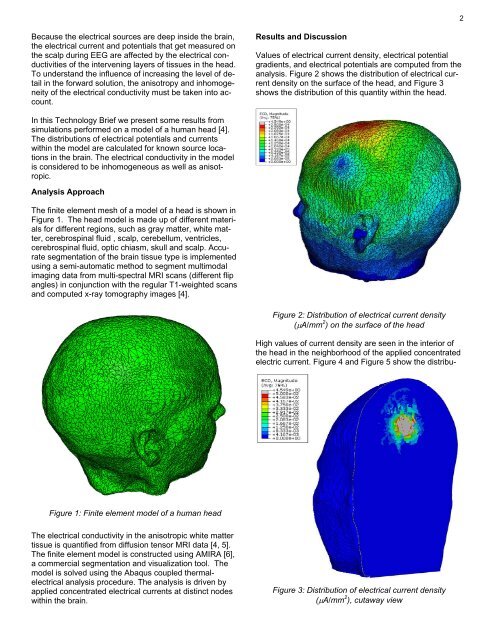
Results and Discussion<br />
Values <strong>of</strong> electrical current density, electrical potential<br />
gradients, and electrical potentials are computed from the<br />
analysis. Figure 2 shows the distribution <strong>of</strong> electrical current<br />
density on the surface <strong>of</strong> the head, and Figure 3<br />
shows the distribution <strong>of</strong> this quantity within the head.<br />
In this Technology Brief we present some results from<br />
simulations performed on a model <strong>of</strong> a human head [4].<br />
The distributions <strong>of</strong> electrical potentials and currents<br />
within the model are calculated for known source locations<br />
in the brain. The electrical conductivity in the model<br />
is considered to be inhomogeneous as well as anisotropic.<br />
Analysis Approach<br />
The finite element mesh <strong>of</strong> a model <strong>of</strong> a head is shown in<br />
Figure 1. The head model is made up <strong>of</strong> different materials<br />
for different regions, such as gray matter, white matter,<br />
cerebrospinal fluid , scalp, cerebellum, ventricles,<br />
cerebrospinal fluid, optic chiasm, skull and scalp. Accurate<br />
segmentation <strong>of</strong> the brain tissue type is implemented<br />
using a semi-automatic method to segment multimodal<br />
imaging data from multi-spectral MRI scans (different flip<br />
angles) in conjunction with the regular T1-weighted scans<br />
and computed x-ray tomography images [4].<br />
Figure 2: Distribution <strong>of</strong> electrical current density<br />
(μA/mm 2 ) on the surface <strong>of</strong> the head<br />
High values <strong>of</strong> current density are seen in the interior <strong>of</strong><br />
the head in the neighborhood <strong>of</strong> the applied concentrated<br />
electric current. Figure 4 and Figure 5 show the distribu-<br />
Figure 1: Finite element model <strong>of</strong> a human head<br />
The electrical conductivity in the anisotropic white matter<br />
tissue is quantified from diffusion tensor MRI data [4, 5].<br />
The finite element model is constructed using AMIRA [6],<br />
a commercial segmentation and visualization tool. The<br />
model is solved using the <strong>Abaqus</strong> coupled thermalelectrical<br />
analysis procedure. The analysis is driven by<br />
applied concentrated electrical currents at distinct nodes<br />
within the brain.<br />
Figure 3: Distribution <strong>of</strong> electrical current density<br />
(μA/mm 2 ), cutaway view