Simulation of Electroencephalography (EEG) Using Abaqus
Simulation of Electroencephalography (EEG) Using Abaqus
Simulation of Electroencephalography (EEG) Using Abaqus
You also want an ePaper? Increase the reach of your titles
YUMPU automatically turns print PDFs into web optimized ePapers that Google loves.
<strong>Abaqus</strong> Technology Brief<br />
TB-07-<strong>EEG</strong>-1<br />
Revised: February 2008<br />
<strong>Simulation</strong> <strong>of</strong> <strong>Electroencephalography</strong> (<strong>EEG</strong>) <strong>Using</strong> <strong>Abaqus</strong><br />
Summary<br />
<strong>Electroencephalography</strong> (<strong>EEG</strong>) is used to obtain information<br />
about the electrical activity in the brain and is routinely<br />
used to diagnose neurological abnormalities. The<br />
inverse problem in <strong>EEG</strong> refers to the procedure <strong>of</strong> locating<br />
electrical sources in the brain from the extracranial<br />
electrical field measured on the scalp. The solution <strong>of</strong> the<br />
inverse problem requires the forward calculation <strong>of</strong> the<br />
electric field for a given source location. The significance<br />
<strong>of</strong> the inclusion <strong>of</strong> inhomogeneity and anisotropy in the<br />
electrical conductivity <strong>of</strong> the brain tissue in the forward<br />
solution is not well understood. In this Technology Brief<br />
we present examples that illustrate the use <strong>of</strong> the electrical<br />
analysis capability in <strong>Abaqus</strong> for calculating the forward<br />
solution based on the finite element method (FEM).<br />
The results from simulations in <strong>Abaqus</strong> can then be used<br />
to solve the inverse problem.<br />
Background<br />
To predict the locations <strong>of</strong> the sources <strong>of</strong> electrical activity<br />
within the brain, neurosurgeons currently depend on<br />
measurements <strong>of</strong> the brain’s electrical fields using electroencephalography<br />
(<strong>EEG</strong>). Knowledge <strong>of</strong> the location <strong>of</strong><br />
sources <strong>of</strong> seizure activity, such as in epilepsy, helps<br />
plan a course <strong>of</strong> treatment.<br />
Currently, the ability to locate the sources <strong>of</strong> electrical<br />
activity is only moderately accurate [1]; increasing the<br />
accuracy will help neurosurgeons to better delineate the<br />
specific regions <strong>of</strong> the brain that need intervention. Subsequently,<br />
surgeries can be targeted to those regions,<br />
and unnecessary damage to healthy neural tissue will be<br />
reduced.<br />
About 30% <strong>of</strong> the patient population suffering from epilepsy<br />
have medically uncontrollable seizures and may be<br />
considered for surgical therapy, based on scalp <strong>EEG</strong><br />
monitoring. In some cases these patients undergo intracranial<br />
<strong>EEG</strong> monitoring for spontaneous seizures [2]. Localization<br />
<strong>of</strong> the seizure focus from intracranial <strong>EEG</strong> is<br />
critical for decreasing or eliminating seizure activity after<br />
surgery.<br />
Studies have shown the clinical utility <strong>of</strong> advanced source<br />
imaging techniques in localizing epileptogenic loci using<br />
boundary element method (BEM) [1] and finite element<br />
method (FEM) models [3]. However, these methods are<br />
Key <strong>Abaqus</strong> Features and Benefits<br />
• Coupled thermal-electrical analysis capability<br />
for the computation <strong>of</strong> electric field and current<br />
density<br />
• Ability to include anisotropic and inhomogeneous<br />
electrical conductivity properties<br />
not yet routinely used in clinical settings due to their high<br />
computational cost and lack <strong>of</strong> direct validations with<br />
experimental data.<br />
Prediction <strong>of</strong> the locations <strong>of</strong> electrical sources using<br />
data obtained through <strong>EEG</strong>s is an inverse problem. The<br />
solution <strong>of</strong> the inverse problem requires the ability to calculate<br />
the electric field for a source at a given location;<br />
we refer to this as the forward problem. Reductions in<br />
forward model errors using FEM could lead to improved<br />
source localizations <strong>of</strong> seizures, with the eventual goal <strong>of</strong><br />
surgery without the need for invasive intracranial <strong>EEG</strong><br />
monitoring.
2<br />
Because the electrical sources are deep inside the brain,<br />
the electrical current and potentials that get measured on<br />
the scalp during <strong>EEG</strong> are affected by the electrical conductivities<br />
<strong>of</strong> the intervening layers <strong>of</strong> tissues in the head.<br />
To understand the influence <strong>of</strong> increasing the level <strong>of</strong> detail<br />
in the forward solution, the anisotropy and inhomogeneity<br />
<strong>of</strong> the electrical conductivity must be taken into account.<br />
Results and Discussion<br />
Values <strong>of</strong> electrical current density, electrical potential<br />
gradients, and electrical potentials are computed from the<br />
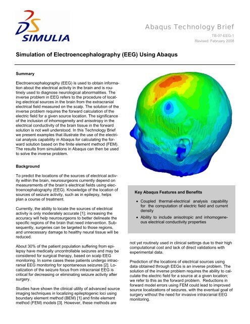
analysis. Figure 2 shows the distribution <strong>of</strong> electrical current<br />
density on the surface <strong>of</strong> the head, and Figure 3<br />
shows the distribution <strong>of</strong> this quantity within the head.<br />
In this Technology Brief we present some results from<br />
simulations performed on a model <strong>of</strong> a human head [4].<br />
The distributions <strong>of</strong> electrical potentials and currents<br />
within the model are calculated for known source locations<br />
in the brain. The electrical conductivity in the model<br />
is considered to be inhomogeneous as well as anisotropic.<br />
Analysis Approach<br />
The finite element mesh <strong>of</strong> a model <strong>of</strong> a head is shown in<br />
Figure 1. The head model is made up <strong>of</strong> different materials<br />
for different regions, such as gray matter, white matter,<br />
cerebrospinal fluid , scalp, cerebellum, ventricles,<br />
cerebrospinal fluid, optic chiasm, skull and scalp. Accurate<br />
segmentation <strong>of</strong> the brain tissue type is implemented<br />
using a semi-automatic method to segment multimodal<br />
imaging data from multi-spectral MRI scans (different flip<br />
angles) in conjunction with the regular T1-weighted scans<br />
and computed x-ray tomography images [4].<br />
Figure 2: Distribution <strong>of</strong> electrical current density<br />
(μA/mm 2 ) on the surface <strong>of</strong> the head<br />
High values <strong>of</strong> current density are seen in the interior <strong>of</strong><br />
the head in the neighborhood <strong>of</strong> the applied concentrated<br />
electric current. Figure 4 and Figure 5 show the distribu-<br />
Figure 1: Finite element model <strong>of</strong> a human head<br />
The electrical conductivity in the anisotropic white matter<br />
tissue is quantified from diffusion tensor MRI data [4, 5].<br />
The finite element model is constructed using AMIRA [6],<br />
a commercial segmentation and visualization tool. The<br />
model is solved using the <strong>Abaqus</strong> coupled thermalelectrical<br />
analysis procedure. The analysis is driven by<br />
applied concentrated electrical currents at distinct nodes<br />
within the brain.<br />
Figure 3: Distribution <strong>of</strong> electrical current density<br />
(μA/mm 2 ), cutaway view
3<br />
Figure 4: Distribution <strong>of</strong> electrical potential gradient<br />
(μV/mm) on the surface <strong>of</strong> the head.<br />
Figure 6: Distribution <strong>of</strong> electrical potential (μV) on the<br />
surface <strong>of</strong> the head.<br />
Figure 5: Distribution <strong>of</strong> electrical potential gradient<br />
(μV/mm), cutaway view.<br />
tion <strong>of</strong> electrical potential gradients on the surface <strong>of</strong> the<br />
head and in the interior <strong>of</strong> the head. Figure 6 and Figure 7<br />
show the distribution <strong>of</strong> electrical potentials on the surface<br />
<strong>of</strong> the head and within the head. These results are compared<br />
to the intracranial and scalp <strong>EEG</strong> measurements<br />
obtained from patients implanted with depth electrodes<br />
using in vivo experiments in [4]. The forward solution can<br />
then be used to estimate inversely the locations <strong>of</strong> electrical<br />
sources from known <strong>EEG</strong> measurements.<br />
Conclusions<br />
Currently, <strong>EEG</strong> is used as a diagnostic tool to determine<br />
the location <strong>of</strong> the source <strong>of</strong> electrical disturbances inside<br />
the brain. However, this localization is not very accurate<br />
Figure 7: Distribution <strong>of</strong> electrical potential (μV), cutaway<br />
view<br />
at present. Numerical simulations <strong>of</strong> electrical conduction<br />
in the brain can help correlate the <strong>EEG</strong> signals to the<br />
electrical sources and increase the understanding <strong>of</strong> the<br />
influence <strong>of</strong> increased complexity on the forward solution.<br />
An improved forward solution can in turn determine the<br />
location <strong>of</strong> sources accurately from measured <strong>EEG</strong> values<br />
by solving the inverse problem. Accurate localization<br />
<strong>of</strong> the electrical disturbances will help neurosurgeons fine<br />
tune their surgical procedures, making such procedures<br />
more accurate and minimally invasive. The coupled thermal-electrical<br />
analysis procedure in <strong>Abaqus</strong> can be used<br />
to simulate intracranial electrical conduction and is well<br />
poised to play a big role in this crucial medical advancement.
4<br />
Acknowledgements<br />
SIMULIA would like to thank Nitin Bangera from Boston University (Biomedical Engineering)/UCSD (Multimodal Imaging<br />
Lab) for providing the <strong>Abaqus</strong> model used in this Technology Brief.<br />
References<br />
1. Cuffin, B. N., “<strong>EEG</strong> Dipole Source Localization,” IEEE Engineering in Medicine and Biology, pp. 118–122,1998.<br />
2. Theodore, W. H., and R. S. Fisher, “Brain Stimulation for Epilepsy,” The Lancet Neurology, vol. 3, issue 2,<br />
pp. 111–18, 2004.<br />
3. Plummer, C., L. Litewka, S. Farish, A. S. Harvey, and M. J. Cook, “Clinical Utility <strong>of</strong> Current-Generation Dipole<br />
Modelling <strong>of</strong> Scalp <strong>EEG</strong>,” Clinical Neurophysiology, vol. 118, issue 11, pp. 2344–2361, 2007.<br />
4. Bangera, N. B., “Development and Validation <strong>of</strong> a Realistic Head Model for <strong>EEG</strong>” (PhD thesis), in Biomedical<br />
Engineering, Boston University, Boston, MA, 2008.<br />
5. Tuch, D. S., V. J. Wedeen, A. M. Dale, J. S. George, and J. W. Belliveau, Conductivity Tensor Mapping <strong>of</strong> the<br />
Human Brain <strong>Using</strong> Diffusion Tensor MRI, Proceedings <strong>of</strong> the National Academy <strong>of</strong> Sciences, vol. 96, no. 20,<br />
pp. 11697–11701, 2001.<br />
6. amira User’s Guide, amira 4.1, Konrad-Zuse-Zentrum f¨ur Informationstechnik Berlin (ZIB) and Mercury<br />
Computer Systems.<br />
<strong>Abaqus</strong> References<br />
For additional information on the <strong>Abaqus</strong> capabilities referred to in this brief, please see the following<br />
<strong>Abaqus</strong> Version 6.7 documentation references:<br />
• <strong>Abaqus</strong> Analysis User’s Manual<br />
− Coupled thermal-electrical analysis, Section 6.6.2.<br />
About SIMULIA<br />
SIMULIA is the Dassault Systèmes brand that delivers a scalable portfolio <strong>of</strong> Realistic <strong>Simulation</strong> solutions including the <strong>Abaqus</strong> product<br />
suite for Unified Finite Element Analysis, multiphysics solutions for insight into challenging engineering problems, and lifecycle<br />
management solutions for managing simulation data, processes, and intellectual property. By building on established technology, respected<br />
quality, and superior customer service, SIMULIA makes realistic simulation an integral business practice that improves product<br />
performance, reduces physical prototypes, and drives innovation. Headquartered in Providence, RI, USA, with R&D centers in<br />
Providence and in Suresnes, France, SIMULIA provides sales, services, and support through a global network <strong>of</strong> over 30 regional<br />
<strong>of</strong>fices and distributors. For more information, visit www.simulia.com<br />
The 3DS logo, SIMULIA, <strong>Abaqus</strong> and the <strong>Abaqus</strong> logo are trademarks or registered trademarks <strong>of</strong> Dassault Systèmes or its subsidiaries, which include <strong>Abaqus</strong>, Inc. Other company, product and service<br />
names may be trademarks or service marks <strong>of</strong> others.<br />
Copyright Dassault Systèmes, 2007


