
Create successful ePaper yourself
Turn your PDF publications into a flip-book with our unique Google optimized e-Paper software.
SIRAJ and JOSHI. Brunei Int Med J. 2012; 8 (3): 146<br />
a<br />
b<br />
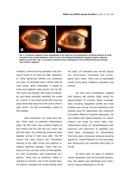
Fig. 1: a) Anterior segment colour photograph of the right eye (at presentation) showing evidence of acute<br />
necrosis of the corneal epithelium (black arrow) and widespread epithelial damage. Anterior segment<br />
details are not clear. Fig. 1; b) Anterior segment colour photograph of the unaffected left eye showing<br />
clear anterior segment.<br />
irritation, severe burning sensation and blurring<br />
of vision in his right eye (RE). Apparently,<br />
while gardening without any protective<br />
eye wear, he stumbled upon a black cobra at<br />
close range. When challenged, it raised its<br />
hood and suddenly spat venom into his RE.<br />
The victim was familiar with snake morphology<br />
and hence promptly identified the snake<br />
as a cobra. It was about three feet long and<br />
about three feet away from the victim when it<br />
spat venom. He was immediately rushed to<br />
the hospital.<br />
Upon evaluation, his vitals were stable.<br />
There were no adnexial inflammatory<br />
signs. His RE vision was counting fingers at<br />
two metres and his Left eye (LE) vision was<br />
6/9 (Pin hole). His intraocular pressures were<br />
digitally normal in both eyes (BE). The RE<br />
cornea was hazy (Figure 1a). Flourescein<br />
staining of the right cornea was positive revealing<br />
epithelial damage. There was evidence<br />
of toxic necrosis of the corneal epithelium<br />
with circumciliary and conjunctival congestion.<br />
There was no chemosis, limbal ischaemia<br />
or necrosis. Due to the corneal haze,<br />
his anterior chamber and fundus details were<br />
not clear. LE evaluation was normal (Figure<br />
1b). Extra-ocular movements and convergence<br />
were intact. There was no associated<br />
cranial nerve palsy. Systemic evaluation was<br />
normal.<br />
His eyes were immediately irrigated<br />
with balance salt solution (BSS, Alcon) for<br />
approximately 20 minutes. Blood investigations<br />
including coagulation profile and renal<br />
profiles were normal. He was admitted to the<br />
medical ward for observation and treatment<br />
of possible effects of systemic absorption. He<br />
was treated with topical antibiotic (G. Ciprofloxacin)<br />
eye drops six times daily, antiinflammatory<br />
drops (G. Dexamethasone with<br />
neomycin and polymyxin B sulphate) four<br />
times daily, cycloplegics (G. Homatropine<br />
2%) twice daily, intraocular pressure lowering<br />
agents (G. Timolol Maleate 0.5 %) twice daily<br />
and Tetracycline eye ointment twice daily to<br />
his RE.<br />
As there were no signs of systemic<br />
venom absorption and normal blood parameters,<br />
the patient was discharged and continued<br />
on topical medications in his RE.