Eosinophilic esophagitis: Updated consensus ... - AInotes
Eosinophilic esophagitis: Updated consensus ... - AInotes
Eosinophilic esophagitis: Updated consensus ... - AInotes

You also want an ePaper? Increase the reach of your titles
YUMPU automatically turns print PDFs into web optimized ePapers that Google loves.
6 LIACOURAS ET AL<br />
J ALLERGY CLIN IMMUNOL<br />
nnn 2011<br />
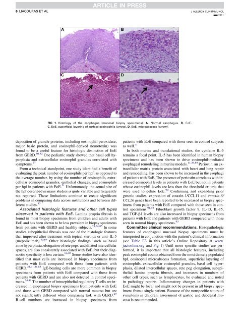
FIG 1. Histology of the esophagus (mucosal biopsy specimens). A, Normal esophagus. B, EoE.<br />
C, EoE, superficial layering of surface eosinophils (arrow). D, EoE, microabscess (arrow).<br />
deposition of granule proteins, including eosinophil peroxidase,<br />
major basic protein, and eosinophil-derived neurotoxin) was<br />
found to be a useful feature for histologic distinction of EoE<br />
from GERD. 80-82 One pediatric study showed that basal cell hyperplasia<br />
and extracellular eosinophil granules correlated with<br />
symptoms. 77<br />
From a technical standpoint, one study identified a benefit of<br />
evaluating the peak number of eosinophils per hpf, as opposed to<br />
the average number, by using the number of eosinophils, extracellular<br />
eosinophil granules, epithelial changes, and eosinophils<br />
per hpf in patients with EoE. 51 Unfortunately, the actual size of<br />
the hpf described in many studies is quite variable and frequently<br />
not reported. These limitations continue to create significant<br />
problems in comparing data across institutions and between different<br />
studies. 83<br />
Associated histologic features and other cell types<br />
observed in patients with EoE. Lamina propria fibrosis is<br />
found in most biopsy specimens from children and adults with<br />
EoE and has been shown to be less prevalent in biopsy specimens<br />
from patients with GERD and healthy subjects. 78,84,85 In some<br />
studies subepithelial fibrosis was one of the histologic features<br />
that improved after treatment with topical steroids or anti–IL-5<br />
(mepolizumab). 40,69 Other histologic findings, such as basal<br />
zone hyperplasia, elongation of rete pegs, and dilated intercellular<br />
spaces, are also consistently associated with EoE, but their diagnostic<br />
specificity is less certain. 86,87 Some studies have also identified<br />
that mast cells are increased in biopsy specimens from<br />
patients with EoE compared with those from patients with<br />
GERD. 24,41,81,88 IgE-bearing cells are more common in biopsy<br />
specimens from patients with EoE compared with those from<br />
patients with GERD and are also not detected in control specimens.<br />
24,41 The number of intraepithelial regulatory T cells are increased<br />
in esophageal biopsy specimens from patients with EoE<br />
and those with GERD compared with normal mucosa but are<br />
not significantly different when comparing EoE with GERD. 29<br />
B-cell numbers are increased in biopsy specimens from<br />
patients with EoE compared with those seen in control subjects<br />
as well. 41<br />
In both murine and translational studies, the cytokine IL-5<br />
remains a focal point. IL-5 has been identified in human biopsy<br />
specimens and has been shown to drive eosinophil-mediated<br />
esophageal remodeling in murine models. 11,26,89 Periostin, an extracellular<br />
matrix protein associated with heart and lung repair<br />
and remodeling, has been shown to be increased in the esophagi<br />
of patients with EoE. The presence of periostin correlates with increased<br />
eosinophil levels in patients with EoE but not in patients<br />
whose eosinophil levels are less than the threshold criteria that<br />
were used to define EoE. 90 Confirming and expanding prior<br />
genetic studies, expression of eotaxin-1/CCL11 and eotaxin-3/<br />
CCL26 genes have been reported to be increased in biopsy specimens<br />
from patients with EoE compared with those seen in control<br />
specimens. 91,92 Fibroblast growth factor 9, IL-13, IL-15,<br />
and TGF-b1 levels are also increased in biopsy specimens from<br />
patients with EoE and patients with GERD compared with those<br />
seen in normal biopsy specimens. 93<br />
Committee clinical recommendations. Histopathologic<br />
features of esophageal mucosal biopsy specimens must be<br />
interpreted in conjunction with the patient’s clinical information<br />
(see Table E3 in this article’s Online Repository at www.<br />
jacionline.org and Fig 1) Until more specific studies are performed,<br />
it is important that all histologic features, including<br />
peak eosinophil counts obtained from the most densely populated<br />
hpf, eosinophil microabscess formation, superficial layering of<br />
eosinophils, extracellular eosinophil granules, basal cell hyperplasia,<br />
dilated intercellular spaces, rete peg elongation, subepithelial<br />
lamina propria fibrosis, and increases in numbers of<br />
other cell types, such as lymphocytes, be evaluated and noted<br />
in pathology reports. Inflammatory changes in patients with<br />
EoE might be focal and might not be present in all biopsy specimens<br />
from a single patient. Because of the nonspecific nature of<br />
symptoms in children, assessment of gastric and duodenal mucosa<br />
is recommended.