Pathology of the endocrine system Endocrine system
Pathology of the endocrine system Endocrine system
Pathology of the endocrine system Endocrine system
You also want an ePaper? Increase the reach of your titles
YUMPU automatically turns print PDFs into web optimized ePapers that Google loves.
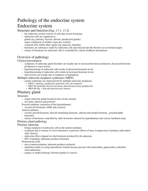
<strong>Pathology</strong> <strong>of</strong> <strong>the</strong> <strong>endocrine</strong> <strong>system</strong><br />
<strong>Endocrine</strong> <strong>system</strong><br />
Structure and function [Figs. 17-1, 17-2]<br />
– <strong>the</strong> <strong>endocrine</strong> <strong>system</strong> consists <strong>of</strong> cells that secrete hormones<br />
– <strong>endocrine</strong> cells are organized as:<br />
glands [eg. pituitary, thyroid, adrenal, parathyroid glands]<br />
major component <strong>of</strong> ano<strong>the</strong>r organ [eg. testicle]<br />
scattered cells within o<strong>the</strong>r organs [eg. pancreas, intestine)<br />
– hormones are substances made by <strong>endocrine</strong> cells and released into <strong>the</strong> blood to act on distant targets<br />
– release <strong>of</strong> hormones by <strong>endocrine</strong> cells is controlled by various feedback mechanisms<br />
Overview <strong>of</strong> pathology<br />
Clinical presentation<br />
– symptoms <strong>of</strong> <strong>endocrine</strong> gland disorders are usually due to increased hormone production, decreased hormone<br />
production or mass lesions<br />
– hyp<strong>of</strong>unctioning <strong>of</strong> <strong>endocrine</strong> cells results in decreased hormone levels<br />
– hyperfunctioning <strong>of</strong> <strong>endocrine</strong> cells results in increased hormone levels<br />
– mass lesions are usually due to neoplasia or hyperplasia<br />
Multiple <strong>endocrine</strong> neoplasia syndromes (MEN)<br />
– certain syndromes are characterized by multiple <strong>endocrine</strong> neoplasms<br />
• MEN I (pituitary, parathyroid, pancreatic islet cell neoplasia)<br />
• MEN IIa (medullary thyroid carcinoma, pheochromocytoma, parathyroid)<br />
• MEN IIb (IIa + skin and mucosal nerve tumors)<br />
Pituitary gland<br />
Structure<br />
– single <strong>endocrine</strong> gland located in base <strong>of</strong> <strong>the</strong> cranium<br />
– two parts, anterior and posterior<br />
Posterior pituitary (extension <strong>of</strong> <strong>the</strong> hypothalamus)<br />
– secretes <strong>the</strong> hormones ADH, and oxytocin<br />
Anterior pituitary<br />
– secretes growth hormone, thyroid stimulating hormone, adrenocorticotropin hormone, gonadotrophin<br />
hormones<br />
– release <strong>of</strong> hormones controlled by o<strong>the</strong>r hormones released by hypothalamus and various feedback loops<br />
Pituitary gland pathology<br />
Pituitary adenoma<br />
– benign neoplasm <strong>of</strong> <strong>endocrine</strong> cells in <strong>the</strong> anterior pituitary<br />
– symptoms due to release <strong>of</strong> excess hormones or pressure effects <strong>of</strong> mass (compression <strong>of</strong> pitutary stalk and/or<br />
optic chiasm)<br />
– <strong>endocrine</strong> effects depend on what hormone produced by <strong>the</strong> adenoma<br />
– 80% <strong>of</strong> pituitary adenomas produce hormones<br />
Prolactinoma (LH)<br />
– most common pituitary adenoma produces prolactin<br />
– identified earlier in young reproductive female because present with amenorrhea, galactorrhea, infertility<br />
(microadenoma)<br />
– surgery or medical <strong>the</strong>rapy (bromocryptine) to remove
<strong>Pathology</strong> <strong>of</strong> <strong>the</strong> <strong>endocrine</strong> <strong>system</strong><br />
Pituitary gland pathology<br />
Pituitary adenoma<br />
Somatotropic adenomas<br />
– neoplastic cells produce growth hormone<br />
– gigantism results from excess growth hormone before growth plates close<br />
• generalized increase in body size with disproportionately long legs, arms<br />
– acromegaly results from excess growth hormone after puberty<br />
• enlargement <strong>of</strong> hands, feet, jaw, tongue, and s<strong>of</strong>t tissue)<br />
Corticotropic adenoma<br />
– neoplastic cells produce adrenocorticotropin hormone<br />
– Cushing’s disease refers to <strong>the</strong> syndrome resulting from excess glucocorticoid release by <strong>the</strong> adrenal cortex<br />
due to excess ACTH<br />
Pituitary hyp<strong>of</strong>unction<br />
– causes <strong>of</strong> pituitary hyp<strong>of</strong>unction include<br />
• congenital defect <strong>of</strong> pituitary gland (primary dwarfism)<br />
• destructive tumor (pituitary adenoma)<br />
• ischemia <strong>of</strong> <strong>the</strong> pituitary gland (Sheehan’s syndrome)<br />
symptoms<br />
– weakness, ↓ appetite, ↓ weight, hypotension, amenorrhea<br />
– secondary hyp<strong>of</strong>unction <strong>of</strong> target organs<br />
Diabetes insipidus<br />
– lack <strong>of</strong> ADH<br />
– usually due to destructive lesion in hypothalamus, pituitary<br />
– unable to resorb water, large amounts <strong>of</strong> hypotonic urine<br />
Thyroid gland<br />
Structure and function<br />
– located in anterior neck, right and left lobes joined by isthmus<br />
– follicular cells produce thyroid hormones (T4, T3)<br />
– release <strong>of</strong> thyroid hormones controlled by TSH (from pituitary)<br />
– C cells produce calcitonin<br />
<strong>Pathology</strong> <strong>of</strong> <strong>the</strong> thyroid gland<br />
Thyroid hyperfunction (hyperthyroidism) [Figs. 17-5, 17-6]<br />
– major causes are Grave’s disease, some multinodular goiters, tumors<br />
symptoms <strong>of</strong> hyperthyroidism<br />
– restless, nervous, emotional lability, sweating, tachycardia, diarrhea, weight loss with increased appetite<br />
Hyperthyroidism<br />
Grave’s disease<br />
– autoimmune disease due to antibodies targeting <strong>the</strong> TSH receptor on thyroid follicular cells<br />
– AB binds to TSH receptor causing release <strong>of</strong> thyroid hormones<br />
– more common in females<br />
– associated with o<strong>the</strong>r autoimmune diseases<br />
– exopthalmos occurs in Grave’s disease<br />
Multinodular goiter<br />
– enlarged, nodular thyroid may produce increased amounts <strong>of</strong> thyroid hormone (some may be euthyroid or<br />
hyp<strong>of</strong>unctioning)
<strong>Pathology</strong> <strong>of</strong> <strong>the</strong> <strong>endocrine</strong> <strong>system</strong><br />
<strong>Pathology</strong> <strong>of</strong> <strong>the</strong> thyroid gland<br />
Thyroid hyp<strong>of</strong>unction (hypothyroidism) [Fig. 17-6]<br />
– major causes are agenesis, surgery, thyroiditis, iodine deficiency<br />
Symptoms <strong>of</strong> hypothyroidism<br />
– cretinism and dwarfism if occurs in perinatal period or infant<br />
– myxedema if occurs in older child or adult<br />
• sleepy, tire easily, cold intolerance, constipation, weak<br />
– treat with thyroid hormone replacement<br />
Nodular goiter<br />
– goiter is general term for enlarged thyroid gland (many causes)<br />
– nodular goiter is a form <strong>of</strong> goiter characterized by multiple nodules<br />
– goiters are usually euthyroid (normal thyroid hormone levels)<br />
– may cause mass effects (surgery if problematic)<br />
Thyroid neoplasms<br />
Adenomas<br />
– benign neoplasms <strong>of</strong> thyroid follicular cells<br />
– may produce symptoms due to mass effect<br />
– treated by surgery (microscopic examination required to rule out cancer)<br />
Carcinoma<br />
– malignant neoplasm <strong>of</strong> thyroid follicular cells<br />
– two major types:<br />
• papillary carcinoma (80%)- good prognosis<br />
• follicular carcinoma (15%)- relatively good prognosis<br />
Parathyroid glands<br />
Structure and function<br />
– usually 4 glands, located in <strong>the</strong> anterior neck<br />
– produce parathyroid hormone (PTH), involved in calcium homeostasis<br />
<strong>Pathology</strong> <strong>of</strong> <strong>the</strong> parathyroid glands<br />
Hyperparathyroidism[Figs. 17-8, 17-10]<br />
– major causes are parathyroid adenoma and parathyroid hyperplasia<br />
symptoms <strong>of</strong> hyperparathyroidism (hypercalcemia)<br />
– bones, stones, moans, abdominal groans<br />
Hypoparathyroidism<br />
– causes <strong>of</strong> include surgery, congenital hypoplasia<br />
symptoms <strong>of</strong> hypoparathyroidism (hypocalcemia)<br />
– muscle spasms, irregular heart beat, cardiac arrest (if severe)<br />
Adrenal glands<br />
Structure and function<br />
– right and left glands, located retroperitoneal<br />
adrenal cortex<br />
– zona glomerulosa (aldosterone)<br />
– zona fasciculata (cortisol)<br />
– zona reticularis (secondary sex steroids)<br />
adrenal medulla<br />
– secrete epinephrine, norepinephrine
<strong>Pathology</strong> <strong>of</strong> <strong>the</strong> <strong>endocrine</strong> <strong>system</strong><br />
<strong>Pathology</strong> <strong>of</strong> <strong>the</strong> adrenal gland<br />
Adrenocortical hyperfunction[Figs. 17-11, 17-13]<br />
Hypercortisolism (Cushing’s syndrome)<br />
– syndrome due to excess glucocorticoid hormones (cortisol)<br />
– most common cause is exogenous steroids, o<strong>the</strong>r causes include<br />
• adrenal hyperplasia or neoplasia<br />
• hypersecretion <strong>of</strong> ACTH by pituitary gland (Cushing’s disease)<br />
• ectopic ACTH (paraneoplastic syndrome)<br />
– dramatic appearance: central obesity, buffalo hump, moon face, striae<br />
Hyperaldosteronism (Conn’s syndrome)<br />
– syndrome due to excess mineralocorticoid hormone (aldosterone)<br />
– causes include adrenocortical adenoma and adrenal hyperplasia<br />
– present with hypertension and hypokalemia<br />
Adrenocortical hyp<strong>of</strong>unction<br />
– usually autoimmune destruction <strong>of</strong> adrenals, also due Tb, malignancy<br />
Addison’s disease<br />
– autoimmune destruction <strong>of</strong> adrenal gland<br />
– fatigue, weight loss, nausea, increased infections, low Na, high K<br />
Diseases <strong>of</strong> Adrenal Medulla<br />
Neuroblastoma<br />
– malignant neoplasm <strong>of</strong> neuroblasts (primitive cells) in neonates, infant<br />
– treatment with chemo<strong>the</strong>rapy, surgery, radiation (90 % cure)<br />
Pheochromocytoma<br />
– a neoplasm (usually benign) derived from adrenal medulla cells<br />
– diagnosed on basis <strong>of</strong> dramatic clinical picture, metabolites in urine<br />
– treated by surgery<br />
<strong>Endocrine</strong> pancreas<br />
Structure<br />
– cells located in islets within <strong>the</strong> pancreas<br />
– several different types <strong>of</strong> cells (Β cells produce insulin)<br />
<strong>Pathology</strong> <strong>of</strong> <strong>the</strong> <strong>endocrine</strong> pancreas<br />
Diabetes mellitus<br />
– heterogeneous group <strong>of</strong> diseases due to inadequate insulin activity<br />
– present with polyuria, polydypsia, weight loss<br />
classification <strong>of</strong> primary diabetes<br />
– type 1 diabetes (insulin dependent, juvenile onset)<br />
– type 2 diabetes (non-insulin dependent, adult onset)<br />
• most common, relative lack <strong>of</strong> insulin<br />
– impaired glucose tolerance<br />
pathogenesis<br />
– insulin released in response to increased blood sugar<br />
• insulin required by certain cells for entry <strong>of</strong> glucose into <strong>the</strong> cell<br />
– insulin decreases blood glucose levels<br />
– lack <strong>of</strong> insulin causes hyperglycemia<br />
• present with polyuria (osmotic diuresis) and resulting polydypsia<br />
• also fatigue due to inability <strong>of</strong> glucose to enter certain cells<br />
– striated muscle cells must use anaerobic glycolysis<br />
• increased lactic acid<br />
• decreased use <strong>of</strong> fats result in increased free fatty acids<br />
– free fatty acids oxidized into ketones (acidosis)
<strong>Pathology</strong> <strong>of</strong> <strong>the</strong> <strong>endocrine</strong> <strong>system</strong><br />
<strong>Pathology</strong> <strong>of</strong> <strong>the</strong> <strong>endocrine</strong> pancreas<br />
Diabetes mellitus<br />
– genetic predisposition<br />
• type 2 > type 1<br />
– environmental factors<br />
Complications ( due to long term uncontrolled hyperglycemia)<br />
– Cardiovascular [increased a<strong>the</strong>rosclerosis (CAD, CVD, distal gangrene)]<br />
– Renal [glomerulosclerosis, pyelonephritis, papillary necrosis]<br />
– Eyes [diabetic microangiopathy <strong>of</strong> retinal vessels, glaucoma, cataracts]<br />
– Nervous <strong>system</strong><br />
Treatment (depends on type )<br />
– type 1 requires insulin<br />
– type 2 diet, oral hypoglycemics, insulin if unable to control


