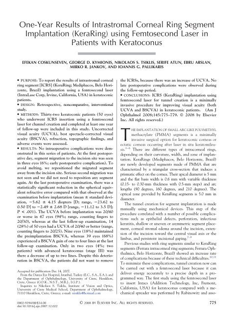
One-Year Results of Intrastromal Corneal Ring Segment - Mediphacos
One-Year Results of Intrastromal Corneal Ring Segment - Mediphacos
One-Year Results of Intrastromal Corneal Ring Segment - Mediphacos

Create successful ePaper yourself
Turn your PDF publications into a flip-book with our unique Google optimized e-Paper software.
<strong>One</strong>-<strong>Year</strong> <strong>Results</strong> <strong>of</strong> <strong>Intrastromal</strong> <strong>Corneal</strong> <strong>Ring</strong> <strong>Segment</strong><br />
Implantation (Kera<strong>Ring</strong>) using Femtosecond Laser in<br />
Patients with Keratoconus<br />
EFEKAN COSKUNSEVEN, GEORGE D. KYMIONIS, NIKOLAOS S. TSIKLIS, SERIFE ATUN, EBRU ARSLAN,<br />
MIRKO R. JANKOV, AND IOANNIS G. PALLIKARIS<br />
● PURPOSE: To report the results <strong>of</strong> intrastromal corneal<br />
ring segment [ICRS] (Kera<strong>Ring</strong>; <strong>Mediphacos</strong>, Belo Horizonte,<br />
Brazil) implantation using a femtosecond laser<br />
(IntraLase Corp, Irvine, California, USA) in keratoconic<br />
patients.<br />
● DESIGN: Retrospective, noncomparative, interventional<br />
study.<br />
● METHODS: Thirty-two keratoconic patients (50 eyes)<br />
who underwent ICRS insertion using a femtosecond<br />
laser for channel creation and completed at least one year<br />
<strong>of</strong> follow-up were included in this study. Uncorrected<br />
visual acuity (UCVA), best spectacle-corrected visual<br />
acuity (BSCVA), refraction, topographic findings, and<br />
adverse events were assessed.<br />
● RESULTS: No intraoperative complications were demonstrated<br />
in this series <strong>of</strong> patients. At the first postoperative<br />
day, segment migration to the incision site was seen<br />
in three eyes (6%; early postoperative complication). To<br />
avoid melting, we repositioned the migrated segment<br />
away from the incision site. Serious second migration was<br />
not seen and we did not need to reposition any segment<br />
again. At the last postoperative examination, there was a<br />
statistically significant reduction in the spherical equivalent<br />
refractive error compared with that observed at the<br />
examination before implantation (mean standard deviation,<br />
5.62 4.15 diopters [D; range, 23.62 to<br />
0.50 D] to 2.49 2.68 D [range, 11.12 to 3.5 D];<br />
P < .001). The UCVA before implantation was 20/40<br />
or worse in 47 eyes (94%; range, counting fingers to<br />
20/30), whereas at the last follow-up examination, 14<br />
(28%) <strong>of</strong> 50 eyes had a UCVA <strong>of</strong> 20/40 or better (range,<br />
counting fingers to 20/25). Nine eyes (18%) maintained<br />
the preimplantation BSCVA, whereas 39 eyes (68%)<br />
experienced a BSCVA gain <strong>of</strong> one to four lines at the last<br />
follow-up examination. Only in two eyes (4%; two<br />
patients) with advanced keratoconus (stage III) was<br />
there a decrease <strong>of</strong> up to two lines. Despite this deterioration<br />
in BSCVA, the patients did not want to remove<br />
Accepted for publication Dec 14, 2007.<br />
From the Dunya Eye Hospital, Istanbul, Turkey (E.C., S.A., E.A.); and<br />
the Department <strong>of</strong> Ophthalmology, University <strong>of</strong> Crete, Heraklion,<br />
Crete, Greece (G.D.K., N.S.T., M.R.J., I.G.P.).<br />
Inquiries to Nikolaos S. Tsiklis, Institute <strong>of</strong> Vision and Optics,<br />
University <strong>of</strong> Crete Medical School, Department <strong>of</strong> Ophthalmology,<br />
71110 Heraklion, Crete, Greece; e-mail: ntsiklis@hotmail.com<br />
the ICRSs, because there was an increase <strong>of</strong> UCVA. No<br />
late postoperative complications were observed during<br />
the follow-up period.<br />
● CONCLUSIONS: ICRS (Kera<strong>Ring</strong>) implantation using<br />
femtosecond laser for tunnel creation is a minimally<br />
invasive procedure for improving visual acuity (both<br />
UCVA and BSCVA) in keratoconic patients. (Am J<br />
Ophthalmol 2008;145:775–779. © 2008 by Elsevier<br />
Inc. All rights reserved.)<br />
THE IMPLANTATION OF SMALL ARC-LIKE POLYMETHYL<br />
methacrylate (PMMA) segments is a minimally<br />
invasive surgical option for keratoconic corneas or<br />
ectatic corneas occurring after laser in situ keratomileusis.<br />
1–6 There are different types <strong>of</strong> intracorneal rings,<br />
depending on their curvature, width, and zone <strong>of</strong> implantation.<br />
Kera<strong>Ring</strong>s (<strong>Mediphacos</strong>, Belo Horizonte, Brazil)<br />
are newly developed segments made <strong>of</strong> PMMA that are<br />
characterized by a triangular cross-section that induces a<br />
prismatic effect on the cornea. Their apical diameter is 5 mm<br />
and the flat basis width is 0.6 mm with variable thickness<br />
(0.15- to 0.30-mm thickness with 0.5-mm steps) and arc<br />
lengths (90 degrees, 160 degrees, and 210 degrees). The<br />
optical zone provided by Kera<strong>Ring</strong> segments is 5.0 mm in<br />
diameter.<br />
The tunnel creation for segment implantation is made<br />
manually using mechanical devices. This step <strong>of</strong> the<br />
procedure correlated with a number <strong>of</strong> possible complications<br />
such as epithelial defects, perforation, infectious<br />
keratitis, shallow or uneven or asymmetric segment placement,<br />
corneal stromal edema around the incision, extension<br />
<strong>of</strong> the incision toward the central visual axis or the<br />
limbus, and persistent incisional gaping. 7–9<br />
Previous studies with ring segments similar to Kera<strong>Ring</strong><br />
segments (Ferrara intracorneal ring segments; Ferrara Ophthalmics,<br />
Belo Horizonte, Brazil) showed an increase rate<br />
<strong>of</strong> complications because <strong>of</strong> these technical difficulties. 10,11<br />
To minimize these complications, tunnel creation now can<br />
be carried out with a femtosecond laser because it can<br />
deliver energy accurately to a precise depth in a programmed<br />
way. The first study using the femtosecond laser<br />
to insert Intacs (Addition Technology, Inc, Fremont,<br />
California, USA) for keratoconus compared with a mechanical<br />
spreader was performed by Rabinowitz and asso-<br />
0002-9394/08/$34.00 © 2008 BY ELSEVIER INC. ALL RIGHTS RESERVED.<br />
775<br />
doi:10.1016/j.ajo.2007.12.022
TABLE. Preoperative and Last Follow-up Examination Data <strong>of</strong> Patients with Keratoconus after <strong>Intrastromal</strong> <strong>Corneal</strong> <strong>Ring</strong><br />
<strong>Segment</strong> (Kera<strong>Ring</strong>) Implantation<br />
Preoperative<br />
Last Postoperative Examination<br />
No. <strong>of</strong> eyes 50<br />
No. <strong>of</strong> patients 32<br />
Gender<br />
Male 18<br />
Female 14<br />
Age (yrs)<br />
Mean SD 28.32 7.28<br />
Range 18 to 44<br />
Follow-up (mos)<br />
Mean SD 15.6 3.2<br />
Range 12 to 24<br />
Spherical equivalent (D)<br />
Mean SD 5.62 4.16 2.50 2.68 P .001<br />
Range 0.50 to 23.62 3.50 to 11.12<br />
Cylinder (D)<br />
Mean SD 4.13 2.02 2.18 1.27 P .001<br />
Range 0.5 to 9.50 0.5 to 6.00<br />
Topographic K values (D)<br />
Mean SD 50.63 3.97 47.56 4.46 P .001<br />
Range 42.05 to 60.95 36.00 to 61.50<br />
D diopters; SD standard deviation; mos months.<br />
ciates, whereas additional studies indicate that Intacs<br />
implantation with the use <strong>of</strong> a femtosecond laser is a safe<br />
and effective procedure for treating keratoconic corneas.<br />
12–15 Recently, Shabayek and Alió published a series <strong>of</strong><br />
keratoconic patients who were followed up six months<br />
after intrastromal corneal ring segment (ICRS) implantation<br />
with Kera<strong>Ring</strong>s and a femtosecond laser. 16 In the<br />
current study, we evaluated the outcomes and long-term<br />
complications (all patients completed at least one year <strong>of</strong><br />
follow-up) <strong>of</strong> this type <strong>of</strong> intracorneal ring segments using<br />
a femtosecond laser for tunnel creation (IntraLase Corp,<br />
Irvine, California, USA) in keratoconic patients.<br />
METHODS<br />
FIFTY EYES OF 32 KERATOCONIC PATIENTS (18 MALES [26<br />
eyes] and 14 females [24 eyes]) who underwent ICRS<br />
implantation <strong>of</strong> Kera<strong>Ring</strong>s using a femtosecond laser for<br />
tunnel creation were included in this study. All procedures<br />
performed by the same surgeon (E.C.) at Dunya Eye<br />
Hospital, Istanbul, Turkey. The mean patient age was<br />
28.32 7.28 years (range, 18 to 44 years). All patients<br />
completed at least one year <strong>of</strong> follow-up. The Table<br />
summarizes patient demographic and refractive data.<br />
All patients demonstrated clear central corneas and<br />
contact lens intolerance. <strong>Corneal</strong> thickness was at least<br />
350 m at the thinnest corneal point and at least 450 m<br />
at the incision side. Patients were excluded if any <strong>of</strong> the<br />
following criteria applied after the preoperative examination:<br />
history <strong>of</strong> herpes, keratitis, corneal dystrophies, diagnosed<br />
autoimmune disease, systemic connective tissue<br />
disease, acute or grade IV keratoconus, and endothelial cell<br />
count <strong>of</strong> less than 1000 cells/mm 2 .<br />
A complete ophthalmologic examination performed<br />
before surgery included uncorrected visual acuity (UCVA)<br />
and best spectacle-corrected visual acuity (BSCVA) assessment,<br />
manifest refraction, biomicroscopy, corneal topography<br />
(using Orbscan IIz [Bausch & Lomb GmbH,<br />
Feldkirchen, Germany] or WaveLight Allegretto Topolyzer<br />
[WaveLight Technologie, Erlangen, Germany]), and<br />
endothelial cell measurement with specular microscopy<br />
(Konan Specular Microscope SP 9000; Noncon Robo<br />
Pachy Konan Medical, Inc, Hyogo, Japan).<br />
● SURGICAL PROCEDURE: The surgical procedure was<br />
carried out under sterile conditions and topical anesthesia.<br />
Purkinje reflex was chosen as the central point and was<br />
marked under Wavelength Allegretto Biomicroscope. A<br />
5-mm marker was used to locate the exact ring channel.<br />
<strong>Corneal</strong> thickness was measured during surgery using<br />
ultrasonic pachymetry (Sonogage, Cleveland, Ohio, USA)<br />
along the ring location markings. Tunnel depth was set at<br />
75% <strong>of</strong> the thinnest corneal thickness on the tunnel<br />
location in the femtosecond laser.<br />
An incision was made on the steepest topographic axis.<br />
<strong>One</strong> or two segments were implanted according to the<br />
distribution <strong>of</strong> the ectatic area on the corneal surface,<br />
776 AMERICAN JOURNAL OF OPHTHALMOLOGY<br />
MAY 2008
FIGURE 1. Bar graphs demonstrating (Left) uncorrected visual acuity and (UCVA) and (Right) best spectacle-corrected visual acuity<br />
(BSCVA) changes (lines <strong>of</strong> Snellen acuity) after intrastromal corneal ring segment (Kera<strong>Ring</strong>) implantation in keratoconic patients.<br />
whereas the thickness <strong>of</strong> the segment was decided according<br />
to the distribution <strong>of</strong> the ectatic area as well as the<br />
spherical equivalent (SE). A 60-khz femtosecond laser was<br />
used to create the ring channels. Special attention was<br />
given in centralizing the disposable suction ring to mark<br />
the central point to minimize decentration. The channel’s<br />
inner diameter was set to 4.4 mm, the outer diameter was<br />
5.6 mm, the entry cut thickness was 1 m (at the steepest<br />
topographic axis), the ring energy used for channel creation<br />
was 1.30 j, and the entry cut energy was 1.30 j.<br />
Channel creation timing with the femtosecond laser was<br />
15 seconds. The intracorneal ring segments were implanted<br />
immediately after channel creation before the<br />
disappearing <strong>of</strong> the bubbles, which reveals the exact tunnel<br />
location. To avoid any injury to the incision area, we<br />
directly implanted the segment with the special Kera<strong>Ring</strong><br />
forceps. The Kera<strong>Ring</strong> segment was implanted uneventfully<br />
in all cases. After surgery, antibiotic steroid eye drops<br />
four times daily for two weeks were prescribed. The<br />
patients were instructed to avoid rubbing the eye and to<br />
use preservative-free artificial tears frequently. On the first<br />
postoperative day, slit-lamp biomicroscopic examination<br />
was performed. Healing <strong>of</strong> the wound and migration <strong>of</strong> the<br />
segments were evaluated. At the last follow-up examination,<br />
manifest refraction, UCVA and BSCVA, slit-lamp,<br />
and topographic examinations were performed.<br />
● STATISTICAL ANALYSIS: Group differences for continuous<br />
variables were tested using the paired Student t test.<br />
<strong>Results</strong> are presented as mean standard deviation (SD). A<br />
P value less than .05 was regarded as statistically significant.<br />
RESULTS<br />
MEAN FOLLOW-UP WAS 15.6 3.2 MONTHS (RANGE, 12 TO 24<br />
months). At the last follow-up examination, SE error was<br />
statistically significantly reduced (preimplantation mean <br />
SD, 5.62 4.15 diopters [D; range, 23.62 to 0.50 D];<br />
postimplantation mean SD, 2.49 2.68 D [range,<br />
11.12 to 3.5 D; P .001; Table).<br />
UCVA and BSCVA were measured using Snellen scale<br />
charts. UCVA was improved in 39 eyes (78%) compared<br />
with the preoperative levels. Preoperative UCVA was<br />
20/40 or worse in 47 eyes (94%; range, counting fingers to<br />
20/30), whereas at the last follow-up examination, 14<br />
(28%) <strong>of</strong> 50 eyes had UCVA <strong>of</strong> 20/40 or better (range,<br />
counting fingers to 20/25). The mean difference between<br />
preoperative and postoperative UCVA was a gain <strong>of</strong> 1.7<br />
lines (range, loss <strong>of</strong> one UCVA Snellen line to gain <strong>of</strong> six<br />
lines; Figure 1, Left).<br />
BSCVA was maintained to the preimplantation level in<br />
nine eyes (18%), whereas 39 eyes (68%) experienced a<br />
BSCVA gain <strong>of</strong> one to up to four lines at the last follow-up<br />
examination. Only in two eyes (4%; two patients) with<br />
advanced keratoconus (stage III) was there a decrease <strong>of</strong> up to<br />
two lines. Despite this deterioration in BSCVA, the patient<br />
did not want to remove the ICRSs because there was an<br />
increase <strong>of</strong> UCVA. The mean difference between preoperative<br />
and postoperative BSCVA was a gain <strong>of</strong> 1.3 lines (range,<br />
loss <strong>of</strong> two lines to a gain <strong>of</strong> four lines; Figure 1, Right).<br />
A significant reduction in keratometric values was found<br />
at the last follow-up examination (Table). Mean preoperative<br />
keratometry was 50.63 3.97 D (range, 42.05 to<br />
60.95 D) and changed significantly to 47.56 4.46 D<br />
(range, 36.00 to 61.50 D; P .001). Similarly, mean<br />
preoperative keratometric astigmatism was 4.71 2.64 D<br />
(range, 0.60 to 12.50 D) and statistically significantly<br />
changed to 3.17 2.27 D (range, 1.50 to 9.80 D;<br />
P .001) at the last follow-up examination (Table).<br />
Furthermore, in patients with more pronounced keratoconus<br />
(stage III; 15 31 eyes), the mean reduction in keratometric<br />
values was 3.62 3.72 D (range, 11.25 to<br />
8.75 D). This reduction was higher compared with the<br />
VOL. 145, NO. 5 KERARINGS USING FEMTOSECOND LASER<br />
777
mean reduction in patients with stage I (eight eyes; 1.88 <br />
3.01 D [range, 6.50 to 1.15 D]) or stage II (11 eyes;<br />
2.37 1.93 D [range, 5.8 to 0.45 D]) keratoconus, but<br />
it did not reached statistically significant levels (P .05).<br />
● ADVERSE EFFECTS AND COMPLICATIONS: At the first<br />
postoperative day, segment migration to the incision site<br />
was seen in three eyes (6%; early postoperative complications).<br />
To avoid melting, we repositioned the migrated<br />
segment away from the incision site, advised the patients<br />
not to rub the eye, and again applied a bandage contact<br />
lens. Serious second migration was not seen and we did not<br />
need to reposition any segment again. No late postoperative<br />
complications occurred in this series <strong>of</strong> patients.<br />
DISCUSSION<br />
INTRACORNEAL RING SEGMENTS WERE FIRST IMPLANTED<br />
in human eyes by Nosé and associates, and a few years<br />
later, Colin and associates reported their preliminary<br />
results regarding the management <strong>of</strong> keratoconus with<br />
Intacs. 4,17 The procedure is effective in most keratoconic<br />
patients, but the predictability is low and the results are<br />
variable. 17–22<br />
Intacs inserts have a crescent-shaped arc length <strong>of</strong> 150<br />
degrees. Their inner diameter is 6.8 mm and the outer<br />
diameter is 8.1 mm when placed in the cornea. However,<br />
Kera<strong>Ring</strong>s have a smaller optical zone (apical diameter, 5.0<br />
mm). Theoretically, Kera<strong>Ring</strong>s may have a greater effect<br />
than Intacs because they are placed more centrally, but the<br />
procedure may be more difficult (they are implanted in<br />
thinner corneal areas).<br />
In the current study, one year after Kera<strong>Ring</strong> implantation,<br />
UCVA was improved in 39 eyes (78%) compared<br />
with the preoperative levels, with a mean gain <strong>of</strong> 1.9 lines<br />
(range, loss <strong>of</strong> one to gain <strong>of</strong> six lines). BSCVA was<br />
maintained to the preimplantation level in nine eyes<br />
(18%), whereas 39 eyes (68%) experienced a BSCVA gain<br />
<strong>of</strong> one to up to three lines at the last follow-up examination<br />
(mean gain, 1.3 lines; range, loss <strong>of</strong> two lines to gain<br />
<strong>of</strong> four lines). Only in two eyes (two patients) with<br />
advanced keratoconus (stage III) was a decrease <strong>of</strong> two<br />
lines with an increase <strong>of</strong> UCVA found.<br />
In comparison with other studies <strong>of</strong> intracorneal rings<br />
implantation with mechanical devices, 7–9 there was a<br />
significant reduction <strong>of</strong> complication rate. Regarding Ferrara<br />
rings (similar to Kera<strong>Ring</strong>s), Siganos and associates<br />
reported an incidence <strong>of</strong> 7.7% <strong>of</strong> improper implantation,<br />
whereas Kwitko and Severo reported a higher incidence <strong>of</strong><br />
complications (such as ring extrusion in 19.6% <strong>of</strong> the<br />
eyes). 10,11 In the current study, no explantation was<br />
performed (in three eyes [6%], segments were repositioned<br />
at the first postoperative day without any further intervention<br />
needed), mainly because <strong>of</strong> the precise depth <strong>of</strong><br />
implantation with femtosecond laser (Figure 2). Recently,<br />
FIGURE 2. Visante OCT AC Imaging demonstrating intrastromal<br />
corneal ring segments (Kera<strong>Ring</strong>) inserted using Femtosecond<br />
laser in a keratoconic eye.<br />
Shabayek and Alió first published a series <strong>of</strong> keratoconic<br />
patients followed up for six months after ICRSs implantation<br />
with Kera<strong>Ring</strong>s using a femtosecond laser. 16 They<br />
concluded that this method is effective and safe for<br />
keratoconus.<br />
A few studies (most <strong>of</strong> them with limited treated eyes)<br />
used a femtosecond laser for segment implantation in<br />
keratoconic patients. 14–16 Ertan and associates published a<br />
series <strong>of</strong> keratoconic patients who underwent Intacs implantation<br />
using a femtosecond laser. 14 Intacs were implanted<br />
successfully in all eyes. At the end <strong>of</strong> the first<br />
postoperative year, 81.3% <strong>of</strong> eyes had improved UCVA<br />
and 73.7% had improved BCVA. Similarly in our study,<br />
UCVA was improved in 78% <strong>of</strong> the eyes compared with<br />
the preoperative levels, whereas 68% <strong>of</strong> the studied eyes<br />
experienced a BSCVA gain <strong>of</strong> one to up to three lines at<br />
the last follow-up examination. All these studies concur<br />
that femtosecond laser-assisted intracorneal ring segments<br />
insertion in most keratoconic eyes improves the UCVA<br />
and BSCVA (gain <strong>of</strong> more than six Snellen lines) and<br />
reduces patient manifest refraction and corneal astigmatism<br />
(up to 4 D). These refractive and topographic results<br />
are comparable with the results <strong>of</strong> mechanical Intacs<br />
channels creation. 1–6,10,11,17–22<br />
The use <strong>of</strong> the femtosecond laser in corneal tunnel<br />
creation made the procedure faster, easier (especially for<br />
inexperienced surgeons), and more comfortable for the<br />
patient. However, the main advantage <strong>of</strong> IntraLaseassisted<br />
channel creation over the mechanical technique<br />
seems to be the precise depth <strong>of</strong> implantation. From the<br />
published data 14–16 and our experience, the safety <strong>of</strong> the<br />
procedure seems to be very high.<br />
A few potential limitations are apparent in this study,<br />
with the small sample <strong>of</strong> treated eyes, the lack <strong>of</strong> a<br />
comparative group (keratoconic eyes after Kera<strong>Ring</strong> insertion<br />
using mechanical devises), and the absence <strong>of</strong><br />
follow-up data in the intermediate period being the major<br />
reservations in concluding sufficient results.<br />
In conclusion, Kera<strong>Ring</strong> implantation using a femtosecond<br />
laser seems to be a minimally invasive treatment with good<br />
visual and refractive outcomes. Future larger, comparative<br />
studies focusing on the nomogram modification and the<br />
possible complications <strong>of</strong> the technique are needed.<br />
778 AMERICAN JOURNAL OF OPHTHALMOLOGY<br />
MAY 2008
THE AUTHORS INDICATE NO FINANCIAL SUPPORT OR FINANCIAL CONFLICT OF INTEREST. DR COSKUNSEVEN IS A SCIENTIFIC<br />
consultant to <strong>Mediphacos</strong>, Belo Horizonte, Brazil. Involved in design and conduct <strong>of</strong> the study (E.C., G.D.K., N.T., M.J., I.G.P.); collection,<br />
management, analysis, and interpretation <strong>of</strong> the data (E.C., E.A., S.A.); and preparation, review, and approval <strong>of</strong> the manuscript (E.C., G.D.K., N.T.,<br />
S.A., E.A., M.J., I.G.P.). All patients were appropriately informed <strong>of</strong> risks and benefits before operation, and they gave their written informed consent<br />
in accordance with the Institutional Review Board <strong>of</strong> Dunya Eye Hospital, Istanbul, Turkey, according to the Declaration <strong>of</strong> Helsinki.<br />
REFERENCES<br />
1. Kymionis GD, Siganos CS, Tsiklis NS, et al. Long-term<br />
follow-up <strong>of</strong> Intacs in keratoconus. Am J Ophthalmol 2007;<br />
143:236–244.<br />
2. Kymionis GD, Tsiklis NS, Pallikaris AI, et al. Long-term<br />
follow-up <strong>of</strong> Intacs for post-LASIK corneal ectasia. Ophthalmology<br />
2006;113:1909–1917.<br />
3. Schanzlin DJ, Abbott RL, Asbell PA, et al. Two-year<br />
outcomes <strong>of</strong> intrastromal corneal ring segments for the<br />
correction <strong>of</strong> myopia. Ophthalmology 2001;108:1688–1694.<br />
4. Nosé W, Neves RA, Schanzlin DJ, Belfort R Jr. <strong>Intrastromal</strong><br />
corneal ring: one-year results <strong>of</strong> first implants in humans: a<br />
preliminary nonfunctional eye study. Refract <strong>Corneal</strong> Surg<br />
1993;9:452–458.<br />
5. Alió JL, Shabayek MH, Artola A. Intracorneal ring segments<br />
for keratoconus correction: long-term follow-up. J Cataract<br />
Refract Surg 2006;32:978–985.<br />
6. Boxer Wachler BS, Christie JP, Chandra NS, et al. Intacs for<br />
keratoconus. Ophthalmology 2003;110:1031–1040.<br />
7. Ruckh<strong>of</strong>er J, Stoiber J, Alzner E, Grabner G. <strong>One</strong>-year results<br />
<strong>of</strong> European multicenter study <strong>of</strong> intrastromal corneal ring<br />
segments. Part 2: complications, visual symptoms, and patient<br />
satisfaction. The Multicenter European <strong>Corneal</strong> Correction<br />
Assessment Study Group. J Cataract Refract Surg<br />
2001;27:287–296.<br />
8. Kanellopoulos AJ, Lawrence H, Perry HD, Donnenfeld ED.<br />
Modified intracorneal ring segment implantations (Intacs) for<br />
the management <strong>of</strong> moderate to advanced keratoconus: efficacy<br />
and complications. Cornea 2006;25:29–33.<br />
9. Bourcier T, Borderie V, Laroche L. Late bacterial keratitis<br />
after implantation <strong>of</strong> intrastromal corneal ring segments.<br />
J Cataract Refract Surg 2003;29:407–409.<br />
10. Siganos D, Ferrara P, Chatzinikolas K, et al. Ferrara intrastromal<br />
corneal rings for the correction <strong>of</strong> keratoconus.<br />
J Cataract Refract Surg 2002;28:1947–1951.<br />
11. Kwitko S, Severo NS. Ferrara intracorneal ring segments for<br />
keratoconus. J Cataract Refract Surg 2004;30:812–820.<br />
12. Rabinowitz YS, Li X, Ignacio TS, Maguen E. Intacs inserts<br />
using the femtosecond laser compared to the mechanical<br />
spreader in the treatment <strong>of</strong> keratoconus. J Refract Surg<br />
2006;22:764–771.<br />
13. Sugar A. Ultra-fast (femtosecond) laser refractive surgery.<br />
Curr Opin Ophthalmol 2002;13:246–249.<br />
14. Ertan A, Kamburoglu G, Bahadir M. Intacs insertion with<br />
the femtosecond laser for the management <strong>of</strong> keratoconus:<br />
one-year results. J Cataract Refract Surg 2006;32:2039–<br />
2042.<br />
15. Ertan A, Bahadir M. Topography-guided vertical implantation<br />
<strong>of</strong> Intacs using a femtosecond laser for the treatment <strong>of</strong><br />
keratoconus. J Cataract Refract Surg 2007;33:148–151.<br />
16. Shabayek MH, Alió JL. <strong>Intrastromal</strong> corneal ring segment<br />
implantation by femtosecond laser for keratoconus correction.<br />
Ophthalmology 2007;114:1643–1652.<br />
17. Colin J, Cochener B, Savary G, Malet F. Correcting keratoconus<br />
with intracorneal rings. J Cataract Refract Surg 2000;<br />
26:1117–1122.<br />
18. Colin J, Cochener B, Savary G, et al. Intacs inserts for<br />
treating keratoconus: one-year results. Ophthalmology 2001;<br />
108:1409–1414.<br />
19. Siganos CS, Kymionis GD, Kartakis N, et al. Management<br />
<strong>of</strong> keratoconus with Intacs. Am J Ophthalmol 2003;135:<br />
64 –70.<br />
20. Hellstedt T, Makela J, Uusitalo R, Emre S, Uusitalo R.<br />
Treating keratoconus with Intacs corneal ring segments.<br />
J Refract Surg 2005;21:236–246.<br />
21. Levinger S, Pokroy R. Keratoconus managed with Intacs:<br />
one-year results. Arch Ophthalmol 2005;123:1308–1314.<br />
22. Alió JL, Artola A, Hassanein A, Haroun H, Galal A. <strong>One</strong> or<br />
two Intacs segments for the correction <strong>of</strong> keratoconus.<br />
J Cataract Refract Surg 2005;31:943–953.<br />
VOL. 145, NO. 5 KERARINGS USING FEMTOSECOND LASER<br />
779
Biosketch<br />
Dr Efekan Coskunseven was born in 1968 in Nazilli, Turkey. He graduated from Medical School <strong>of</strong> Ege University and<br />
specialized in Ophthalmology in the Medical School <strong>of</strong> Dicle University. Dr Coskunseven is the Director <strong>of</strong> Refractive<br />
Surgery Department in Dunya Eye Hospital, Istanbul, Turkey since 2003.<br />
779.e1 AMERICAN JOURNAL OF OPHTHALMOLOGY<br />
MAY 2008