One-Year Results of Intrastromal Corneal Ring Segment - Mediphacos
One-Year Results of Intrastromal Corneal Ring Segment - Mediphacos
One-Year Results of Intrastromal Corneal Ring Segment - Mediphacos

Create successful ePaper yourself
Turn your PDF publications into a flip-book with our unique Google optimized e-Paper software.
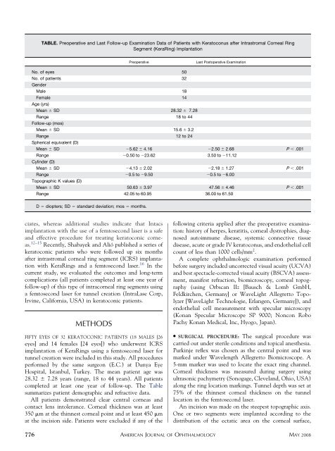
TABLE. Preoperative and Last Follow-up Examination Data <strong>of</strong> Patients with Keratoconus after <strong>Intrastromal</strong> <strong>Corneal</strong> <strong>Ring</strong><br />
<strong>Segment</strong> (Kera<strong>Ring</strong>) Implantation<br />
Preoperative<br />
Last Postoperative Examination<br />
No. <strong>of</strong> eyes 50<br />
No. <strong>of</strong> patients 32<br />
Gender<br />
Male 18<br />
Female 14<br />
Age (yrs)<br />
Mean SD 28.32 7.28<br />
Range 18 to 44<br />
Follow-up (mos)<br />
Mean SD 15.6 3.2<br />
Range 12 to 24<br />
Spherical equivalent (D)<br />
Mean SD 5.62 4.16 2.50 2.68 P .001<br />
Range 0.50 to 23.62 3.50 to 11.12<br />
Cylinder (D)<br />
Mean SD 4.13 2.02 2.18 1.27 P .001<br />
Range 0.5 to 9.50 0.5 to 6.00<br />
Topographic K values (D)<br />
Mean SD 50.63 3.97 47.56 4.46 P .001<br />
Range 42.05 to 60.95 36.00 to 61.50<br />
D diopters; SD standard deviation; mos months.<br />
ciates, whereas additional studies indicate that Intacs<br />
implantation with the use <strong>of</strong> a femtosecond laser is a safe<br />
and effective procedure for treating keratoconic corneas.<br />
12–15 Recently, Shabayek and Alió published a series <strong>of</strong><br />
keratoconic patients who were followed up six months<br />
after intrastromal corneal ring segment (ICRS) implantation<br />
with Kera<strong>Ring</strong>s and a femtosecond laser. 16 In the<br />
current study, we evaluated the outcomes and long-term<br />
complications (all patients completed at least one year <strong>of</strong><br />
follow-up) <strong>of</strong> this type <strong>of</strong> intracorneal ring segments using<br />
a femtosecond laser for tunnel creation (IntraLase Corp,<br />
Irvine, California, USA) in keratoconic patients.<br />
METHODS<br />
FIFTY EYES OF 32 KERATOCONIC PATIENTS (18 MALES [26<br />
eyes] and 14 females [24 eyes]) who underwent ICRS<br />
implantation <strong>of</strong> Kera<strong>Ring</strong>s using a femtosecond laser for<br />
tunnel creation were included in this study. All procedures<br />
performed by the same surgeon (E.C.) at Dunya Eye<br />
Hospital, Istanbul, Turkey. The mean patient age was<br />
28.32 7.28 years (range, 18 to 44 years). All patients<br />
completed at least one year <strong>of</strong> follow-up. The Table<br />
summarizes patient demographic and refractive data.<br />
All patients demonstrated clear central corneas and<br />
contact lens intolerance. <strong>Corneal</strong> thickness was at least<br />
350 m at the thinnest corneal point and at least 450 m<br />
at the incision side. Patients were excluded if any <strong>of</strong> the<br />
following criteria applied after the preoperative examination:<br />
history <strong>of</strong> herpes, keratitis, corneal dystrophies, diagnosed<br />
autoimmune disease, systemic connective tissue<br />
disease, acute or grade IV keratoconus, and endothelial cell<br />
count <strong>of</strong> less than 1000 cells/mm 2 .<br />
A complete ophthalmologic examination performed<br />
before surgery included uncorrected visual acuity (UCVA)<br />
and best spectacle-corrected visual acuity (BSCVA) assessment,<br />
manifest refraction, biomicroscopy, corneal topography<br />
(using Orbscan IIz [Bausch & Lomb GmbH,<br />
Feldkirchen, Germany] or WaveLight Allegretto Topolyzer<br />
[WaveLight Technologie, Erlangen, Germany]), and<br />
endothelial cell measurement with specular microscopy<br />
(Konan Specular Microscope SP 9000; Noncon Robo<br />
Pachy Konan Medical, Inc, Hyogo, Japan).<br />
● SURGICAL PROCEDURE: The surgical procedure was<br />
carried out under sterile conditions and topical anesthesia.<br />
Purkinje reflex was chosen as the central point and was<br />
marked under Wavelength Allegretto Biomicroscope. A<br />
5-mm marker was used to locate the exact ring channel.<br />
<strong>Corneal</strong> thickness was measured during surgery using<br />
ultrasonic pachymetry (Sonogage, Cleveland, Ohio, USA)<br />
along the ring location markings. Tunnel depth was set at<br />
75% <strong>of</strong> the thinnest corneal thickness on the tunnel<br />
location in the femtosecond laser.<br />
An incision was made on the steepest topographic axis.<br />
<strong>One</strong> or two segments were implanted according to the<br />
distribution <strong>of</strong> the ectatic area on the corneal surface,<br />
776 AMERICAN JOURNAL OF OPHTHALMOLOGY<br />
MAY 2008