One-Year Results of Intrastromal Corneal Ring Segment - Mediphacos
One-Year Results of Intrastromal Corneal Ring Segment - Mediphacos
One-Year Results of Intrastromal Corneal Ring Segment - Mediphacos

Create successful ePaper yourself
Turn your PDF publications into a flip-book with our unique Google optimized e-Paper software.
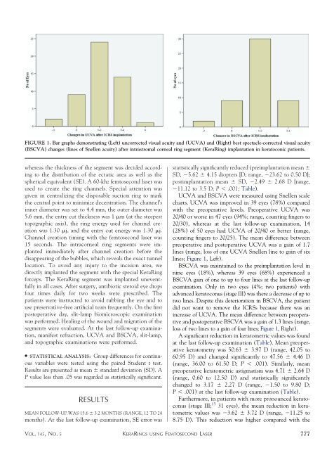
FIGURE 1. Bar graphs demonstrating (Left) uncorrected visual acuity and (UCVA) and (Right) best spectacle-corrected visual acuity<br />
(BSCVA) changes (lines <strong>of</strong> Snellen acuity) after intrastromal corneal ring segment (Kera<strong>Ring</strong>) implantation in keratoconic patients.<br />
whereas the thickness <strong>of</strong> the segment was decided according<br />
to the distribution <strong>of</strong> the ectatic area as well as the<br />
spherical equivalent (SE). A 60-khz femtosecond laser was<br />
used to create the ring channels. Special attention was<br />
given in centralizing the disposable suction ring to mark<br />
the central point to minimize decentration. The channel’s<br />
inner diameter was set to 4.4 mm, the outer diameter was<br />
5.6 mm, the entry cut thickness was 1 m (at the steepest<br />
topographic axis), the ring energy used for channel creation<br />
was 1.30 j, and the entry cut energy was 1.30 j.<br />
Channel creation timing with the femtosecond laser was<br />
15 seconds. The intracorneal ring segments were implanted<br />
immediately after channel creation before the<br />
disappearing <strong>of</strong> the bubbles, which reveals the exact tunnel<br />
location. To avoid any injury to the incision area, we<br />
directly implanted the segment with the special Kera<strong>Ring</strong><br />
forceps. The Kera<strong>Ring</strong> segment was implanted uneventfully<br />
in all cases. After surgery, antibiotic steroid eye drops<br />
four times daily for two weeks were prescribed. The<br />
patients were instructed to avoid rubbing the eye and to<br />
use preservative-free artificial tears frequently. On the first<br />
postoperative day, slit-lamp biomicroscopic examination<br />
was performed. Healing <strong>of</strong> the wound and migration <strong>of</strong> the<br />
segments were evaluated. At the last follow-up examination,<br />
manifest refraction, UCVA and BSCVA, slit-lamp,<br />
and topographic examinations were performed.<br />
● STATISTICAL ANALYSIS: Group differences for continuous<br />
variables were tested using the paired Student t test.<br />
<strong>Results</strong> are presented as mean standard deviation (SD). A<br />
P value less than .05 was regarded as statistically significant.<br />
RESULTS<br />
MEAN FOLLOW-UP WAS 15.6 3.2 MONTHS (RANGE, 12 TO 24<br />
months). At the last follow-up examination, SE error was<br />
statistically significantly reduced (preimplantation mean <br />
SD, 5.62 4.15 diopters [D; range, 23.62 to 0.50 D];<br />
postimplantation mean SD, 2.49 2.68 D [range,<br />
11.12 to 3.5 D; P .001; Table).<br />
UCVA and BSCVA were measured using Snellen scale<br />
charts. UCVA was improved in 39 eyes (78%) compared<br />
with the preoperative levels. Preoperative UCVA was<br />
20/40 or worse in 47 eyes (94%; range, counting fingers to<br />
20/30), whereas at the last follow-up examination, 14<br />
(28%) <strong>of</strong> 50 eyes had UCVA <strong>of</strong> 20/40 or better (range,<br />
counting fingers to 20/25). The mean difference between<br />
preoperative and postoperative UCVA was a gain <strong>of</strong> 1.7<br />
lines (range, loss <strong>of</strong> one UCVA Snellen line to gain <strong>of</strong> six<br />
lines; Figure 1, Left).<br />
BSCVA was maintained to the preimplantation level in<br />
nine eyes (18%), whereas 39 eyes (68%) experienced a<br />
BSCVA gain <strong>of</strong> one to up to four lines at the last follow-up<br />
examination. Only in two eyes (4%; two patients) with<br />
advanced keratoconus (stage III) was there a decrease <strong>of</strong> up to<br />
two lines. Despite this deterioration in BSCVA, the patient<br />
did not want to remove the ICRSs because there was an<br />
increase <strong>of</strong> UCVA. The mean difference between preoperative<br />
and postoperative BSCVA was a gain <strong>of</strong> 1.3 lines (range,<br />
loss <strong>of</strong> two lines to a gain <strong>of</strong> four lines; Figure 1, Right).<br />
A significant reduction in keratometric values was found<br />
at the last follow-up examination (Table). Mean preoperative<br />
keratometry was 50.63 3.97 D (range, 42.05 to<br />
60.95 D) and changed significantly to 47.56 4.46 D<br />
(range, 36.00 to 61.50 D; P .001). Similarly, mean<br />
preoperative keratometric astigmatism was 4.71 2.64 D<br />
(range, 0.60 to 12.50 D) and statistically significantly<br />
changed to 3.17 2.27 D (range, 1.50 to 9.80 D;<br />
P .001) at the last follow-up examination (Table).<br />
Furthermore, in patients with more pronounced keratoconus<br />
(stage III; 15 31 eyes), the mean reduction in keratometric<br />
values was 3.62 3.72 D (range, 11.25 to<br />
8.75 D). This reduction was higher compared with the<br />
VOL. 145, NO. 5 KERARINGS USING FEMTOSECOND LASER<br />
777