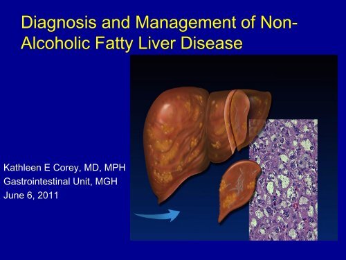
Diagnosis and Management of Non- Alcoholic Fatty Liver Disease
Diagnosis and Management of Non- Alcoholic Fatty Liver Disease
Diagnosis and Management of Non- Alcoholic Fatty Liver Disease

You also want an ePaper? Increase the reach of your titles
YUMPU automatically turns print PDFs into web optimized ePapers that Google loves.
<strong>Diagnosis</strong> <strong>and</strong> <strong>Management</strong> <strong>of</strong> <strong>Non</strong>-<br />
<strong>Alcoholic</strong> <strong>Fatty</strong> <strong>Liver</strong> <strong>Disease</strong><br />
Kathleen E Corey, MD, MPH<br />
Gastrointestinal Unit, MGH<br />
June 6, 2011
This Talk Will Guide You Through the<br />
<strong>Diagnosis</strong> <strong>and</strong> Treatment <strong>of</strong> NAFLD<br />
What is NAFLD<br />
How Do I Diagnose NAFLD<br />
Treating NAFLD
Case Presentation<br />
• AM is a 63M with obesity <strong>and</strong> hyperlipidemia<br />
who presents for evaluation <strong>of</strong> abnormal LFTs<br />
• AST 57, ALT 52, AP 80, TB 0.4<br />
• No history <strong>of</strong> alcohol use, no risk factors for viral<br />
hepatitis
Case Presentation<br />
• Viral serologies, iron<br />
studies, AIAT, celiac<br />
studies, TSH normal<br />
• ANA positive at 1:40,<br />
ASMA positive at 1:80<br />
• US shows fatty<br />
infiltration, no ascites,<br />
splenomegaly or<br />
abdominal varices
Case Presentation: What is<br />
the most likely diagnosis<br />
• Autoimmune hepatitis<br />
• A1AT<br />
• NAFLD<br />
• Celiac disease
Case Presentation: What is<br />
the most likely diagnosis<br />
• Autoimmune hepatitis<br />
• A1AT<br />
• NAFLD<br />
• Celiac disease
NAFLD: Spectrum <strong>of</strong> Hepatic Pathology<br />
Steatohepatitis<br />
Steatosis<br />
Cirrhosis<br />
Hepatocellular<br />
carcinoma
<strong>Non</strong>-<strong>Alcoholic</strong> <strong>Fatty</strong> <strong>Liver</strong><br />
(NAFL) Steatosis<br />
<strong>Fatty</strong> hepatocytes<br />
Intracellular<br />
fat deposition
<strong>Non</strong>-<strong>Alcoholic</strong><br />
SteatoHepatitis (NASH)<br />
Intracellular<br />
fat deposition<br />
Necrosis<br />
Fibrosis<br />
Fat deposits<br />
Inflammation<br />
Fibrosis with necrosis
Cirrhosis<br />
Regenerative<br />
nodule<br />
Fibrosis<br />
Nodules<br />
surrounded by<br />
fibrosis
How Many Americans Have<br />
NAFLD<br />
• 10%<br />
• 25%<br />
• 40%<br />
• 60%<br />
• 75%
How Many Americans Have<br />
NAFLD<br />
• 10%<br />
• 25%<br />
• 40%<br />
• 60%<br />
• 75%
Impact <strong>of</strong> NAFLD<br />
Wanless IR Hepatology 1990; Thursz. EASL Berlin 2011
NAFLD Prevalence<br />
Dallas Heart Study<br />
(~1100 African Americans, 700 Caucasians, 400 Hispanics)<br />
No risk factors<br />
(n = 375)<br />
Assess risk factors<br />
for fatty liver<br />
H 1 -NMR spectroscopy<br />
Define normal<br />
liver fat content<br />
Assess prevalence <strong>of</strong> increased liver fat (steatosis)<br />
in entire population & ethnic subgroups
Dallas Heart Study Results<br />
<strong>Liver</strong> fat<br />
< 5.5%<br />
<strong>Liver</strong> fat<br />
> 5.5%<br />
Steatosis = 31%<br />
<strong>Liver</strong> enzymes NORMAL in most (79%) with steatosis
Prevalence <strong>of</strong> Steatosis<br />
Varies with Ethnicity<br />
<strong>Fatty</strong> liver<br />
45%<br />
33%<br />
24%<br />
Hispanics Whites Blacks
NAFLD in Type 2 Diabetes<br />
Mellitus<br />
•Prevalence <strong>of</strong> NAFLD is high<br />
ultrasound detects fatty liver in 50%<br />
• NASH unusually common<br />
NAFL: 12%<br />
NASH: 87%<br />
• Fibrosis or cirrhosis documented in 20%<br />
Gupter et al. J Gastro Hepatol 2004;19:854-859<br />
Tolman et al. Ann Intern Med 2004; 141:946-956
What percentage <strong>of</strong> NASH<br />
patients have cirrhosis<br />
• 1.5%<br />
• 3%<br />
• 20%<br />
• 33%<br />
• 55%
What percentage <strong>of</strong> NASH<br />
patients have cirrhosis<br />
• 1.5%<br />
• 3%<br />
• 20%<br />
• 33%<br />
• 55%
Impact <strong>of</strong> NAFLD<br />
• Cirrhosis complicates
Impact <strong>of</strong> NAFLD<br />
• NAFLD is associated with an increased all<br />
cause mortality<br />
• NAFLD is associated with increased risk <strong>of</strong><br />
cardiovascular disease in diabetic <strong>and</strong><br />
non-diabetic patients<br />
Tarher Diabetes Care 2007;Ekstedt M<br />
Hepatolgy 2006
How Do I Diagnose<br />
NAFLD<br />
• Goal #1: Determine the etiology <strong>of</strong> liver<br />
disease as NAFLD<br />
• Goal #2: Determine the type <strong>of</strong> NAFLD<br />
• Goal #3: Determine the severity <strong>of</strong><br />
NAFLD
• Who might have NAFLD<br />
Goal #1: Determine<br />
the Etiology<br />
– Anyone with an elevated AST or ALT<br />
– Anyone with the Metabolic Syndrome<br />
– Anyone with fatty liver on imaging<br />
– Anyone with cryptogenic cirrhosis
Goal #1: Determine the<br />
Etiology<br />
Exclude other causes <strong>of</strong> abnormal LFTs<br />
– Alcohol use<br />
– Celiac disease: TTG<br />
– Medications<br />
<strong>and</strong> IgA<br />
– Viral hepatitis: HbSAg & HBV DNA <strong>and</strong> HCV AB &<br />
– Thyroid disease<br />
RNA<br />
– Iron deposition<br />
– Autoimmune liver disease (PBC, PSC, AIH)<br />
disease: iron<br />
• AMA, ANA, ASMA, SPEP<br />
saturation > 45%<br />
ferritin <strong>and</strong> if needed<br />
HFE<br />
– Alpha 1 anti-trypsin<br />
level <strong>and</strong> phenotype
Goal #1: Determine<br />
the Etiology<br />
• AST, ALT < 10x ULN <strong>and</strong> may be normal<br />
• Evaluate for presence <strong>of</strong> metabolic syndrome<br />
• No specific serologic markers <strong>of</strong> NAFLD exist<br />
• Autoimmune markers are frequently seen in<br />
patients with steatosis <strong>and</strong> NASH<br />
• ASMA positive in 15%, ANA in 24%, AMA 1-6%
Goal #1: Determine<br />
the Etiology<br />
• Imaging for NAFLD<br />
– US, MRI <strong>and</strong> CT can<br />
assess for fat<br />
– Other liver disease<br />
can cause steatosis<br />
– Absence <strong>of</strong> fat does<br />
not exclude NAFLD<br />
especially in setting <strong>of</strong><br />
advanced fibrosis
Goal #1: Determine the<br />
Etiology<br />
• Distinguish <strong>Alcoholic</strong> <strong>Liver</strong> <strong>Disease</strong> from<br />
NAFLD<br />
– NAFLD implies no EtOH or “safe” EtOH levels<br />
– 1 drink or less per day for women, 2 drinks or<br />
less per day for men
Goal #2: Determine the Type <strong>of</strong><br />
NAFLD<br />
• <strong>Liver</strong> function tests are insufficient to distinguish<br />
steatosis <strong>and</strong> NASH<br />
– In a cohort <strong>of</strong> diabetics 86% <strong>of</strong> both steatosis <strong>and</strong> NASH had<br />
normal LFTs<br />
– ALT is not a reliable marker to differentiate steatosis from NASH<br />
• Biopsy is the only definitive way to differentiate steatosis<br />
<strong>and</strong> NASH<br />
Tarher Diabetes Care 2007
Goal #3: Determine<br />
Severity <strong>of</strong> <strong>Disease</strong><br />
• Goal is to identify patients with advanced<br />
fibrosis or cirrhosis<br />
– Fibrosis stage >=2<br />
• Normal physical exam, labs <strong>and</strong> imaging<br />
are not sufficient to exclude cirrhosis<br />
• <strong>Liver</strong> biopsy is needed
Goal #3: Determine<br />
Severity <strong>of</strong> <strong>Disease</strong><br />
• Risk Factors for Advanced Fibrosis/<br />
Cirrhosis<br />
• Age > 50 years<br />
• Obesity<br />
• Diabetes<br />
• 66% prevalence <strong>of</strong> bridging fibrosis if<br />
age > 50 years <strong>and</strong> patient obese or<br />
diabetic
Who Needs a <strong>Liver</strong><br />
Biopsy<br />
• Anyone with risk factors for advanced<br />
fibrosis<br />
• Age > 50 years plus<br />
• Obesity or<br />
• Diabetes<br />
• Anyone with unexplained LFT elevations<br />
• When in doubt, refer for evaluation
Percutaneous <strong>Liver</strong> Biopsy<br />
• Performed by GI <strong>and</strong><br />
radiology<br />
• Uses ultrasound<br />
guidance or real time US<br />
• Same day procedure<br />
Risks <strong>of</strong> <strong>Liver</strong> Biopsy<br />
• Pain<br />
• Bleeding<br />
• Organ perforation<br />
• Death
• Steatosis<br />
• NASH<br />
– Lobular inflammation<br />
– Ballooning degeneration<br />
• Fibrosis<br />
What to look for on liver<br />
biopsy reports<br />
– Stage 2-4 (out <strong>of</strong> 4) is advanced fibrosis = cirrhosis
Back to our Patient:<br />
What is the next step<br />
in diagnosis<br />
• Weight loss <strong>and</strong> exercise<br />
• Watchful waiting – repeat LFTs in 1 year<br />
• Start Vitamin E<br />
• <strong>Liver</strong> Biopsy
Back to our Patient:<br />
What is the next step<br />
in diagnosis<br />
• Weight loss <strong>and</strong> exercise<br />
• Watchful waiting – repeat LFTs in 1 year<br />
• Start Vitamin E<br />
• <strong>Liver</strong> Biopsy
<strong>Liver</strong> Biopsy<br />
Findings<br />
Steatosis<br />
Lobular<br />
inflammation<br />
Regenerative<br />
nodules<br />
consistent with<br />
cirrhosis
Weight loss & exercise<br />
are core <strong>of</strong> NAFLD<br />
Therapy<br />
• Weight loss: 7 % weight loss has been associated with a<br />
significant reduction in NAS<br />
• Exercise even in the absence <strong>of</strong> weight loss improves<br />
steatosis<br />
• <strong>Management</strong> <strong>of</strong> metabolic syndrome components<br />
– Statins are safe <strong>and</strong> may be beneficial<br />
Ekstedt J <strong>of</strong> Hepatology 2007 (statin);<br />
Pomrat Hepatology 2010 (7%)
Vitamin E is Beneficial<br />
in NASH<br />
• Placebo controlled RTC <strong>of</strong> 247 patients<br />
w/biopsy proven NASH w/o DM<br />
• Daily pioglitazone 30mg vs vitamin E 800U<br />
for 96 weeks<br />
• Vitamin E improved lobular inflammation &<br />
steatosis compared to placebo<br />
PIVENS Trial Sanyal NEJM 2010
Pioglitazone is Beneficial<br />
In NASH<br />
• RTC <strong>of</strong> patients with impaired glucose<br />
tolerance or T2DM with biopsy proven<br />
NASH<br />
• Pioglitazone 45mg daily for 48 weeks<br />
resulted in improved necroinflammation<br />
Belfort NEJM 2006
Advanced fibrosis<br />
patients should be treated<br />
as cirrhotic patients<br />
• HCC screening<br />
– US or MRI Q6 months<br />
– AFP Q6 months<br />
• EGD for variceal<br />
screening Q1-3 years
Advanced fibrosis<br />
patients should be treated<br />
as cirrhotic patients<br />
• Monitoring for the<br />
complications <strong>of</strong> liver<br />
disease<br />
– Ascites<br />
– Encephalopathy<br />
• MELD labs to assess<br />
synthetic function<br />
– Cr, TB, INR<br />
• Referral to<br />
hepatologist
Take Home Messages<br />
What is NAFLD Leading Cause <strong>of</strong> <strong>Liver</strong><br />
<strong>Disease</strong> in the US<br />
How Do I <strong>Diagnosis</strong> NAFLD<br />
Low threshold for biopsy <strong>and</strong><br />
referral<br />
Treating NAFLD:<br />
Diet, Exercise <strong>and</strong><br />
Vitamin E
• Please contact me with questions at<br />
kcorey@partners.org
Secondary Causes <strong>of</strong> NAFLD<br />
• Disorders <strong>of</strong> lipid<br />
metabolism<br />
– Abetalipoproteinemia<br />
– Hypobetalipoproteinemia<br />
– Andersen’s disease<br />
– Weber-Christian syndrome<br />
• Total parenteral nutrition<br />
• Severe weight loss<br />
– Jejunoileal bypass<br />
• Severe starvation<br />
• Iatrogenic<br />
– Amiodarone<br />
– Diltiazem<br />
– Tamoxifen<br />
– Steroids<br />
– Highly active antiretroviral<br />
therapy<br />
• Refeeding syndrome<br />
• Toxic exposure<br />
• Environmental
NAFLD: Diagnostic Approach<br />
NAFLD: Diagnostic Approach<br />
Metabolic<br />
syndrome<br />
FL on<br />
imaging<br />
Elevated AST, ALT<br />
or cryptogenic cirrhosis<br />
Image liver<br />
Check AST, ALT,<br />
EtoH intake<br />
R/o other<br />
liver dz<br />
Image<br />
<strong>Liver</strong> fat<br />
Unsafe<br />
EtOH<br />
Safe<br />
EtOH<br />
Neg Pos Neg<br />
AFLD<br />
NAFLD<br />
<strong>Liver</strong> biopsy*<br />
* If safe, plus high risk <strong>of</strong> bridging fibrosis by clinical parameters