MRI of anorectal malformations and relationship of the ...
MRI of anorectal malformations and relationship of the ...
MRI of anorectal malformations and relationship of the ...

Create successful ePaper yourself
Turn your PDF publications into a flip-book with our unique Google optimized e-Paper software.
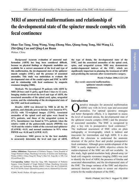
<strong>MRI</strong> <strong>of</strong> ARM <strong>and</strong> <strong>relationship</strong> <strong>of</strong> <strong>the</strong> developmental state <strong>of</strong> <strong>the</strong> SMC with fecal continence<br />
<strong>MRI</strong> <strong>of</strong> <strong>anorectal</strong> <strong>malformations</strong> <strong>and</strong> <strong>relationship</strong> <strong>of</strong><br />
<strong>the</strong> developmental state <strong>of</strong> <strong>the</strong> spincter muscle complex with<br />
fecal continence<br />
Shao-Tao Tang, Yong Wang, Yong-Zhong Mao, Qiang-Song Tong, Shi-Wang Li,<br />
Zhi-Qing Cao <strong>and</strong> Qing-Lan Ruan<br />
Wuhan, China<br />
Background: Accurate evaluation <strong>of</strong> <strong>anorectal</strong> malformation<br />
(ARM) has long been considered difficult,<br />
because no physical findings or diagnostic modalities are<br />
available for a correct assessment <strong>of</strong> <strong>the</strong> level <strong>and</strong> type <strong>of</strong><br />
this malformation, <strong>the</strong> developmental state <strong>of</strong> <strong>the</strong> sphincter<br />
muscle complex (SMC), <strong>and</strong> <strong>the</strong> presence <strong>of</strong> associated<br />
anomalies. This study was undertaken to evaluate <strong>the</strong><br />
developmental state <strong>of</strong> <strong>the</strong> caudal region <strong>and</strong> SMC in ARM<br />
<strong>and</strong> its <strong>relationship</strong> with fecal continence by magnetic<br />
resonance imaging (<strong>MRI</strong>).<br />
Methods: We investigated 39 patients with ARM by<br />
<strong>MRI</strong> (28 boys <strong>and</strong> 11 girls), aged from 3 days to 12 years.<br />
Imaging studies involved <strong>the</strong> level <strong>and</strong> type <strong>of</strong> ARM, <strong>the</strong><br />
associated anomalies <strong>of</strong> <strong>the</strong> spinal cord, spine, urogenital<br />
system, <strong>and</strong> <strong>the</strong> <strong>relationship</strong> <strong>of</strong> <strong>the</strong> developmental state <strong>of</strong><br />
<strong>the</strong> SMC <strong>and</strong> fecal continence.<br />
Results: ARM was detected by <strong>MRI</strong> in all <strong>the</strong> 39<br />
patients, <strong>and</strong> clinically proven fistulae were found in 97%<br />
(31/32) with T2-weighted images (T2WI). Associated<br />
anomalies <strong>of</strong> <strong>the</strong> spinal cord <strong>and</strong> spine were found in<br />
41% patients, <strong>and</strong> those <strong>of</strong> <strong>the</strong> urogenital system in<br />
21%. Incontinence was found in 71% patients when <strong>the</strong><br />
relative width <strong>of</strong> <strong>the</strong> puborectalis muscle (PRWR) was<br />
0.15.<br />
Conclusions: <strong>MRI</strong> proves to be <strong>the</strong> best modality<br />
to accurately determine <strong>the</strong> level <strong>and</strong> type <strong>of</strong> ARM,<br />
Author Affiliations: Department <strong>of</strong> Pediatric Surgery, Union Hospital <strong>of</strong><br />
Tongji Medical College, Huazhong University <strong>of</strong> Science <strong>and</strong> Technology,<br />
Wuhan 430022, China (Tang ST, Wang Y, Mao YZ, Tong QS, Li SW, Cao<br />
ZQ <strong>and</strong> Ruan QL)<br />
Corresponding Author: Shao-Tao Tang, MD, Department <strong>of</strong> Pediatric<br />
Surgery, Union Hospital <strong>of</strong> Tongji Medical College, Huazhong University<br />
<strong>of</strong> Science <strong>and</strong> Technology, Wuhan 430022, China (Tel: 86-27-65604520,<br />
86-27-85726396; Email: tshaotao83@yahoo.com.cn, tshaotao83@126.com)<br />
©2006, World J Pediatr. All rights reserved.<br />
<strong>the</strong> type <strong>of</strong> fistula, <strong>the</strong> developmental state <strong>of</strong> <strong>the</strong><br />
SMC, <strong>and</strong> <strong>the</strong> associated anomalies <strong>of</strong> <strong>the</strong> spinal cord,<br />
spine, <strong>and</strong> urogenital system. <strong>MRI</strong> may demonstrate<br />
maldevelopmental indexes <strong>of</strong> <strong>the</strong> SMC, which are<br />
significantly important in planning <strong>the</strong> treatment <strong>of</strong> ARM<br />
<strong>and</strong> predicting <strong>the</strong> outcome after reconstructive surgery.<br />
World J Pediatr 2006;3:223-230<br />
Key words: <strong>anorectal</strong> malformation;<br />
sphincter muscle complex;<br />
continence;<br />
<strong>MRI</strong><br />
Introduction<br />
Original article<br />
Operative strategies for <strong>anorectal</strong> malformation 223<br />
(ARM) vary with its level, type <strong>and</strong> associated<br />
anomalies. For optimal operative strategies<br />
<strong>and</strong> better <strong>the</strong>rapeutic effects, it is important to assess<br />
<strong>the</strong> level <strong>of</strong> neonatal atresia, <strong>the</strong> developmental state <strong>of</strong><br />
<strong>the</strong> sphincter muscle complex (SMC) <strong>and</strong> <strong>the</strong> presence<br />
<strong>of</strong> associated anomalies. The SMC is recognized to<br />
play a key role in postoperative fecal continence. [1]<br />
The traditional assessment <strong>of</strong> SMC relies on plain<br />
radiography or invertography, which is indirect <strong>and</strong><br />
inaccurate for intermediate <strong>and</strong> high ARM. It is also<br />
difficult for clinicians to underst<strong>and</strong> <strong>the</strong> anatomic<br />
structure <strong>of</strong> <strong>the</strong> pelvic floor <strong>and</strong> predict postoperative<br />
fecal continence. Although gross maldevelopment <strong>of</strong> <strong>the</strong><br />
SMC is easily depicted in ARM, objective criteria for<br />
maldevelopment <strong>of</strong> <strong>the</strong> SMC are still not available. In<br />
this study, high spatial-resolution body phased-array (or<br />
head coil) magnetic resonance imaging (<strong>MRI</strong>) was used<br />
to identify <strong>the</strong> level <strong>of</strong> ARM, <strong>the</strong> presence <strong>of</strong> fistulae <strong>and</strong><br />
associated abnormalities, <strong>and</strong> <strong>the</strong> developmental state <strong>of</strong><br />
<strong>the</strong> SMC. The image variance <strong>of</strong> <strong>the</strong> SMC <strong>and</strong> objective<br />
<strong>anorectal</strong> function were taken to screen out objective<br />
criteria for <strong>the</strong> maldevelopment <strong>of</strong> <strong>the</strong> SMC.<br />
World J Pediatr, Vol 2 No 3 . August 15, 2006 .
World Journal <strong>of</strong> Pediatrics<br />
Original article<br />
224<br />
Methods<br />
Patients<br />
Thirty-nine patients with ARM (28 boys <strong>and</strong> 11 girls)<br />
were enrolled in this study. Their age ranged from<br />
3 days to 12 years (average 2.7 years). High ARM<br />
developed in 12 patients (including 1 patient with<br />
cloacal malformation), intermediate ARM in 11, <strong>and</strong><br />
low ARM in 16 (including 1 patient with Currarino<br />
triad). All patients were subjected to preoperative <strong>MRI</strong><br />
<strong>of</strong> <strong>the</strong> abdomen <strong>and</strong> pelvis. In 30 patients aged more<br />
than 5 years who received postoperative <strong>MRI</strong>, 9 had<br />
high ARM, 8 intermediate ARM, <strong>and</strong> 13 low ARM.<br />
The patients with preoperative SMC dysplasia detected<br />
by <strong>MRI</strong> <strong>and</strong> postoperative fecal continence were<br />
followed up.<br />
<strong>MRI</strong> studies<br />
Scanning method <strong>and</strong> parameters<br />
<strong>MRI</strong> was done with a 1.5T magnet (Semens). All<br />
patients were placed in a supine position with <strong>the</strong> pelvis<br />
concentrated on <strong>the</strong> coil. The head coil was applied<br />
in babies, <strong>and</strong> <strong>the</strong> body-phased array was suitable for<br />
elder children. Chloral hydrate (100 mg/kg) was used<br />
for newborns <strong>and</strong> infants, <strong>and</strong> thiopental (30 mg/kg)<br />
per rectum for elder children. In patients who had not<br />
received bowel preparation, a ca<strong>the</strong>ter was deposited to<br />
empty <strong>the</strong> bladder before <strong>the</strong> study.<br />
Sagittal, coronal, transverse tubo spin-echo (SE) T1-<br />
weighted <strong>and</strong> fast spin-echo (FSE) T2-weighted images<br />
<strong>of</strong> <strong>the</strong> abdominal <strong>and</strong> pelvic region were obtained in all<br />
patients, <strong>and</strong> short T1 inversion recovery (STIR) T1-<br />
weighted images were obtained in babies. First, <strong>the</strong> T1-<br />
weighted sequences were performed in <strong>the</strong> transverse<br />
plane from <strong>the</strong> superior border <strong>of</strong> <strong>the</strong> pubic symphysis<br />
to <strong>the</strong> anus, <strong>and</strong> <strong>the</strong>n <strong>the</strong> T2-weighted sequences were<br />
performed in <strong>the</strong> sagittal, coronal, <strong>and</strong> <strong>the</strong> transverse<br />
planes. The sagittal <strong>and</strong> coronal planes included <strong>the</strong><br />
pelvis, lumbosacral spinal cord, spine <strong>and</strong> kidneys.<br />
The transverse T1-weighted, sagittal <strong>and</strong> coronal T2-<br />
weighted sequences served for accurate planning <strong>of</strong> <strong>the</strong><br />
transverse T2-weighted sequences, because <strong>the</strong>y were<br />
important to angle <strong>the</strong>se planes exactly perpendicular<br />
to <strong>the</strong> long axis <strong>of</strong> <strong>the</strong> anal canal. The thickness <strong>of</strong> <strong>the</strong><br />
slice was kept as thin as possible (3 to 5 mm) with a<br />
small interslice gap (usually 0-0.25 mm). The total time<br />
for <strong>MRI</strong> was about 20 minutes.<br />
Image analysis<br />
The level <strong>of</strong> atresia, <strong>the</strong> developmental state <strong>of</strong> <strong>the</strong><br />
SMC, <strong>the</strong> spinal cord <strong>and</strong> spine, <strong>and</strong> <strong>the</strong> length <strong>of</strong> <strong>the</strong><br />
puborectalis <strong>and</strong> external anal sphincter were evaluated<br />
on <strong>the</strong> sagittal plane. The <strong>relationship</strong> <strong>of</strong> <strong>the</strong> level <strong>of</strong><br />
atresia to <strong>the</strong> SMC, <strong>and</strong> <strong>the</strong> thickness <strong>of</strong> puborectalis<br />
<strong>and</strong> external anal sphincter were evaluated on <strong>the</strong><br />
coronal plane. Transverse position on <strong>the</strong> pubococcygeal<br />
plane <strong>and</strong> I plane mainly displayed <strong>the</strong> width <strong>of</strong><br />
puborectalis <strong>and</strong> external anal sphincter, <strong>and</strong> <strong>the</strong><br />
direct sign <strong>of</strong> <strong>the</strong> level <strong>of</strong> <strong>anorectal</strong> atresia. The width<br />
was measured at 3 <strong>and</strong> 9 o'clock at <strong>the</strong> midanal level<br />
<strong>of</strong> <strong>the</strong> anal canal. The measurements used s<strong>of</strong>twere<br />
callipers on a 0.01-mm scale. Values were expressed in<br />
millimeters. MR images were evaluated independently<br />
by an experienced radiologist or pediatric surgeon.<br />
Diameter line measurement <strong>and</strong> muscular<br />
development index<br />
The half distance <strong>of</strong> ischial tuberosities was defined as<br />
half <strong>of</strong> distance between <strong>the</strong> inner border <strong>of</strong> two ischial<br />
tuberosities. The length <strong>of</strong> pubococcygeal line was <strong>the</strong><br />
distance from <strong>the</strong> inferior border <strong>of</strong> <strong>the</strong> pubic symphysis<br />
to <strong>the</strong> sacrococcygeal joint. The muscle length was<br />
measured along <strong>the</strong> pubococcygeal line on <strong>the</strong> sagittal<br />
plane, whereas <strong>the</strong> muscle width was measured along<br />
<strong>the</strong> lateral axis on <strong>the</strong> transverse plane. The total width<br />
<strong>of</strong> muscle was defined as <strong>the</strong> sum <strong>of</strong> both left <strong>and</strong> right<br />
muscle width <strong>of</strong> <strong>the</strong> rectum or anal canal.<br />
The relative width <strong>of</strong> puborectal muscle (RWPR) =<br />
<strong>the</strong> total width <strong>of</strong> puborectal muscle / <strong>the</strong> half distance <strong>of</strong><br />
ischial tuberosities, whereas <strong>the</strong> relative width <strong>of</strong> external<br />
anal sphincter (RWEAS) = <strong>the</strong> total width <strong>of</strong> external<br />
anal sphincter / <strong>the</strong> half distance <strong>of</strong> ischial tuberosities.<br />
The relative length <strong>of</strong> puborectal muscle (RLPR) =<br />
<strong>the</strong> length <strong>of</strong> puborectal muscle / <strong>the</strong> length <strong>of</strong> <strong>the</strong><br />
pubococcygeal line. The relative length <strong>of</strong> external anal<br />
sphincter (RLEAS) = <strong>the</strong> length <strong>of</strong> posterior external anal<br />
sphincter / <strong>the</strong> length <strong>of</strong> <strong>the</strong> pubococcygeal line.<br />
According to <strong>the</strong> different characteristics <strong>of</strong> imaging<br />
<strong>of</strong> <strong>the</strong> SMC in ARM, <strong>the</strong> developmental state <strong>of</strong> <strong>the</strong><br />
SMC was divided into three degrees as excellent, good,<br />
<strong>and</strong> poor development.<br />
Evaluation <strong>of</strong> <strong>the</strong> objective <strong>anorectal</strong> function<br />
Manometry<br />
Synectics (Polygraf) dynamic manometry was<br />
performed in all patients. A prototype perfusion ca<strong>the</strong>ter<br />
was placed in <strong>the</strong> rectum <strong>and</strong> anus to detect <strong>the</strong> length<br />
<strong>of</strong> <strong>anorectal</strong> high pressure region, <strong>anorectal</strong> contraction<br />
pressure difference, as well as <strong>the</strong> compliance <strong>and</strong><br />
sensibility <strong>of</strong> <strong>the</strong> rectum.<br />
Defecography<br />
Twenty-five to eighty ml contrast medium was<br />
infused into <strong>the</strong> rectum through a Foly's ca<strong>the</strong>ter.<br />
. World J Pediatr, Vol 2 No 3 . August 15, 2006
<strong>MRI</strong> <strong>of</strong> ARM <strong>and</strong> <strong>relationship</strong> <strong>of</strong> <strong>the</strong> developmental state <strong>of</strong> <strong>the</strong> SMC with fecal continence<br />
Barium spillage <strong>and</strong> retention, which reflected <strong>the</strong><br />
controllability <strong>of</strong> <strong>the</strong> anorectum, were observed.<br />
Anorectal angulation was measured by <strong>MRI</strong>: <strong>the</strong><br />
included angle <strong>of</strong> <strong>the</strong> rectal approximal axial line (<strong>the</strong><br />
posterior wall <strong>of</strong> <strong>the</strong> rectum) <strong>and</strong> anal axial line.<br />
surgically (97%) (Fig. 1). The remaining 7 patients had<br />
no fistula: high ARM (1 patient), intermediate ARM<br />
(2), <strong>and</strong> low ARM (4). At <strong>the</strong> low level ARM patients,<br />
<strong>the</strong> position <strong>of</strong> <strong>the</strong> anal canal <strong>and</strong> its <strong>relationship</strong> with<br />
<strong>the</strong> superficial transverse muscle <strong>of</strong> <strong>the</strong> perineum were<br />
shown in Fig. 4.<br />
225<br />
Statistical analysis<br />
The data were expressed as mean ± SD. Statistical analysis <strong>MRI</strong> index <strong>of</strong> SMC development<br />
was made using Fisher's exact test. The <strong>relationship</strong>s The development <strong>of</strong> <strong>the</strong> puborectalis <strong>and</strong> external anal<br />
between <strong>the</strong> levels <strong>of</strong> ARM <strong>and</strong> <strong>the</strong> developmental sphincter in different types <strong>of</strong> ARM on traverse <strong>and</strong><br />
state <strong>of</strong> <strong>the</strong> muscles were analyzed by linear correlation sagittal planes was determined (Tables 1 <strong>and</strong> 2). The<br />
analysis. Ninety-five percent confidence intervals for more plentiful layers were displayed in T2WI, <strong>the</strong><br />
<strong>the</strong> difference between medians were calculated. The character <strong>of</strong> muscle shape was clearly visible on <strong>the</strong><br />
statistical analyses were made by a s<strong>of</strong>tware package STIR sequence (Fig. 5).<br />
(SPSS for Windows, release 10.0). A P value <strong>of</strong> less than The 95% confidence intervals for <strong>the</strong> difference<br />
0.05 was considered statistically significant.<br />
between medians <strong>of</strong> <strong>the</strong> relative width <strong>of</strong> SMC were<br />
calculated as mean±1.96SD. The 95% confidence<br />
intervals <strong>of</strong> medians for poor development <strong>of</strong> <strong>the</strong><br />
puborectalis <strong>and</strong> external anal sphincter were 0.15±0.03<br />
Results<br />
<strong>and</strong> 0.13±0.02, respectively (Table 3).<br />
<strong>MRI</strong> classification <strong>of</strong> ARM<br />
The relative widths <strong>of</strong> puborectalis <strong>and</strong> external anal<br />
Thirty-nine patients with ARM were investigated sphincter were compared in normal children <strong>and</strong> those<br />
with <strong>MRI</strong>. Eleven patients had high ARM (Fig. 1), 11 with ARM <strong>of</strong> different types. There was a significant<br />
intermediate ARM, 15 low ARM (Fig. 2), 1 low ARM difference between <strong>the</strong> relative width in patients with<br />
with Currarino syndrome (Fig. 2), <strong>and</strong> 1 high cloacal high ARM <strong>and</strong> intermediate ARM (PRWR q=11.327,<br />
malformation (rectovesical fistula, Fig. 3). All patients P
World Journal <strong>of</strong> Pediatrics<br />
Original article<br />
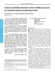
1 2 3<br />
4 5<br />
6 7 8<br />
226<br />
Fig. 1. Sagittal FSE T2-weightted image in a 9-month-old boy with a transverse colon stoma (long white arrow), a high ARM, <strong>and</strong> a rectourethral<br />
fistula. The rectal pouch ends above <strong>the</strong> levator ani muscle (black arrow), <strong>and</strong> <strong>the</strong> fistula (white arrowhead) extends anteriorly to <strong>the</strong> urethra (short<br />
white arrow), BL, <strong>and</strong> bladder.<br />
Fig. 2. Sagittal FSE T2-weighted image in a 1-year-old girl with low ARM <strong>and</strong> rectovaginal fistula (white arrow), who had Currarino triad including<br />
three correlative processes <strong>of</strong> <strong>anorectal</strong> malformation, sacral bony defect, <strong>and</strong> presacral mass (black arrow). The rectal pouch ends below <strong>the</strong> levator<br />
ani muscle, BL, <strong>and</strong> bladder.<br />
Fig. 3. Sagittal FSE T2-weighted image in a 2-year-old girl with cloacal deformity (high type) showing a 3-cm length co-tube (arrowhead), <strong>the</strong><br />
maldeveloped puborectalis muscle <strong>and</strong> external anal sphincter (arrow), BL, <strong>and</strong> bladder (scaphoid).<br />
Fig. 4. Traverse SE T1-weighted image in a 3-month-old girl. The anal canal at <strong>the</strong> level <strong>of</strong> <strong>the</strong> superficial transverse perineal muscle (small dark<br />
arrow) showed an ectopic anterior location (white arrow), ventral to <strong>the</strong> external sphincter (large dark arrow). The external anal sphincter depicted as<br />
normal development (EASWR=2.2/5.6=0.39).<br />
Fig. 5. Traverse STIR T1-weighted image in a 5-month-old boy with high ARM failed to show rectal lumen structures on <strong>the</strong> PC plane. The<br />
puborectalis muscle was easily depicted as maldevelopment (PRWR=0.3/2.1=0.14).<br />
Fig. 6. Sagittal SE T1-weighted image in a 13-year-old boy with complete incontinenece after reconstructive surgery for a high imperforate anus. PR<br />
<strong>and</strong> EASTR cloudiness indicate <strong>the</strong> maldevelopment (black arrow), <strong>and</strong> <strong>the</strong> <strong>anorectal</strong> angle <strong>of</strong> 118º.<br />
Fig. 7. Sagittal FSE T2-weighted image in a 5-year-old girl with a high ARM <strong>and</strong> rectovaginal fistulae, who has incontinence after reconstructive<br />
surgery. A te<strong>the</strong>red spinal cord (long arrow) <strong>and</strong> a syringomyelia on T12-L4 (short arrow). The vertebral column terminated at L5-S1.<br />
Fig. 8. Coronal FSE T2-weighted image in an 8-month-old boy with high ARM. Arrows indicate right hydronephrosis.<br />
. World J Pediatr, Vol 2 No 3 . August 15, 2006
<strong>MRI</strong> <strong>of</strong> ARM <strong>and</strong> <strong>relationship</strong> <strong>of</strong> <strong>the</strong> developmental state <strong>of</strong> <strong>the</strong> SMC with fecal continence<br />
Table 3. Developmental quantization indexes for patients with SMC in different imaging<br />
Degree<br />
Puborectalis<br />
External sphincter<br />
n Relative magnitude <strong>of</strong> width (PRWR) n Relative magnitude <strong>of</strong> width (EASWR)<br />
Excellent (I) 19 0.51±0.04 19 0.45±0.03<br />
Good (II) 13 0.48±0.03 11 0.42±0.02<br />
Poor (III) 7 0.15±0.03 9 0.13±0.02<br />
I <strong>and</strong> II: q=1.641, P>0.05; I <strong>and</strong> III: q=26.247, P0.05;<br />
EASWR q=1.493, P>0.05) (Table 4).<br />
Postoperative follow-up<br />
Thirty patients with ARM were followed up<br />
postoperatively for 1-6 years. When PRWR was 0.15, clinical score for 70% <strong>of</strong> <strong>the</strong><br />
patients was excellent, 21% good, <strong>and</strong> 9% poor in fecal<br />
continence (Table 5).<br />
Developmental index <strong>of</strong> <strong>the</strong> SMC, <strong>anorectal</strong><br />
manometry <strong>and</strong> barium enema<br />
Anorectal contraction pressure in 7 patients with poor<br />
development <strong>of</strong> <strong>the</strong> SMC (PRWR
World Journal <strong>of</strong> Pediatrics<br />
Original article<br />
228<br />
<strong>of</strong> <strong>the</strong> genitourinary system were found in 8 <strong>of</strong> <strong>the</strong> 39<br />
patients including unilateral renal hypoplasia (2 patients),<br />
renal ectopia (1), renal cyst (2), hydronephrosis (1) (Fig.<br />
8), cystic agenesis (1), <strong>and</strong> uterovaginal agenesis (1). Two<br />
<strong>of</strong> <strong>the</strong>se 8 patients demonstrated multiple associated<br />
anomalies <strong>of</strong> <strong>the</strong> genitourinary system.<br />
Discussion<br />
Traditional X-ray film <strong>and</strong> retrograde contrast<br />
examination fail to directly display <strong>the</strong> <strong>relationship</strong><br />
between <strong>the</strong> blind end <strong>of</strong> <strong>the</strong> rectum <strong>and</strong> <strong>the</strong> SMC.<br />
Ultrasonography can detect <strong>the</strong> distance between <strong>the</strong><br />
blind end <strong>of</strong> <strong>the</strong> rectum <strong>and</strong> anal pit, but can not depict<br />
distinct image <strong>of</strong> <strong>the</strong> SMC. [2,3] CT can directly reveal<br />
<strong>the</strong> developmental state <strong>of</strong> <strong>the</strong> SMC, but is limited<br />
to <strong>the</strong> transverse plane <strong>and</strong> difficult to demonstrate<br />
atresia level <strong>and</strong> fistulae. [4-7] Endoanal <strong>MRI</strong> possesses<br />
a higher resolution on muscles, but <strong>the</strong> signal intensity<br />
would decrease rapidly in <strong>the</strong> region beyond 3 cm<br />
from <strong>the</strong> coil, making it impossible to display o<strong>the</strong>r<br />
anomalies in patients with ARM. Meanwhile, infants<br />
may feel uncomfortable to some extent using endoanal<br />
coil, which is not suitable for <strong>the</strong> inspection <strong>of</strong> infants<br />
<strong>and</strong> patients with ARM. [8,9]<br />
Phased-array <strong>MRI</strong> (head<br />
coil in babies) however can perspicuously display all<br />
anomalies <strong>of</strong> ARM.<br />
Application <strong>of</strong> <strong>MRI</strong> techniques<br />
At present, no defined sequences or parameters on<br />
<strong>MRI</strong> are available for patients with ARM. Recent<br />
reports [4,9-12]<br />
have indicated that <strong>MRI</strong> <strong>of</strong> ARM can<br />
be done in spin-echo T1-weighted, fast spin-echo<br />
T2-weighted, <strong>and</strong> short T1 inversion recovery T1-<br />
weighted sequences. The sagittal plane includes <strong>the</strong><br />
pelvis, lumbosacral vertebrae, <strong>and</strong> kidneys, whereas<br />
<strong>the</strong> axial <strong>and</strong> coronal planes are initially limited to<br />
<strong>the</strong> pelvis angulated parallelly <strong>and</strong> perpendicularly to<br />
<strong>the</strong> pelvic floor, respectively. When <strong>malformations</strong> <strong>of</strong><br />
<strong>the</strong> spinal cord, spine, <strong>the</strong> high level <strong>of</strong> ARM, <strong>and</strong>/or<br />
kidneys are detected on <strong>the</strong> sagittal images, additional<br />
axial <strong>and</strong>/or coronal images in <strong>the</strong> region <strong>of</strong> interest<br />
were obtained. In this study, <strong>the</strong> level <strong>of</strong> ARM was<br />
depicted correctly in all patients with T1WI <strong>and</strong><br />
T2WI. The detection rate for fistula was 50% for T1-<br />
weighted images, but <strong>the</strong> higher signal intensity <strong>of</strong><br />
fistular mucosa on FSE T2-weighted sagittal <strong>and</strong> axial<br />
images could increase <strong>the</strong> detection rate, despite <strong>the</strong><br />
difficulty in delineating <strong>the</strong> exact site <strong>of</strong> entrance in<br />
<strong>the</strong> genitourinary system. The fistulae were depicted<br />
correctly in all patients but one who had a minimal<br />
. World J Pediatr, Vol 2 No 3 . August 15, 2006<br />
rectovesical fistula, giving a diagnostic rate <strong>of</strong> 97%.<br />
On muscular images, however, <strong>the</strong> sphincters were<br />
clearly demonstrated by routine T1WI <strong>and</strong> T2WI<br />
sequences. The anatomic characteristics <strong>of</strong> <strong>the</strong> SMC<br />
were depicted more affluently on T2-weighted images.<br />
Hence, if <strong>the</strong> sphincters cannot be demonstrated<br />
clearly with routine <strong>MRI</strong> in newborns or infants with<br />
<strong>the</strong> poor developmental state <strong>of</strong> <strong>the</strong> sphincters, short<br />
T1 inversion recovery T1-weighted sequences can be<br />
applied. As a result, fat signal is restrained <strong>and</strong> <strong>the</strong><br />
shape <strong>and</strong> border <strong>of</strong> muscles can be detected more<br />
distinctly.<br />
Relationship between <strong>the</strong> blind end <strong>of</strong> <strong>the</strong> rectum<br />
<strong>and</strong> SMC<br />
In <strong>the</strong> blind end <strong>of</strong> <strong>the</strong> rectum in newborns, meconium<br />
containing mixture <strong>of</strong> mucus <strong>and</strong> lipid has a higher<br />
intensity on both T1WI <strong>and</strong> T2WI sequences in sharp<br />
contrast to <strong>the</strong> surrounding tissues as a good contrast<br />
medium <strong>of</strong> <strong>MRI</strong>. [10,13]<br />
T2WI image is particularly<br />
helpful in <strong>the</strong> evaluation <strong>of</strong> low <strong>and</strong> intermediate ARM,<br />
because <strong>the</strong> high signal intensity <strong>of</strong> <strong>anorectal</strong> mucosa<br />
allows better delineation <strong>of</strong> <strong>the</strong> anorectum, SMC <strong>and</strong><br />
perineum. The level <strong>of</strong> ARM can be defined as two<br />
classical planes, PC plane <strong>and</strong> I plane, in transverse<br />
sequences. It is a low level <strong>of</strong> ARM if <strong>the</strong> rectum can<br />
be seen from <strong>the</strong> two planes. If <strong>the</strong> rectum can be seen<br />
on PC plane but not on I plane, <strong>the</strong> level <strong>of</strong> ARM is<br />
intermediate. It is a high level <strong>of</strong> ARM if <strong>the</strong> rectum<br />
can not be seen on both planes. The position <strong>of</strong> <strong>the</strong> anal<br />
canal <strong>and</strong> its <strong>relationship</strong> with SMC can be detected<br />
correctly at <strong>the</strong> low level ARM patients, with <strong>the</strong><br />
superficial transverse muscle <strong>of</strong> <strong>the</strong> perineum as a mark<br />
for defining <strong>the</strong> dorsal surface <strong>of</strong> <strong>the</strong> genitourinary<br />
diaphragm. Normally <strong>the</strong> anal canal is located behind<br />
this muscle. When it is at <strong>the</strong> level <strong>of</strong> or ventral to<br />
this muscle, <strong>the</strong> anal canal has an ectopic anterior<br />
location outside <strong>of</strong> <strong>the</strong> SMC. On <strong>the</strong> sagittal plane, it is<br />
high, intermediate, <strong>and</strong> low ARM if <strong>the</strong> atresia <strong>of</strong> <strong>the</strong><br />
distal rectum is above, through, <strong>and</strong> below <strong>the</strong> SMC,<br />
respectively. On <strong>the</strong> coronal plane, <strong>the</strong> <strong>relationship</strong><br />
between <strong>the</strong> levator ani muscle, <strong>the</strong> puborectalis,<br />
external anal sphincter, <strong>and</strong> anorectum can be<br />
demonstrated clearly. These findings suggest that <strong>the</strong><br />
level <strong>of</strong> anal atresia can be depicted more accurately on<br />
sagittal <strong>and</strong> coronal planes.<br />
Muscular developmental state <strong>and</strong> developmental<br />
index<br />
Our previous <strong>MRI</strong> study [14]<br />
showed that <strong>the</strong> absolute<br />
length <strong>and</strong> width <strong>of</strong> <strong>the</strong> puborectalis <strong>and</strong> external<br />
anal sphincter significantly increased with age, but
<strong>MRI</strong> <strong>of</strong> ARM <strong>and</strong> <strong>relationship</strong> <strong>of</strong> <strong>the</strong> developmental state <strong>of</strong> <strong>the</strong> SMC with fecal continence<br />
<strong>the</strong> relative length <strong>and</strong> width <strong>of</strong> <strong>the</strong> muscles were not<br />
correlated with age below 14 years. Therefore, this<br />
relative values might be taken as objective criteria for<br />
<strong>the</strong> measurement <strong>of</strong> <strong>the</strong> SMC in children during <strong>the</strong><br />
development period. In this study, excellent or good<br />
developmental state <strong>of</strong> muscles was found in all patients<br />
with low ARM except one patient with intermediate<br />
ARM, <strong>and</strong> in some patients with high ARM.<br />
Fur<strong>the</strong>rmore, <strong>the</strong> width <strong>and</strong> length <strong>of</strong> <strong>the</strong> two muscles<br />
developed uniformly <strong>and</strong> were positively correlated<br />
(r=0.987, 0.995, P
World Journal <strong>of</strong> Pediatrics<br />
Original article<br />
230<br />
<strong>the</strong> final version. RQL is <strong>the</strong> guarantor.<br />
Acknowledgements<br />
We are grateful to Pr<strong>of</strong>. Xiang-Quan Kong <strong>and</strong> Pr<strong>of</strong>. Zheng-<br />
Jun Peng from Department <strong>of</strong> Radiology, Union Hospital <strong>of</strong><br />
Tongji Medical College, Huazhong University <strong>of</strong> Science <strong>and</strong><br />
Technology for reading pelvic MR images.<br />
References<br />
1 Pena A, Devries PA. Posterior sagittal anorectoplasty:<br />
important technical considerations <strong>and</strong> new applications. J<br />
Pediatr Surg 1982;17:796-811.<br />
2 Oppenheimer DA, Carroll BA, Shochat SJ. Sonography <strong>of</strong><br />
imperforate anus. Radiology 1983;148:127-128.<br />
3 Jones NM, Humphreys MS, Goodman TR, Sullivan PB,<br />
Grant HW. The value <strong>of</strong> anal endosonography compared with<br />
magnetic resonance imaging following <strong>the</strong> repair <strong>of</strong> <strong>anorectal</strong><br />
<strong>malformations</strong>. Pediatr Radiol 2003;33:183-185.<br />
4 Nievelstein RA, Vos A, Valk J. MR imaging <strong>of</strong> <strong>anorectal</strong> <strong>malformations</strong><br />
<strong>and</strong> associated anomalies. Eur Radiol 1998;8: 573-581.<br />
5 Arnbjornsson E, Laurin S, Mikaelsson C. Computed tomography<br />
<strong>of</strong> <strong>anorectal</strong> anomalies. Correlation between radiologic<br />
findings <strong>and</strong> clinical evaluation <strong>of</strong> faecal incontinence. Acta<br />
Radiol 1989;30:25-28.<br />
6 Arnbjornsson E, Malmgren N, Mikaelsson C, Laurin S,<br />
Okmian L. Computed tomography <strong>and</strong> magnetic resonance<br />
tomography findings in children operated for anal atresia. Z<br />
Kinderchir 1990;45:178-181.<br />
7 Wang DY, Qiu XH, Li L, Wang YX, Sun GQ. The approach <strong>of</strong><br />
<strong>anorectal</strong> <strong>malformations</strong>, <strong>the</strong> morphological development <strong>of</strong><br />
musculus puborectalis <strong>and</strong> musculus sphincter. Chin J Pediatr<br />
Surg 1999;1:15-17.<br />
8 Kubota M, Suita S. Assessment <strong>of</strong> sphincter muscle function<br />
before <strong>and</strong> after posterior sagittal anorectoplasty using a<br />
magnetic spinal stimulation technique. J Pediatr Surg 2002;37:<br />
617-622.<br />
9 Beets-Tan RG, Morren GL, Beets GL, Kessels AG, el Naggar<br />
K, Lemaire E, et al. Measurement <strong>of</strong> anal sphincter muscles:<br />
endoanal US, endoanal MR imaging, or phased-array MR<br />
imaging A study with healthy volunteers. Radiology 2001;<br />
220:81-89.<br />
10 McHugh K, Dudley NE, Tam P. Pre-operative <strong>MRI</strong> <strong>of</strong><br />
<strong>anorectal</strong> abnormality in <strong>the</strong> newborn period. Pediatr Radiol<br />
1995;25(Suppl 1):S33-36.<br />
11 Hettiarachchi M, Garcea G, deSouza NM, Williams AD,<br />
Clayden GS, Ward HC. Evaluation <strong>of</strong> dysfunction following<br />
reconstruction <strong>of</strong> an <strong>anorectal</strong> anomaly. Pediatr Surg Int 2002;<br />
18:405-409.<br />
12 Nievelstein RA, Vos A, Valk J, Vermeij-Keers C. Magnetic<br />
resonance imaging in children with <strong>anorectal</strong> <strong>malformations</strong>:<br />
embryologic implications. J Pediatr Surg 2002;37:1138-1145.<br />
13 Sachs TM, Applebaum H, Touran T, Taber P, Darakjian A,<br />
Colleti P. Use <strong>of</strong> <strong>MRI</strong> in evaluation <strong>of</strong> <strong>anorectal</strong> abnormality.<br />
J Pediatr Surg 1990;25:817-821.<br />
14 Tang ST, Mao YZ, Wang Y, Tong QS, Li SW. Quantification<br />
<strong>of</strong> striated muscle complex in normal children with magnetic<br />
resonance imageing. Chin J Pediatr Surg 2005;6:314-318.<br />
15 Levitt MA, Pena A. Outcomes from <strong>the</strong> correction <strong>of</strong><br />
<strong>anorectal</strong> <strong>malformations</strong>. Curr Opin Pediatr 2005;17:394-401.<br />
16 Hul<strong>the</strong>n de Medina V, Mellstam L, Amark P, Amark P,<br />
Frenckner B. Neurovesical dysfunction in children after<br />
surgery for high or intermediate <strong>anorectal</strong> <strong>malformations</strong>.<br />
Acta Paediatr 2004;93:43-46.<br />
17 Boemers TM, Beek FJ, van Gool JD, de Jong TP, Bax KM.<br />
Urologic problems in <strong>anorectal</strong> <strong>malformations</strong>. Part 1:<br />
Urodynamic findings <strong>and</strong> significance <strong>of</strong> sacral abnormality. J<br />
Pediatr Surg 1996;31:407-410.<br />
18 Davies MC, Creighton SM, Wilcox DT. Long-term outcomes<br />
<strong>of</strong> <strong>anorectal</strong> <strong>malformations</strong>. Pediatr Surg Int 2004;20:567-572.<br />
19 Mosiello G, Capitanucci ML, Gatti C, Adorisio O, Lucchetti<br />
MC, Silveri M, et al. How to investigate neurovesical<br />
dysfunction in children with <strong>anorectal</strong> <strong>malformations</strong>. J Urol<br />
2003;170:1610-1613.<br />
20 Tsuji H, Okada A, Nakai H, Azuma T, Yagi M, Kubota A.<br />
Follow-up studies <strong>of</strong> <strong>anorectal</strong> <strong>malformations</strong> after posterior<br />
sagittal anorectoplasty. J Pediatr Surg 2002;37:1529-1533.<br />
21 Jones NM, Humphreys MS, Goodman TR, Sullivan PB,<br />
Grant HW. The value <strong>of</strong> anal endosonography compared with<br />
magnetic resonance imaging following <strong>the</strong> repair <strong>of</strong> <strong>anorectal</strong><br />
<strong>malformations</strong>. Pediatr Radiol 2003;33:183-185.<br />
22 Vliegen RF, Beets-Tan RG, van Heurn LW, van Engelshoven<br />
JM. High resolution <strong>MRI</strong> <strong>of</strong> <strong>anorectal</strong> malformation in<br />
<strong>the</strong> newborn: case reports <strong>of</strong> Currarino syndrome <strong>and</strong><br />
anocutaneous fistula. Abdom Imaging 2002;27:344-346.<br />
23 Riebel T, Maurer J, Teichgraber UK, Bassir C. The spectrum<br />
<strong>of</strong> imaging in Currarino triad. Eur Radiol 1999;9:1348-1353.<br />
24 Lee SC, Chun YS, Jung SE, Park KW, Kim WK. Currarino<br />
triad: <strong>anorectal</strong> malformation, sacral bony abnormality, <strong>and</strong><br />
presacral mass—a review <strong>of</strong> 11 cases. J Pediatr Surg 1997;32:<br />
58-61.<br />
Received May 23, 2006<br />
Accepted after revision May 31, 2006<br />
. World J Pediatr, Vol 2 No 3 . August 15, 2006