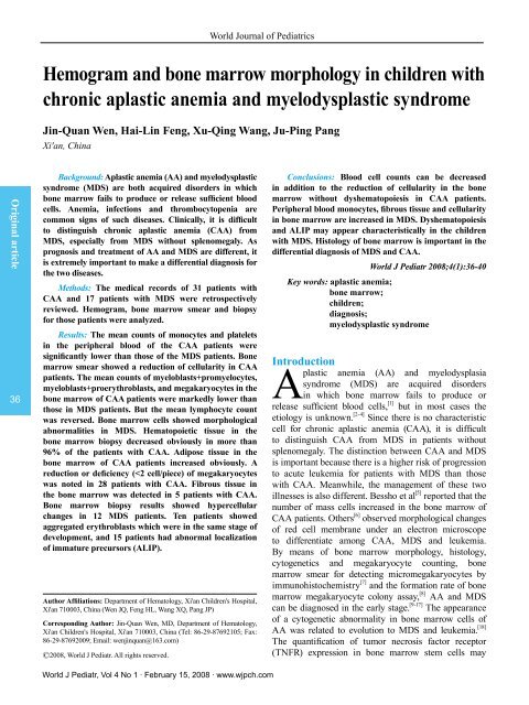
Hemogram and bone marrow morphology in children with chronic ...
Hemogram and bone marrow morphology in children with chronic ...
Hemogram and bone marrow morphology in children with chronic ...
You also want an ePaper? Increase the reach of your titles
YUMPU automatically turns print PDFs into web optimized ePapers that Google loves.
World Journal of PediatricsTable 3. Dyshematopoiesis of <strong>bone</strong> <strong>marrow</strong> <strong>in</strong> the CAA <strong>and</strong> MDS groupsGroupnMicromegakaryocyte(%)S<strong>in</strong>gle-round-nucleusmegakaryocyte (%)Multi-round-nucleusmegakaryocyte (%)Pelger-Huet Megaloblastic changes <strong>in</strong>abnormality (%) granulocytic l<strong>in</strong>eage (%)CAA 31 0 0 0 0 0 0MDS 17 94.1 29.4 64.7 17.6 47.0 88.29430 bpMegaloblastic changes <strong>in</strong>erythrocytic l<strong>in</strong>eage (%)Orig<strong>in</strong>al articleTable 4. Bone <strong>marrow</strong> biopsy results <strong>in</strong> the CAA <strong>and</strong> MDS groupsGroupnStage of proliferationHypercellularity Normal cellularity Hypocellularity Extreme hypocellularityCAA 31 0/31 1/31 23/31 7/31HemotopoietictissueObviousreductionAdiposetissueObvious<strong>in</strong>ductionMDS 17 2/17 10/17 5/17 0 Normal range Normal range 17/17Table 5. Bone <strong>marrow</strong> biopsy results <strong>in</strong> the CAA <strong>and</strong> MDS groupsGroupnErythroblasts <strong>in</strong> thesame develop<strong>in</strong>g stageALIPProliferation offibr<strong>in</strong> tissueMicromegakaryocyteS<strong>in</strong>gle-round-nucleusmegakaryocyteCAA 31 0 0 5/31 0 0 0MDS 17 7/17 15/17 17/17 17/17 9/17 13/17ALIP: abnormal localization of immature precursors.Existence ofmegakaryocyte3/31Multi-round-nucleusmegakaryocyte38Fig. 1. Bone <strong>marrow</strong> smear <strong>in</strong> the CAA group show<strong>in</strong>g hypocellularity<strong>and</strong> normal <strong>morphology</strong> (Wright, 10×100).Fig. 2. Dyshematopoiesis <strong>in</strong> the MDS group shown by <strong>bone</strong> <strong>marrow</strong>smear. Arrow: erythroblasts dysplasia; arrowhead: Pegler-Hunt(Wright, 10×100).Fig. 3. Bone <strong>marrow</strong> biopsy <strong>in</strong> the CAA group show<strong>in</strong>g hypocelluarity<strong>and</strong> normal <strong>morphology</strong> (HGE, 10×40).Fig. 4. MDS <strong>in</strong> <strong>bone</strong> <strong>marrow</strong> biopsy. Arrow: abnormal localization ofimmature precursors; arrowhead: s<strong>in</strong>gle-round-nucleus megakaryocytes(HGE, 10×40).World J Pediatr, Vol 4 No 1 . February 15, 2008 . www.wjpch.com
Chronic aplastic anemia <strong>and</strong> myelodysplastic syndromeBone <strong>marrow</strong> smear <strong>in</strong> CAA <strong>and</strong> MDS patientsBone <strong>marrow</strong> of the MDS patients showed activeor marked proliferation. The counts of myeloblasts,promyelocytes, proerythroblasts, basophilic erythroblasts<strong>and</strong> megakaryocytes <strong>in</strong> the <strong>bone</strong> <strong>marrow</strong> of theMDS patients were significantly higher than thoseof the CAA patients, <strong>and</strong> the count of lymphocyteswas lower than that <strong>in</strong> the CAA patients (P
World Journal of PediatricsOrig<strong>in</strong>al article40<strong>children</strong>. Bone <strong>marrow</strong> biopsy for MDS can reveal<strong>in</strong>significant deficiency of hemotopoietic tissue [7]<strong>in</strong>contrast to fibr<strong>in</strong> tissue because of the productionof megakaryocytes <strong>with</strong> no function. [25]Abnormalmegakaryocytes, ALIP <strong>and</strong> dyshematopoiesis arethe features of MDS. [26]Erythroblast islets <strong>in</strong> the<strong>bone</strong> <strong>marrow</strong> of MDS <strong>and</strong> CAA patients <strong>in</strong> the samedevelop<strong>in</strong>g stage have not been reported so far.Various cl<strong>in</strong>ical features of CAA <strong>and</strong> MDS are perhapsbased on different changes of molecular biology, buthemogram, <strong>bone</strong> <strong>marrow</strong> smear <strong>and</strong> biopsy are alwaysessential to the diagnosis of blood diseases.Fund<strong>in</strong>g: None.Ethical approval: Not needed.Compet<strong>in</strong>g <strong>in</strong>terest: No benefits <strong>in</strong> any form have been receivedor will be received from a commercial party related directly or<strong>in</strong>directly to the subject of this article.Contributors: Wen JQ proposed the study <strong>and</strong> wrote the firstdraft. Feng HL analyzed the data. All authors contributed to thedesign <strong>and</strong> <strong>in</strong>terpretation of the study <strong>and</strong> to further drafts. WangXQ is the guarantor.References1 Yang CL. Aplastic anemia, 2nd ed. Tianj<strong>in</strong>: Tianj<strong>in</strong>Technology Translat<strong>in</strong>g Publish<strong>in</strong>g Corporation, 2000: 1-2.2 Gewirtz AM, Hoffman R. Current considerations of theetiology of aplastic anemia. Crit Rev Oncol Hematal1985;4:1-30.3 Aggio MC, Alvarez RV, Bartomioli MA, Maguitman O.Incidence <strong>and</strong> etiology of aplastic anemia <strong>in</strong> a def<strong>in</strong>edpopulation of Argent<strong>in</strong>a (1966-1977). Medic<strong>in</strong>a (B Aires)1988;48:231-233.4 Marti J, Garcia-Mart<strong>in</strong> C. Aplastic anemia caused bycarbamazep<strong>in</strong>e. Neurologia 1989;4:221-222.5 Bessho F, Imashuku SH, Tsuchida M, Nakata T, Mivazaki S.Serial morphologic observation of <strong>bone</strong> <strong>marrow</strong> <strong>in</strong> aplasticanemia <strong>in</strong> <strong>children</strong>. Int J Hematol 2005;81:400-404.6 Majumder D, Banerjee D, Ch<strong>and</strong>ra S, Banerjee S,Chakerabarti A. Red cell <strong>morphology</strong> <strong>in</strong> leukemia, hypoplasticanemia <strong>and</strong> myelodysplastic syndrome. Pathophysiology2006;13:217-225.7 Das R, Hayer J, Dey P, Garewal G. Comparative study ofmyelodysplastic syndromes <strong>and</strong> normal <strong>bone</strong> <strong>marrow</strong> biopsies<strong>with</strong> conventional sta<strong>in</strong><strong>in</strong>g <strong>and</strong> immunocytochemistry. AnalQuant Cytol Histol 2005;27:152-156.8 Hu T, Shi XD, Feng YL, Liu R, Li JH, Chen J. Comparativestudy on <strong>bone</strong> <strong>marrow</strong> megakaryocytes <strong>in</strong> <strong>children</strong><strong>with</strong> thrombocytopenic purpura, aplastic anemia <strong>and</strong>myelodysplastic syndromes. Ch<strong>in</strong> J Pediatr 2005;43:183-187.9 Sakuma T, Hayashi Y, Kanomata N, Murayama T, Matsui T,Kajimoto K. Histological <strong>and</strong> cytogenetic characterizationof <strong>bone</strong> <strong>marrow</strong> <strong>in</strong> relation to prognosis <strong>and</strong> diagnosis ofmyelodysplastic syndromes. Pathol Int 2006;56:191-199.10 Saad ST, Vassallo J, Arruda VA, Lor<strong>and</strong>-Metze I. Theroleof <strong>bone</strong> <strong>marrow</strong> study <strong>in</strong> diagnosis <strong>and</strong> prognosis ofmyelodysplastic syndromes. Pathologica 1994;86:47-51.11 Barrett J, Saunthararajah Y, Molldrem J. Myelodysplasticsyndromes <strong>and</strong> aplastic anemia:dist<strong>in</strong>ct entities or diseasesl<strong>in</strong>ked by a common pathophysiology. Sem<strong>in</strong> Hematol2000;37:15-29.12 Luraschi A, Buscaglia P, Fedeli P, Montanara S, Uccelli E,Antonietti MP. Myelodysplastic syndromes. Cl<strong>in</strong>ico-pathologicanalysis of 54 cases. Recenti Prog Med 2001;92:521-529.13 Zhang H, Hu B. Application of bioptic <strong>bone</strong> <strong>marrow</strong> impr<strong>in</strong>t<strong>in</strong> diagnosis of anemia. Zhongguo Shi Yan Xue Ye Xue ZaZhi 2002;10:131-132.14 Rigol<strong>in</strong> GM, Bigoni R, Milani R, Cavazz<strong>in</strong>i F, Roberti MG,Bardi A. Cl<strong>in</strong>ical importance of <strong>in</strong>terphase cytogeneticsdetect<strong>in</strong>g occult chromosome lesions <strong>in</strong> myelodysplasticsyndromes <strong>with</strong> normal karyotype. Leukemia 2001;15:1841-1847.15 Rossi G, Pelizzari AM, Bellotti D, Tonelli M, Barlati S.Cytogenetic analogy between myelodysplastic syndromes<strong>and</strong> acute myeloid leukemia of elderly patients. Leukemia2000;14:47-51.16 Marisavljevic D, Cemerikic V, Rolovic Z, Boskovic D,Colovic M. Hypocellular myelodysplastic syndromes: cl<strong>in</strong>ical<strong>and</strong> biological significance. Med Oncol 2005;22:169-175.17 Lawrence LW. Refractory anemia <strong>and</strong> the myelodysplasticsyndromes. Cl<strong>in</strong> Lab 2004;17:178-186.18 Maciejewski JP, Risitano A, Slo<strong>and</strong> EM, Nunez O, YoungNS. Dist<strong>in</strong>ct cl<strong>in</strong>ical outcomes for cytogenetic abnormalitiesevolv<strong>in</strong>g from aplastic anemia. Blood 2002;99:3129-3135.19 Kasahara S, Hara T, Itoh H, Ando K, Tsurumi H, SawadaM, et al. Hypoplastic myelodysplastic syndromes can bedist<strong>in</strong>guished from acquired aplastic anemia by <strong>bone</strong> <strong>marrow</strong>stem cell expression of the tumour necrosis factor receptor. BrJ Haematol 2002;118:181-188.20 Zhang ZN. Diagnosis <strong>and</strong> curative effect st<strong>and</strong>ards for blooddiseases, 2nd ed. Beij<strong>in</strong>g: Science & Technology Publish<strong>in</strong>gCorporation, 1998: 33.21 Bennett JM, Catovsky D, Daniel MT, Fl<strong>and</strong>r<strong>in</strong> G, GaltonDA, Gralnick HR, et al. Proposals for the classification of themyelodysplastic syndromes. Br J Haematol 1982;51:189-199.22 Ji MR, Xie Y. The morphological diagnosis of complicatedblood diseases. Shanghai: Shanghai Science & TechnologyPublish<strong>in</strong>g Corporation, 2002: 280.23 Beutler E, Lichtman MA, Coller T, Kipps TJ. Williamshematology, 5th ed. Xi'an: Xi'an The World Publish<strong>in</strong>gCorporation, 1998: 238-247, 257-266.24 Wu MY, Huang SL. Modern Pediatric Hematology. Fuzhou:Fujian Science & Technology Publish<strong>in</strong>g Corporation, 2003:432-442.25 Shen ZX, OuYang RR. Hematology Oncology. Beij<strong>in</strong>g:People's Medical Publish<strong>in</strong>g House, 1999: 482.26 Pu Q. Color atlas of histopathology <strong>in</strong> hematology. Tianj<strong>in</strong>:Tianj<strong>in</strong> Technology Publish<strong>in</strong>g Corporation, 1990: 67.Received July 25, 2006Accepted after revision October 12, 2007World J Pediatr, Vol 4 No 1 . February 15, 2008 . www.wjpch.com


