1 Chapter 8: Skin Flap Physiology George S. Goding ... - Famona Site
1 Chapter 8: Skin Flap Physiology George S. Goding ... - Famona Site
1 Chapter 8: Skin Flap Physiology George S. Goding ... - Famona Site

Create successful ePaper yourself
Turn your PDF publications into a flip-book with our unique Google optimized e-Paper software.
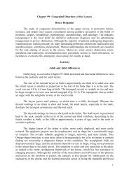
Neurovascular supply to skin<br />
The arterial supply to the skin can be divided into three functional units (Daniel and<br />
Williams, 1973): segmental vessels, which function to distribute blood from the aorta to the<br />
undersurface of muscle; perforating vessels, which provide nutritional support to muscle; and<br />
cutaneous vessels, which allow for thermoregulation and nutritional support of skin (Fig. 8-2).<br />
The segmental vessels originate as branches of the paired dorsal aortas during the<br />
embryologic period (Langman, 1975). Characteristics of the segmental vessels include (1) a<br />
perfusion pressure closely related to that found in the aorta, (2) a location deep to muscle, and<br />
(3) a common association with a nerve and vein (Daniel and Williams, 1973).<br />
The perforator vessels are branches of the segmental vessels. These vessels travel via<br />
one of two main routes to terminate in the cutaneous circulation. Musculocutaneous arteries<br />
pass through the overlying muscle to which they provide nutrition whereas direct cutaneous<br />
or septocutaneous arteries travel through fascial septa dividing muscular segments (Daniel and<br />
Kerrigan, 1990).<br />
The cutaneous portion of direct cutaneous (septocutaneous) arteries typically runs<br />
parallel to the skin surface providing nutrition to a large area of skin. Direct cutaneous<br />
arteries typically are accompanied by a pair of veins and run above the superficial muscular<br />
fascia (Webster, 1937). The more common musculocutaneous arteries leave the muscle and<br />
directly penetrate the subcutaneous tissue to supply a smaller region of skin.<br />
Both direct cutaneous and musculocutaneous arteries empty into a diffuse,<br />
interconnecting vascular network often referred to as the dermal and subdermal plexi. This<br />
network provides a redundancy in the vascular supply to the skin with the formation of<br />
collaterals at the periphery of the vascular territory formed by each musculocutaneous artery.<br />
The cutaneous microcirculation consists of a nutrient capillary network in the reticular dermis<br />
and arteriovenous shunts in the more superficial papillary dermis (Sherman, 1963). Arterioles,<br />
which act as preshunt and precapillary sphincters, regulate the flow through each vascular<br />
network (Greene, 1962). Lymphatic vessels form a plexus running parallel and deep to the<br />
network of blood capillaries (Bloom and Fawcett, 1975). The lymphatic capillaries end in<br />
blind sacs and conduct extracellular fluid back into the bloodstream.<br />
The neural supply to the skin originates from both sensory and sympathetic nerves.<br />
The sensory nerves are distributed in segmental fashion, forming dermatomes, and participate<br />
in the skin's protective function. The postganglionic terminals of cutaneous sympathetic nerves<br />
contain the neurotransmitter norepinephrine and are found in the area of cutaneous arterioles<br />
(Anden et al, 1969; Guyton, 1976; Mellander and Johansson, 1968).<br />
<strong>Skin</strong> <strong>Physiology</strong><br />
The skin serves as a sensory and a protective organ. The thick epidermal layers are<br />
largely impermeable to gases and to most liquids. Because of this, many agents that could<br />
have beneficial effects are ineffective when applied topically to intact skin. Preservation of<br />
sensation in transferred cutaneous flaps is desirable, but its effects on the physiology of flaps<br />
is unclear.<br />
2