
You also want an ePaper? Increase the reach of your titles
YUMPU automatically turns print PDFs into web optimized ePapers that Google loves.
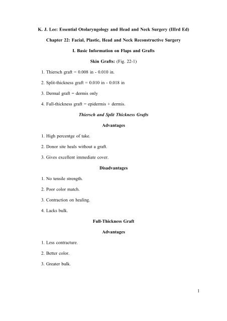
K. J. Lee: Essential Otolaryngology and Head and Neck Surgery (IIIrd Ed)<strong>Chapter</strong> <strong>22</strong>: Facial, Plastic, Head and Neck Reconstructive SurgeryI. Basic Information on Flaps and GraftsSkin Grafts: (Fig. <strong>22</strong>-1)1. Thiersch graft = 0.008 in - 0.010 in.2. Split-thickness graft = 0.010 in - 0.018 in3. Dermal graft = dermis only4. Full-thickness graft = epidermis + dermis.Thiersch and Split Thickness GraftsAdvantages1. High percentge of take.2. Donor site heals without a graft.3. Gives excellent immediate cover.Disadvantages1. No tensile strength.2. Poor color match.3. Contraction on healing.4. Lacks bulk.Full-Thickness GraftAdvantages1. Less contracture.2. Better color.3. Greater bulk.1
Disadvantages1. Small areas.2. Poorer take.Dermal GraftsAdvantages1. Strong - will take in irradiated tissue.2. High resistance to infections.Uses1. For carotid artery cover following neck dissection in irradiated patients.2. For pharyngeal suture line cover in irradiated patients.Physiology of Graft Nutritionbed.0-12 hours Plasma circulation, nutrition to graft by imbibition of exudate from host12-24 hours Inosculation - direct connection of graft and blood vessels.24-48 hours Host vessels grow into graft.48 hours on Peripheral cellular union of graft and host with epithelialization.Plasmatic circulation works as follows: The hydrostatic pressure at the capillary endof the skin's capillary-venule glomerulus drives crystalloids through the interendothelialcement substance into the tissues where they become tissue fluid. This tissue fluid flowsbetween the fibers of collagen and elastin, embedded in mucopolysaccharides of the groundsubstance. To recirculate or to return to the donor circulation, the tissue fluid can do so bylymphatic circulation or by the blood circulatory system directly. An open wound does nothavee a plasmatic circulation, and hence the tissue fluid escapes through the portal of thewound and is not recirculated. A wound covered with a skin graft permits restoration ofvascular and tissue-fluid balance, returning plasmatic and hemic circulation to normal.Hemic circulation of the graft depends on capillary buds that grow from the donortowards the graft's circumference by seemingly purposeful orientation. Blood flow can bedetected microscopically in new capillary buds by the fourth day.Once the transplant has a vascular purchase on the host bed, peripheral cellular unionof graft and host is accomplished by epithelization. The rate of epithelization is about 0.5mm/day.2
Vascularization and graft take depend on:1. Healthy host bed with adequate blood supply.2. Adequate fixation and immobilization.3. No infection.4. Graft over bone or cartilage requires intact periosteum or perichondrium.Causes of failure of graft take:1. Graft:a. Tension.b. Inadequate immobilization.c. Inaccurate approximation of graft to host bed and the margins.2. Host bed:a. Infection.b. Poor hemostasis (hematoma seroma).c. Fibrosis of bed.d. Irradiation of host bed.e. Lack of perichondrium or periosteum over cartilage and the bone.Major Flaps in Head and Neck Reconstructive Surgery (Pedicle Flaps)A. Classification:1. Forehead.2. Medially-based chest flap.3. Laterally-based chest flap.4. Nape of neck.B. Definition: Pedicle flp (vascular attachment to the body at all times by transferredtissue).3
C. Basic concepts:1. Based on arterial supply.2. A flap is an island. The width of the base of the flap is unimportant beyond thenecessity to contain a vascular pedicle.3. Tubing of flap is useful if only the distal portion is used in the transfer (preventsinfection and granulation).4. Delay of flaps:a. Decision based on need.b. Will increase chances of survival.delay.c. Effects dermal and subdermal vessel hypertrophy and hyperplasia with a 7-14 dayd. Allows greater length of flap for use.5. Transfer of blood supply from graft site distal, useful end of flap - approximately14 days.6. Flaps die of congestion, rarely from anemia.7. The patient's blood pressure, hemoglobin, and hematocrit are vital for flap survival.8. A clean, well-vascularized bed required for transfer.9. No tension at suture line.10. No twisting of pedicle or pressure on pedicle allowed or impairment of flapcirculation will occur.Advantages and Disadvantages of the Flap1. Capable of carrying tissues other than skin.2. Carries its own blood supply, therefore, more likely to "take".3. Less tendency to discolor, more resistant, more elastic, more movable, and lesslikely to contract. Although a 25% leeway should be kept in mind in the planning of a pedicleflap.4. More adaptable to weight bearing.5. Capable of bridging a defect.4
6. Can be used on a host bed of questionable nutrition.7. No pressure dressing necessary.8. One disadvantage is that it usually needs many stages.Forehead FlapArterial base: Superficial temporal artery and postauricular artery. Note: Flap can bebased on scalp vessels should the external carotid be tied in a previous major procedur.Delay: Rarely necessary but can be achieved by isolation and ligation of thesupratrochlear and supraorbital vessels on both sides.Uses: Buccal cavity, oropharynx, hypopharyngeal reconstruction, chin and neck skinreconstruction.Methods of entry:1. Beneath or above the zygoma.2. Through a cheek incision to the buccal cavity.Disadvantage: Disfiguring in females (nipple in the neck).Special uses: A bipedicle bucket handle forehead flap for chin reconstruction.Donor site: Closed by immediate or delayed skin grafting.Specific Forehead Flaps1. Forehead island flap: The flap is based on the supraorbital or supratrochlear artery.It is a full-thickness skin flap with a subcutaneous pedicle carrying the artery. It is transferredsubcutaneously and is useed as a full thickness skin graft over the nasal bridge.2. Indian forehead flap: It is based on the superficial temporal artery. It is used fornasal reconstruction. The disadvantage is a minor cosmetic defect. Closure of the donor siteis with a split-thickness skin graft.3. Median forehead flap: Based on supratrochlear-supraorbital artery, its donor siteeis closed primarily.Medially-Based Chest FlapArterial base: Includes four perforated arteries of the internal mammary artery.Delay: Advisable. Allows greater length and greater reliability.5
Staged delay: Increases length possibilities and reliability.Uses: Buccal cavity, orohypopharyngeal, and cervical esophageal reconstructionfollowing major head and neck reconstruction. Reliable, particularly following delay.Donor site: Closed with split-thickness skin graft, immediate or delayed.Arterial base: Acromiothoracic artery.Laterally-Based Chest FlapDelay: Preferable but not necessary. Will increase both length and reliability.Disadvantages: Disfiguring in females (nipple in the neck).Uses: Buccal-oral cavity, hypopharyngeal, and laryngopharyngeal recostruction.Donor site: Covered by split-thickness skin graft, immediate or delayed.Arterial base: Occipital artery.Delay: Essential.Nape of NeckUses: Neck skin replacement, cervical esophageal reconstruction, oral cavity andhypopharyngeal reconstruction.Disadvantges: One of the least reliable of major head and neck flaps.Donor site: Covered with split-thickness skin graft, delayed or immediate.The Usee of Flaps and Pharyngostomes in Irradiated PatientsIn using flaps for head and neck reconstructive surgery, careful planning is essential.Such planning allows preparation for possible complications. In initial surgery on irradiatedpatients, discretion is always the better part of valor. It often is desirabl to create apharyngostome to be sure of adequate carotid coverage and healing before plannedreconstruction at a later date. Prepared delayed chest flaps with greater length and reliabilityprior to definitive surgery decreases the risks of failure of the flap on reconstruction.6
Transposed, Advancement, and Interposed FlapsDefinition: Use of local viable tissue to close a defect, with simultaneous staggeringof the inevitable scar line.A. Types:1. Z-plasty.2. Simple advancement.3. Bilobe advancement.4. V-Y closure.B. Advantages:1. Viable graft tissue with excellent blood supply.2. Tissue close to the defect, subsequently excellent color and texture match.3. Good scar camouflage.4. Adequate thickness, hence less contracture.5. Necrosis rare with correct planning.C. Disadvantages: Limited by local available tissue.D. Uses:1. To correct contracted linear scar.2. To reposition malposed tissue.3. To close facial cutaneous defects.Z-PlastyA. Definition: Classically, the Z-plasty is based on a Z figure. The central limb andarms are of equal length. The arms are at opposite nds of the central limb and parallel.Therefore, the two angles so formed are equal.B. Principle:1. The greater the angle the greater the amount of gain. Inevitably, the greater theangle the more difficult the flap transposition.7
2. In general, the 60° Z-plasty is the most widly used, giving good lengthening anda reasonable margin of safety for flap survival.3. When facd with a long scar revision, multiple Z-plasties are preferable to large Z-plasties.4.A60°angle Z-plasty gives a 73% increase in scar length.C. Uses:1. To prevnt scar contracture.2. To change plane of scar.3. To reposition poorly placed tissue.4. To remove webbing.5. To close cutaneous defects.6. To remove "dog ears".7. To augment tissue.8. To correct defects at the commissure of the mouth, eye, etc.9. To enlarge a constricting tracheostome.Composite GraftsA. Definition: Graft which contains two layers of tissue, i.e. skin and cartilage.B. Uses: Ear, nose, eyelid, and trachea.C. Principles:1. Handle with care.2. Always use sharp instruments when taking a graft.3. Use no electrocautery to graft or vessel ligation.4. Never allow the periphery of the graft to be at a greater distance than 1.5 cm fromthe center. The length is irrelevant.5. Fine sutures (6-0) when suturing in place.8
D. Bed:1. To have well-nourished recipient site.2. To have no infection.Methods of Increasing Graft Survival1. Cooling of graft to decrease metabolic requirements. This appears to allow increasein size of periphery-to-center of up to 2 cm.Disadvantages: Edema of graft appears to last for a longer period.2. Treatment of donor site with histamine dichloride.3. Galvanic current stimulation to donor site one day before surgery.Crane Principle1. Use of pedicle flap of skin and subcutaneous tissue.2. The bulk of the flap is returned to the donor site leaving the subcutaneous tissueat the graft site.3. Skin graft is then used to cover the grafted area.4. Applicable to head and neck surgery for large areas of exposed skull afterperiosteum has been removed in the treatment of scalp cancers.II. Myocutaneous FlapIn recent years the myocutaneous flap has become the major method of reconstructionemployed by head and neck surgeons. Cadaver dissections and subsequent operativeexperience have permitted surgeons to better understand and apply the fundamental principlesof flap design, muscular and cutaneous circulation, and appropriate indications and limitationsof these flaps. As experience has increased and more series published, it has become clearthat optimal results are obtained only when strict adherence to fundamental surgical tenets isobserved.DefinitionMyocutaneous Flap: A myocutaneous flap is a flap that incorporates as a single unittmuscle and envloping fascia, subcutanenous elements, skin, and vascular suppor. They areaxial in design, incorporating a major blood vessel which runs along the entire undersurfaceand provides perforators to the muscle and skin along its course. A random flap extendingbeyond the myocutaneous flap can be incorporated providing the subcutaneous tissue andfascia are carried with the overlying skin (Fig. 32-2). Currently, four flaps are used in headand neck reconstruction. These include:9
1. Sternocleidomastoid flap (SCM).2. Trapezius flap (TPZ).3. Pectoralis major (PM).4. Latissimus dorsi (LD).General Considerations Regarding Myocutaneous Flaps1. The skin overlying the muscle bulk is supplied by both cutaneous arteries andmusculocutaneous arteries.2. Myocutaneous flaps may be used without delay.3. Myocutaneous flaps add bulk when extirpative surgery has left a large (and oftendeep) defect.10).4. Myocutaneous flaps may be used in a field previously treated with radiation (see5. The myocutaneous flap may become a "functional flap" as it is employed inesophageal reconstruction.6. Myocutaneous flaps may be used to transfer underlying bone which can be used forfacial reconstruction.7. Reported epithelial loss in skin paddles has been associated with minimal morbidity;fistulization is uncommon.8. Myocutaneous flaps can be performed with relative ease by the skilled surgeon.9. Myocutaneous flaps are readily available and reliable.10. These flaps may be radiated postoperatively.Sternocleidomastoid Myocutaneous FlapsAnatomyThis muscle arises from two heads: a round tendonous head that takes off from themanubrium on its anterosuperior surface and a muscular head from the medial upper third ofthe clavicle. The anterior cervical triangle fascia splits to envelop the muscle. This is of majorimportance in conservation surgery. The two heads unite to form a common body whichpasses superiorly and posteriorly to insert on the mastoid process. Small slips of muscle attachto the superior nuchal line of the occipital bone.10
Arterial Supply1. Occipital artery.2. Superior thyroid artery.3. Thyrocervical trunk.Nerve Supply1. Accessory nerve: motor.2. Anterior rami of the second and third cervical nerve: sensory.SCM Action1. Extension of the head at the atlanto-occipital joint.2. Flexion of the cervical vertebrae.3. Pulls the ipsilateral ear to the shoulder when the face is rotated in the oppositedirection.Operative Technique1. Outline the paddle of skin to be employed, either superiorly or inferiorly based.2. Elevate the skin muscle paddle from the deep fascia before performing the neckdissection.3. Using 3-0 chromic sutures fix the underlying fascia muscle to the skin to avoidavulsion (dermal layer - not epidermis).4. Rotate the paddle under the mandible to rest it in the defect. NB. There should beno tension at any point.5. Suture the muscle tip to the wound to anchor the pedicle.6. Suture the skin margins, again without tension.7. The donor site is closed primarily, or using a rhomboid rotation flap.11
A. SCM flap reconstruction of:1. Tongue.2. Floor of mouth.3. Tonsillar fossa.4. Esophageal patency.1. Limited range and size.Indications for UsageDrawbacks2. Variable covering ability secondary to loss of either superior or inferior bloodsupply.3. Debate exists as to safety of this flap in conservation neck surgery. However, if thefascia is not violated by tumor, and nodes are "self-contained", the muscle may be used.4. Partial epithelial loss over skin paddle is common (50%0.TrapeziusAnatomyThis muscle has a broad base of origin. Fibers originate from the medial third of thesuperior nuchal line of the occipital bone, the external occipital protuberance, and theligamentum nuchae. Further, fibers originate from the seventh cervical vertebrae and thespinous and supraspinous ligaments of all the thoracic vertebrae. The muscle fans far out toinsert in essentially three bundles. The upper portion directs to the lateral third of the clavicle.The middle third fibers are directed to the acromion process horizontally, and the inferiorfibers are directed upward and laterally to insert on the medial end of the spine of the scapula.1. Ascending cervical artery.2. Transverse cervical artery.3. Suprascapular artery.Arterial Supply12
Nerve Supply1. Spinal accessory nerve.2. Third and fourth cervical nerve: sensory.Trapezius ActionThe various muscle bundles act in a coordinated effort. Subdivided, the upper fiberselevate the scapula, the middle fibers pull the scapula medially, and the lower fibers pull themedial border of the scapula downward. These efforts assist the serratus anterior muscle inthe elevation of the arm.Operative Technique1. Positioning is very important. Place the ipsilateral arm across the chest with the sideof the chest elevated.2. Measure the defect carefully. Too much flap is rarely a problem.3. The distance from the thyrocervical trunk to the defect will indicate the lengthavailable.4. Skin and subcutaneous tissue are incised and trapezius muscle with fascia (includingtransverse cervical vessels) are elevated. NB: Full thickness of muscle must be elevated sincethe blood supply enters from the deep surface.5. Blunt dissection will separate muscle bundles. NB: The venous drainage issuperficial and should be well identified and preserved before arterial dissection. The vein(TCV) is found superficial to the omohyoid, while the artery is deep to it.6. The donor site is undermined and the flap is swung forward. At this point thevascular pedicle to the thyrocervical trunk is the sole point of attachment. HANDLE WITHCARE!7. The muscle and skin are sutured into place in the defect.8. The donor site is usually closed primarily.Reconstruction of:1. Tongue.2. Floor of the mouth.3. Lateral pharynx.Indications for Usage13
4. Hypopharynx.5. Neck skin (and opposite neck).6. Mandible and soft tissue.7. Esophagus.1. Sacrifice of the accessory nerve.2. Bulkiness.Drawbacks3. Donor site may require a skin graft.4. Often the base of the flap is too thick for primary intraoral covering and requiresa controlled orocutaneous fistula.Pectoralis Major (PM)AnatomyThis broad, flat, and fan-shaped muscle has its origin from the medial portion of theclavicle (horizontal fibers), the sternum, and the upper six costal cartilages. The fibers, afterreaching maximum span over the chest wall, converge into a large tendon that attaches to thelateral tip of the bicipital groove of the humerus.1. Thoracoacromial artery.Arterial Supply2. Lateral thoracic artery (nondominant).1. Medial pectoral nerve.2. Lateral pectoral nerve.Nerve SupplyPM ActionThe PM muscle bundles adduct the arm and medially rotate it. The clavicular fibersassist in the flexion of the arm.14
Operative Technique1. The paddle is outlined on the chest wall along the vascular strip. Place marks onthe shoulder tip and xyphoid and join these points. The artery is drawn exiting from underthe midportion of the clavicle perpendicular to this line.2. Skin and subcutaneous tissue is incised along the lateral border of the paddle.3. Muscle bundles are split and dissection is carried deep to the muscle to avoid injuryto the vascular pedicle (include fascia).4. Using a retractor, elevate the muscle and deep fascia for direct visualization of theneurovascular bundle.5. Suture deep dermal layers to the underlying fascia with 3-0 chromic suture to avoidavulsion of the flap.6. As the dissection is carried superiorly, keep the neurovascular pedicle in view atall times.7. The infraclavicular attachments are separated.8. The pectoral nerve is cut.9. The flap is turned over the clavicle and placed in the reconstructive site.10. The donor site is closed primarily and drained.Reconstruction of:1. Base of tongue/anterior tongue.2. Floor of mouth.3. Pyriform sinus.4. Esophageal defects.5. Mandible and soft tissue.6. Temporal bone resection site.7. Orbital defects.8. Neck skin defects.Indications for Use15
9. Palate.10. Tonsillar fossa.Note: Paddle can be divided and folded onto itself to form an inner and outer closure.Drawbacks1. Pedicle rests on the clavicle which may cause decrease in vascularity.2. Bulkiness.3. Change in chest wall appearance, especially in younger women.4. McFee's incision is necessary for tunneling flap - this may be hazardous in postirradiatednecks following previous radical neck dissection.Latissimus Dorsi (LD)AnatomyThe largest and first utilized of the myocutaneous flaps, this fan-shaped muscle arisesfrom the posterior portion of the iliac crest, the lumbar fascia, and the spines of the lower sixthoracic vertebrae (deep to the trapezius). Other muscle bundles arise from the lower threeor four ribs and fibers have been recognized from the inferior angle of the scapula. All ofthese bundles converge to a single tendinous insertion. The tendon wraps around the teresmajor muscle and inserts to the floor of the bicipital groove of the humerus.Arterial Supply1. Thoracodorsal branch from the subscapular artery.Nerve Supply1. Thoracodorsal nerve: from the posterior cord of the brachial plexus.LD ActionThis broad, fan-shaped muscle is best appreciated in trained athletes (especiallyswimmers). It extends, adducts, and medially rotates the arm.Operative Technique for Latissimus Dorsi Flap1. Place the patient in a lateral decubitus position.2. Draw the paddle obliquely along the long axis of the muscle with the lateral borderon the lateral margin of the muscle.16
3. Initial incision is begun in the upper lateral portion of the flap defining a placebetween the latissimus dorsi and the serratus anterior.4. Dissect toward the axilla to identify the neurovascular bundle on the deep surfaceof the muscle.5. Once the neurovascular pedicle is identified, free the remaining portion of thedesigned myocutaneous unit.NB: Bleeding may be a problem if vessels to the teres major and serratus anterior arenot identified and ligated.6. The deep border of the pectoralis major is appreciated and a tunnel is createdbetween the pectoralis major and pectoralis minor up to the clavicle.7. The flap is pulled through this tunnel, over the clavicle, and deep to the neck flap.8. If intraoral use is intended, the flap is directed deep to the mandible.9. Suture the pectoralis fascia to the latissimus fascia to avoid sliding and potentialcompromise of the vascular pedicle.Reconstruction of:1. Intraoral defects.Indications for Use2. Intraoral and extraoral combined defects.3. Temporal bone resection.4. Orbital defects.5. Nasal defects.6. Scalp defects.7. Frontoethmoid defects.Drawbacks and Disadvantages1. Requires change in patient position to reach the donor site.2. The flap is tunneled through an area of the anterior chest wall which may be usedas a donor site.3. Attempts to incorporate rib (ninth) may lead to pneumothorax. NB: Any17
myocutaneous flap placed in a heavily irradiated field may break down at the point ofattachment of the skin paddle and the radiated tissue. This, however, rarely has requiredsurgical correction.Complications of Myocutaneous Flaps1. Loss of flap due to disruption of the vascular pedicle.2. Loss of random skin flap (island) attached to an axial myocutaneous flap.3. Skin slough.4. Separation of suture line.5. Infection: donor and recipient site.6. Fistulae.7. Flap too small to fill defect. NB: Most flaps that appear bulky initially after surgery,with the nerve cut, will shrink as the muscle atrophies (6-9 months).8. Loss of muscle form and function from donor site.9. Hematoma at the donor site: should be drained using Hemovac.III. Cosmetic SurgeryBlepharoplastyPreoperative Evaluation1. Minimal ophthalmic evaluation includes visual acuity with a Snellen's eye chart,ocular motility, and ocular tension by palpation. In patients who have had previous eyedisease or systemic disease suggesting retinal pathology an ophthalmology consultation ismandatory.2. Upper lid:a. Amount of redundant skin of the upper eyelid and inequality between each uppereyelid should be noted and mentioned to the patient. At the same time, hooding and browptosis should be evaluated and identified for the patient, counseling them that a blepharoplastymay not correct hooding and may exacerbate brow ptosis.b. The eyelid crease from the supratarsal fold must measure 7-10 mm above theeyelash margin. If this crease in the central part of the eyelid is less than 7 mm above thelash margin, consideration must be given to performing an upper lid fixation blepharoplasty.c. The presence or absence of fat in the medial and pretarsal compartments is18
evaluated for subsequent removal at surgery.d. A bulge in the lateral upper eyelid usually is due to ptosis of the lacrimal gland.3. Lower lid:a. The pinch test to evaluate tonicity of the lower lid is performed by pulling the lowerlid from the globe and observing its return to the original position. If the lid settles back anddoes not snap back, then consideration must be given to performing a lid-shorteningprocedure.b. The presence or absence of scleral show and inequality between scleral show ofeach lower lid is mentioned to the patient if it should exist preoperatively. Should aninequality exist postoperatively, the patient will have been prepared for this particularcosmetic result.c. The presence of pseudo-herniated fat in the three compartments of the lower lid iselevated by having the patient elevate the eyes and look superiorly. Palpation of the globe andobservation of the transmitted pulse through the pseudo-herniated fat also will identify the fatthat must be removed.4. A four-lid blepharoplasty will not correct brow ptosis, hooding, or crows'-feet.Surgical ProceduresUpper Lid1. The lower incision is made in the supratarsal fold and may run laterally in a crow'sfoota distance of not more than 15-20 mm from the lateral canthus. Preferably it should liewithin the margin of the bony orbit.2. The upper incision lies at the junction in the change in texture of the upper eyelidskin and brow. Pinching the skin to be excised along the proposed incisions will help preventinjudicious skin excision and postoperative lagophthalmos.3. Thick upper eyelids are treated by removing a strip of the orbicularis muscle thatmay be as wide as the width of skin excised. Fat is then removed from the pretarsal andmedial pockets. The fat of the medial pocket is whiter and of different consistency than thepretarsal fat.4. A bulge in the lateral quadrant of the upper eyelid is due to a ptotic lacrimal gland.The fullness may be corrected by suturing the lacrimal gland with a 5-0 clear nylon sutureto the periosteum of the lacrimal fossa.19
1. Indications:a. Males.Aponeurosis Fixation Upper Lid Blepharoplastyb. Young females 45 or younger with thick skin.c. Somtimes in middle-aged females 45-60.d. Never in elderly females.2. Preoperative counseling must include information about transient ptosis anddifficulty looking upward for up to 3 months after surgery.3. The upper incision is made 12 mm above the lash line in the central part of the lidin females, and 10 mm above the lash line in males. After opening the septum and removingthe preaponeurotic fat, the levator aponeurosis is identified. The orbicularis oculi muscle isthen trimmed flush with the skin of the lower eyelid incision and is not resected below theskin margin. Five to seven sutures of 6-0 clear nylon are made through the orbicularis oculimuscle and levator aponeurosis. The skin is then closed with a running suture.4. Extreme care must be paid to marking and creating equal and symmetricalsupratarsal folds.Lower Lid Blepharoplasty - Skin Flap1. Indicated in patients with excessive skin laxity. An incision is made 2 mm belowthe lash line and is carried laterally in a crow's-foot to the bony orbital margin.2. A minimum of 5 mm of skin must lie between the upper and lower eyelid incisionsto prevent webbing and flap edema.3. The skin is redraped and the patient is asked to open the mouth and look upwardas the skin is redraped to the new concavity of the lower eyelid following fat removal. Aftermarking out the amount of skin to be excised, slightly less than the expected amount of skinexcision should be carried out to reduce the likelihood of a postoperative ectropion.Lower Lid Blepharoplasty - Skin Muscle Flap1. This operation is useful for patients with pseudo-herniated fat and relatively smoothlower eyelid skin.2. The incision is made as before, 2 mm below the lash line, and laterally carrieddown to the periosteum of the orbit. The orbicularis muscle is separated by blunt dissectionfrom the soft tissues of the orbit. The skin muscle flap is separated from the previous skinincision with scissors beveled to preserve the tarsal plate and lash follicles. Gentle pressureon the globe will reveal the pockets of fat that need to be resected. Following resection of the20
orbital fat, the skin muscle flap is redraped and again the patient is asked to open the mouthwidely and look upward as the flap is repositioned prior to marking for excision. Afterexcision of the full thickness skin and muscle, a second 1-2 mm of orbicularis oculi muscleis removed from the skin flap to prevent postoperative bulging of the muscle along the sutureline.ComplicationsSerious complications following blepharoplasty are rare.1. Usually clears in time.Hematoma2. If clot liquefies, drainag through a small stab incision and milking the clot willsuffice.3. Large hematomas with loss of visual acuity will produce deep unilateral eye pain.This must be handled with immediate removal of sutures from the incision to facilitatedecompression of the orbit.a. Immediate ophthalmology consultation.b. 2 g mannitol IV, and IV acetazolamide (Diamox).c. May develop up to 1 week after surgery, but usually have been repaired within 12-24 hours after surgery.d. Incidence is 1:25,000-50,000 cases.e. There has been one reported case of bilateral blindness.1. Most frequent complication.Milia2. They develop along the suture tracks and are treated by pinpoint cautery ormarsupialization with a 25 gauge needle.Lagophthalmos1. Inability to completely close the eyes is relatively common for a period of severalweeks following blepharoplasty. As the lids and brows gradually settle, this usually resolvesspontaneously.2. Severe lagophthalmos with corneal drying is due to overzealous excision of theupper eyelid skin and should be treated with a full-thickness skin graft.21
Ectropion1. Minimal eversion of the lower eyelid skin is also relatively common followinglower lid blepharoplasty and will resolve as the edema and induration subside within the firstweek or two following blepharoplasty.2. Severe ectropion due to scarring or overzealous excision of the lower eyelid istreated conservatively for several months with taping and forceful eyelid closure.a. If the ectropion fails to resolve, full-thickness skin graft from the upper eyelid skin(if sufficient quantities exist) is the donor site of choice.b. Full-thickness skin grafts from the postauricular area and supraclavicular fossaprovide the next best texture and tissue match for the lower eyelid skin.c. A horizontal lid shortening procedure with skin grafting may be necessary to correctthe ectropion in atonic lids.Blindness1. This complication almost always follows fat removal and occurs in 1:25,000-1:50,000 cases.2. Postoperative instructions should include frequent checking of the visual acuity bypatient, nurses, and family.3. If the patieent complains of deep, unilateral eye pain that is similar to the painproduced with teasing the fat from the orbit, the surgeon should be notified immediately.4. Sudden onset of pain and blindness has been reported up to 5 days following ablepharoplasty. Therefore, the patient should be instructed to rest and recover from surgerywithout returning to normal activities for a minimum of 2 weeks.Hooding of the Lateral Canthus1. Develops when the upper and lower eyelid incisions are too close together.2. The incisions should be separated by at least 5 mm.Dry Eye1. Blepharoplasty may exacerbate a preexisting lacrimal disorder.2. Schirmer's testing preoperatively will identify those patients who should have aconservative skin excision of the upper eyelid skin.<strong>22</strong>
FaceliftPreoperative EvaluationA. The facelift operation needs to be tailored to the pathologic anatomy causing thecosmetic deformity for which the patient seeks consultation.B. A preoperative classification of the cervical area enables the surgeon to tailor theoperation to each patient.1. Class I is a patient with minimal cosmetic deformity who should either be advisedagainst having cosmetic surgery or may have a "prophylactic" facelift with skin underminingto maintain the present visage.2. Class II is a patient with skin laxity only. Treatment involves wide undermining andplication of the subcutaneous muscular aponeurotic system (SMAS) and posterior border ofthe platysma muscle.3. Class III patient has excessive fat in the submandibular and/or submental areas thatrequire sculpturing.a. A submental incision is required.b. Submental lipectomy which is removal of fat deep to the platysma muscle in thesubmental triangle to expose the mylohyoid muscle should be done very carefully to avoidthe hollowed-out appearance in the submental area. Fat usually is removed on top of theplatysma and between its medial borders.c. Removal of fat deep to the platysma muscle any distance from the midline increasesthe likelihood of injuring the marginal branch of the facial nerve.4. Class IV is the patient with anterior banding of the platysma muscle that is eitherpresent in repose or accentuated by voluntary contraction.a. Plication, excision, and creation into flaps of the medial borders of the platysmamuscle will remove this stigmata of the aging neck.b. Complete horizontal sectioning of the platysma muscle at the level of the hyoidbone, or lower, is advocated by some to create a sling of the platysma muscle as it is suturedin the midline and created into flaps and sutured posteriorly along its posterior border to thesternocleidomastoid fascia.1) Complete horizontal sectioning of the platysma muscle, if done at a level higherthat the hyoid bone, may result in rolled-up borders of the platysma muscle which are difficultto camouflage.2) The platysma muscle may be safely sectioned by first undermining it and thensquezing the muscle with the hemostat prior to clamp closure while observing the corner of23
the mouth. If no movement occurs, sectioning below the clamp after it is closed will preventinjury to the mandibular branch of the facial nerve.c. Ptosis of the submaxillary glands may be accentuated and increased by completehorizontal sectioning of the platysma muscle.d. Plication of the medial borders of the platysma muscle with a permanent buriedsuture will not only give support to the submental area but will prevent the hollowed-outappearance in the submental area whn a submental lipectomy has been done.5. Class V neck is the patient with acquired retrognathia or congenital microgenia whoneeds a chin implant at the same time as the cervical facial rhytidectomy.a. The implant may be placed through the submental incision.b. The increase in projection of the pognion will greatly enhance the postoperativecosmetic result and the cervicomental contour.6. Class VI is the patient with a low-lying hyoid bone.a. Ideally the hyoid bone should lie at or above the fourth cervical vertebra.b. Preoperative identification of this patient will enhance preoperative counseling towarn the patient of a less-than-optimum postoperative cervicomental contour.C. Patients who are heavy smokers should have a conservative operation as they tendto bleed, heal poorly, and are susceptible to skin slough.D. The facelift operation will improve the lower third of the face and upper one-halfof the neck. It will not improve the nasolabial fold area.Operative TechniqueA. The facelift operation is basically divided into four flaps: temporal, preauricular,cervical, and postauricular.1. These flaps are at different levels with temporal and occipital flaps being deep topreserve the hair follicles while the preauricular and cervical flaps are superficial to theSMAS.B. The danger zone are where vital structures may be injured.1. The zygomaticotemporal branch of the facial nerve begins to run very superficiallyover the zygomatic arch and runs in a line from 0.5 cm below the tragus diagonally to a point1.5 cm lateral to the eyebrow.a. Elevation of the temporal flap anterior to the hairline must be deep to the frontalismuscle and on top of the periosteum to avoid injury to the superficial level of the temporal24
nerve.b. Below the zygomatic arch, elevation of flaps to the lateral canthal area are possible,but must be superficial to the SMAS to avoid injuring the nerve.2. The marginal mandibular branch of the facial nerve lies in the fascia of thesubmaxillary gland. At the level of the notch in the mandible for the facial artery and vein,the nerve begins to course superiorly to, and subsequently lie above, the horizontal ramus ofthe mandible deep to the platysma muscle.a. Posterior to the notch of the facial artery there is a relatively thick cushion of tissueprotecting the facial nerve and its branches.b. Anterior to the notch of the facial artery, the nerves run much more superficiallybeneath the SMAS and the platysma muscle.3. The greater auricular nerve lies 6.5 cm below the sternal meatus of the ear in themidportion of the sternocleidomastoid muscle.a. As dissection begins posteriorly and runs over the sternocleidomastoid muscle,dissection must be superficial to leave the fascia overlying the sternocleidomastoid muscleintact to protect the greater auricular nerve.b. Severing this nerve will result in a numb ear postoperatively and if recognized atsurgery should be reanastomosed and buried in the sternocleidomastoid muscle to preventneuroma formation.C. The conservative or minimally undermined facelift.1. Flap elevation is limited to 2.0-2.5 cm.2. Ethibond 2-0 sutures are used to plicate the SMAS and posterior border of theplatysma muscle posterosuperiorly on the perichondrium of the tragus and fascia of thesternocleidomastoid muscle.3. The skin flaps are then redraped and trimmed with slightly more tension thanallowed with wider undermining since the flaps have such an excellent blood supply.4. This technique has fewer complications with bleeding, skin slough, and nervedamage.5. Patients who have a class III, IV, or V neck will require submental andsubmandibular sculpturing in addition to the minimally undermined facelift to adequatelycorrect the cosmetic deformity.D. The widely undermined facelift.1. Flap elevation and undermining may be extended to the nasolabial folds and25
connected to the undermining in the submental area.2. The SMAS and posterior border of the platysma muscle are either plicatedposterosuperiorly with buried 2-0 Ethibond sutures or the SMAS is excised beginning 1 cmbelow the zygomatic arch and 1 cm in front of the tragus and carried inferiorly to connectwith the platysma muscle.a. Following elevation of the SMAS layer to the anterior border of the parotid, the flapis redraped posterosuperiorly, trimmed, and then sutured to the pretragal and postauricularfascia with 4-0 Mersilene and/or 4-0 Dexon sutures.3. When indicated, fat is removed along the inferior border of the mandible on top ofthe platysma muscle to define the horizontal ramus of the mandible.a. Patients who have an ill-defined platysma muscle must be handled conservativelyto prevent damage to the mandibular nerve.b. Obese individuals who require submental and submandibular sculpturing will requiretapering of the fat excision in the inferior aspect of the neck to prevent a sharp cutoff.4. The skin flaps then are redraped in a posterosuperior direction that is parallel to aline drawn from the tragus to Darwin's tubercle.5. Excessive tension on the flaps must be avoided in these widely undermined faceliftsto prevent ischemia and skin necrosis.6. If a submental incision is used, little if any skin is excised since the skin is redrapedsuperiorly and is under marked tension.a. The submental incision is placed posterior to the first submental crease, but not anylower than the hyoid bone.b. The witch's chin deformity with a ptotic chin may be alleviated by elevating theskin from the first submental crease and filling the area with a fat flap.ComplicationHematoma1. The incidence of hematoma varies from 3.0-15.9%.a. Major hematomas requiring evacuation occur approximately 3-4% of the time.b. Minor hematomas that will resolve spontaneously or require aspiration may run ashigh as 15% in selected series.c. The average incidence of hematoma formation is 7%.26
2. The diagnosis of hematoma is based upon unilateral facial pain, swelling, andbuccal ecchymosis.a. When suspected, immediate removal of the dressing and inspection of the flaps ismandatory.b. When diagnosed, the sutures are removed immediately to take tension off the skinflaps and the clot extracted.1) Major hematomas will require general anesthesia and return of the patient to theoperating room.2) Minor hematomas may be managed by intravenous sedation and irrigation of thewound with normal saline until clear.Skin Necrosis1. The incidence of large and small skin necrosis may run as high as 25%, but usuallyis reported to occur in 1-3% of the cases.2. The most common site is postauricular due to excessive tension on the flaps.3. WHen diagnosed, conservative therapy without excision is the treatment of choiceas the wound will contract and most often give a satisfactory cosmetic result.4. When wide scars or large areas of skin slough persist, serial scar excision willadequately handle the cosmetic deformity. Skin grafting rarely is required.Nerve Injury1. The most frequently injured nerve is the greater auricular nerve.a. If the patient develops delayed postoperative pain due to severance of the greaterauricular nerve, re-exploration and anastomosis of the severed ends frequently will alleviatethe trigger-type pain.b. Immediate, severe postoperative auricular pain may be due to plication sutures thatlie in or across the greater auricular nerve and usually will resolve in 6-8 weeks.Infection1. The excellent vasculature of the face fortunately makes this an unusualcomplication.2. It has been reported with a slightly higher incidence with Penrose drains as opposedto suction drains.3. Treatment with incision and drainage and appropriate antibiotics usually will resolve27
the infection without serious sequelae.Auricular Deformities1. Excessive tension on the lobule at closure will result in a "pixie ear".2. Correction of this deformity is managed by redraping the skin superiorly in frontof and behind the ear so that the lobule hangs free.Submental Depression1. Injudicious removal of fat deep to the platysma muscle may leave the cobradeformity.2. A layer of fat left on the skin flap will prevent this from developing and plicationof the anterior borders of the platysma muscle similarly will avoid the sunken look.Alopecia1. Superficial undermining of the flaps in the temporal and postauricular area willdamage the hair follicles.2. Excessive tension on these flaps will result similarly in hair loss.3. Male patients must be told preoperatively that the sideburns will be reduced inwidth as the incision is made halfway between the tragus and hair-bearing skin of the face.In addition, they must be told they will have to shave behind their ears postoperatively.4. When large areas exist, hair transplantation or localized advancement flaps willcorrect the bald areas.Scar RevisionGeneral PrinciplesA. Relaxed skin tension lines (RSTL) and their direction is of paramount importancein the revision of linear scars.1. The RSTL are different from Langer's lines which are the lines of skin tension inrigor mortis. Langer's lines in a living person are found when the supine position is assumedwith the extremities in extension.2. Relaxed skin tension lines are the grooves and ridges that follow the direction ofthe greatest pull in a relaxed attitude, hence these may be different from Langer's lines.3. When the body is placed in a relaxed attitude, the skin tension will follow only onespecific direction, that of the RSTL direction.28
a. Relaxed skin tension lines are similar to wrinkle lines in most, but not all instances.b. Wrinkle lines are influenced by muscle pull.c. Muscle pull may accentuate the relaxed skin tension lines by relaxing the skinperpendicular to them or it may produce folds that do not follow the RSTL.4. The long axis of excision of tissue should followe the RSTL.5. The RSTL can be found by relaxing the skin of the region.a. Passive manipulation by pinching the skin will form ridges and furrows that extendfor a greater distance in the direction of the RSTL than they do when a similar maneuver ismade at angles to the RSTL.b. Pinching in the wrong direction will result in distorted crumpling.6. The RSTL in body apertures (the ear, nose, mouth) extend perpendicularly orradially from their centers.B. A scar reaches maturation ion 9-12 months.1. An incision that is sutured will reach its maximum breaking strength at six weeks.2. While the strength of the wound will not change after 6 weeks, there is a continualdegradation and synthesis of collagen in the scar as the tension of the wound edgesdetermines the width and thickness of the scar that subsequently develops.3. Therefore, revision of scars should not be undertaken prior to a minimum of 6months, and preferably 1 year to allow complete maturation.Operative TechniqueSimple Excision with Fusiform Revision1. Scars no longer than 2 cm in length.2. Scars that run along the RSTL.3. Beveling of the incision away from the wound as it extends to the deeper layers willaid in subsequent eversion of the wound edges on closure.a. A permanent, nonabsorbable suture of 5-0 clear nylon placed through the dermisto evert the skin edges by closing the dermal layer will guarantee the edges of the wound tobe approximated without tension for several weeks or months.b. A subcuticular 6-0 nylon suture that approximates the everted skin edges will yieldmaximum cosmetic result.29
c. Interrupted sutures that are removed in 3-5 days to prevent permanent suture marksalso will give a satisfactory result.Serial Excision of Wide Scars1. This method involves resecting a wide scar or lesion or skin graft in stages.2. The area to be excised is reduced with each stage excision until a satisfactory linearscar remains.3. This method takes advantage of the elasticity of the skin and subsequent stages areplanned at 3-6 month intervals when clinically indicated.W-Plasty and Zigzag Plasty1. Linear scars over 2 cm in length that do not lie in the relaxed skin tension linesmay be conspicuous because the eye as it perceives a long scar is able to follow its directionand predict its course.a. By breaking up a long scar into short, broken-line closures, the eye is not able tofollow these breaks and the scar is thus camouflaged.2. The W-plasty is the simplest of the broken-line closure techniques.a. Small triangular flaps are incised on each side of the scar so that they interdigitatefollowing excision and subsequent closure.b. The sides of the individual triangular flaps should not be longer than 6.5 mm andthe base no wider than 6 mm.c. All incisions are made at right angles to the skin.d. Undermining of the skin is made so that the thickness of the flaps at the tip is thesame as the base.e. Subcutaneous and intradermal interrupted sutures are used to close the dead spaceand to relieve tension on the skin suture line.f. Small, half-buried mattress sutures secure the tips of the flaps to their receptorangles.3. A zigzag-plasty.a. In addition to the W-plasty described, small rectangles and squares mixed betweenthe triangles of the W-plasty will break the continuity of the triangle.b. Rectangles and squares on one side correspond with those in size on the oppositeside of the skin to allow them to interdigitate when advanced at closure.30
c. Because of the marked irregularity of the closure line, a better camouflage isobtained.d. Flaps of the zigzag-plasty should not be larger than 4-5 mm and are readily madewith a number 11 knife blade.e. Undermining and wound closure is similar to the W-plasty.1. A type of transposition flap.Z-Plasty2. The Z-plasty consists of a central limb with arms extending from each end inopposite directions, forming an angle with a central limb of 60° or less.3. The central limb in both arms are of equal length.4. Two triangular flaps are created which when transposed result in a change of thedirection of the central limb and a redistribution of tension.5. The change of direction depends upon the size of the angles of the Z-plasty.a. If both angles of the Z-plasty are 60°, the central limb will rotate 90° when the flapsare transposed.b. The smaller the angle of the Z-plasty, the smaller the rotation of the central limb.6. To predict the direction of the central limb after transposition, an imaginary lineconnecting the ends of both arms to the Z-plasty is made.7. The correct direction for the arms of the Z-plasty would be one that parallels thetension lines.8. The advantages of the Z-plasty over the broken line closures.a. It can release or prevent linear scar contractions.b. It can change the long axis of the scar.c. It can redistribute tension in an area.d. It uses all available tissue.9. The primary disadvantages of a Z-plasty.a. It increases total scar length 200%.b. The tip areas of the triangle or flaps tend to become depressed.31
c. The angles tend to elevate causing differences in contour of the wound edges.10. The W-plasty, zigzag plasty and other broken line closure techniques give betterresults than the Z-plasty for most facial scars.1. Dermabrasion.Ancillary Procedures for Scar Revisiona. Minor elevations or depressions along incision lines may be corrected by superficialdermabrasion.2. Shave excision with a razor blade to "plane" the skin also may be used to reviseminor elevations and depressions.3. Collagen injections subsequently may prove to be another tool in thearmamentarium for correcting and augmenting small irregular depressions.4. Hypertrophic scars and keloids are treated with intralesional steroids of 10 mg/mL.32