1 Chapter 8: Skin Flap Physiology George S. Goding ... - Famona Site
1 Chapter 8: Skin Flap Physiology George S. Goding ... - Famona Site
1 Chapter 8: Skin Flap Physiology George S. Goding ... - Famona Site

You also want an ePaper? Increase the reach of your titles
YUMPU automatically turns print PDFs into web optimized ePapers that Google loves.
<strong>Chapter</strong> 8: <strong>Skin</strong> <strong>Flap</strong> <strong>Physiology</strong><br />
<strong>George</strong> S. <strong>Goding</strong>, Jr.<br />
The creation of a cutaneous flap applies specific stresses to otherwise normal skin.<br />
These stresses include local tissue trauma and reduced neurovascular supply to the affected<br />
tissue. The extent to which skin can survive these injuries is a reflection of the anatomy and<br />
physiology of skin as well as the cutaneous response to injury. Knowledge of these principles<br />
has led to improvement in skin flap survival by means of flap design and flap delay. Further<br />
attempts to augment cutaneous flap survival have been directed at taking advantage of<br />
cutaneous physiology by minimizing the deleterious effects of flap creation and combating<br />
the metabolic and cellular events that ultimately lead to tissue death.<br />
Anatomy<br />
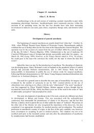
<strong>Skin</strong> (Fig. 8-1)<br />
The epidermis of the skin is derived from ectoderm in the early embryo. The glandular<br />
appendages of the skin (sebaceous glands, hair follicles, etc) develop from tubes and solid<br />
cords that invaginate from the covering ectoderm (Langman, 1975). The epidermis is made<br />
up of stratified squamous epithelium that consists of two categories of cells. The majority of<br />
cells undergo keratinization and form the various epithelial layers. The superficial keratinized<br />
cells of the skin are replaced continuously by cells arising as a result of the mitotic activity<br />
in the basal layer of the epidermis (Bloom and Fawcett, 1975). Melanocytes derived from<br />
neural crest cells are also found in the epithelium of skin and comprise a second cell type.<br />
The dermis is derived from embryonic mesoderm and has an average thickness of 1<br />
to 2 mm (Bloom and Fawcett, 1975). The outer surface of the dermis has an uneven border<br />
contacting the epidermis and is known as the papillary layer. The remainder of the dermis is<br />
called the reticular layer.<br />
Deep to the reticular layer of the dermis the anatomy of loose-skinned and fixedskinned<br />
animals diverges. In fixed-skinned animals (man and swine), the subcutaneous layer<br />
consists of loose connective tissue and a varying amount of fat cells and is a deeper<br />
continuation of the dermis and collagenous fibers continuous with those in the dermis (Bloom<br />
and Fawcett, 1975). The density of the collagenous fibers is related to the degree of cutaneous<br />
mobility over the underlying structures. In the palms and soles, for example, these fibers are<br />
particularly numerous. The deep surface of the subcutaneous layer is attached to the<br />
superficial fascia of underlying muscle where it is present.<br />
In loose-skinned animals (rat, rabbit, and dog) the panniculus carnosus muscle is<br />
firmly attached to the reticular dermis and is separated from the superficial fascia of<br />
underlying muscles by a loose areolar tissue layer. This layer allows for increased mobility<br />
of the superficial cutaneous-panniculus carnosus complex relative to the underlying tissue.<br />
This mobility afforded by the loose areolar tissue layer creates a greater dependence on direct<br />
cutaneous arterial supply than is seen in man.<br />
1
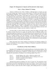
Neurovascular supply to skin<br />
The arterial supply to the skin can be divided into three functional units (Daniel and<br />
Williams, 1973): segmental vessels, which function to distribute blood from the aorta to the<br />
undersurface of muscle; perforating vessels, which provide nutritional support to muscle; and<br />
cutaneous vessels, which allow for thermoregulation and nutritional support of skin (Fig. 8-2).<br />
The segmental vessels originate as branches of the paired dorsal aortas during the<br />
embryologic period (Langman, 1975). Characteristics of the segmental vessels include (1) a<br />
perfusion pressure closely related to that found in the aorta, (2) a location deep to muscle, and<br />
(3) a common association with a nerve and vein (Daniel and Williams, 1973).<br />
The perforator vessels are branches of the segmental vessels. These vessels travel via<br />
one of two main routes to terminate in the cutaneous circulation. Musculocutaneous arteries<br />
pass through the overlying muscle to which they provide nutrition whereas direct cutaneous<br />
or septocutaneous arteries travel through fascial septa dividing muscular segments (Daniel and<br />
Kerrigan, 1990).<br />
The cutaneous portion of direct cutaneous (septocutaneous) arteries typically runs<br />
parallel to the skin surface providing nutrition to a large area of skin. Direct cutaneous<br />
arteries typically are accompanied by a pair of veins and run above the superficial muscular<br />
fascia (Webster, 1937). The more common musculocutaneous arteries leave the muscle and<br />
directly penetrate the subcutaneous tissue to supply a smaller region of skin.<br />
Both direct cutaneous and musculocutaneous arteries empty into a diffuse,<br />
interconnecting vascular network often referred to as the dermal and subdermal plexi. This<br />
network provides a redundancy in the vascular supply to the skin with the formation of<br />
collaterals at the periphery of the vascular territory formed by each musculocutaneous artery.<br />
The cutaneous microcirculation consists of a nutrient capillary network in the reticular dermis<br />
and arteriovenous shunts in the more superficial papillary dermis (Sherman, 1963). Arterioles,<br />
which act as preshunt and precapillary sphincters, regulate the flow through each vascular<br />
network (Greene, 1962). Lymphatic vessels form a plexus running parallel and deep to the<br />
network of blood capillaries (Bloom and Fawcett, 1975). The lymphatic capillaries end in<br />
blind sacs and conduct extracellular fluid back into the bloodstream.<br />
The neural supply to the skin originates from both sensory and sympathetic nerves.<br />
The sensory nerves are distributed in segmental fashion, forming dermatomes, and participate<br />
in the skin's protective function. The postganglionic terminals of cutaneous sympathetic nerves<br />
contain the neurotransmitter norepinephrine and are found in the area of cutaneous arterioles<br />
(Anden et al, 1969; Guyton, 1976; Mellander and Johansson, 1968).<br />
<strong>Skin</strong> <strong>Physiology</strong><br />
The skin serves as a sensory and a protective organ. The thick epidermal layers are<br />
largely impermeable to gases and to most liquids. Because of this, many agents that could<br />
have beneficial effects are ineffective when applied topically to intact skin. Preservation of<br />
sensation in transferred cutaneous flaps is desirable, but its effects on the physiology of flaps<br />
is unclear.<br />
2
The blood supply to the skin serves two important functions: it provides nutritional<br />
support and is a thermoregulatory mechanism for the body. Primarily because of its<br />
thermoregulatory function, the rate of blood flow through the skin is one of the most variable<br />
in the body. Under ordinary skin temperatures the amount of blood flowing through the skin<br />
(0.25 L/m 2 of body surface area) is approximately ten times the flow required for nutritional<br />
support (Guyton, 1976). Blood flow can increase up to seven times this value with maximal<br />
vasodilatation. When the body is exposed to extreme cold, blood flow can be reduced to<br />
levels that are marginal for cutaneous nutrition.<br />
The two vascular patterns found in skin, the nutrient capillary network and<br />
arteriovenous shunts, are integral in performing the two functions of cutaneous circulation.<br />
The amount of blood flow to the skin depends ultimately on arteriolar pressure and flow.<br />
Under conditions of adequate systemic vascular pressure, however, the distribution of the<br />
cutaneous blood flow is regulated by precapillary and preshunt sphincters (Greene, 1962).<br />
The sphincters in the two vascular systems respond to different stimuli. The<br />
precapillary sphincter, which controls the amount of nutritive blood flow to the skin, responds<br />
to local hypoxemia and increased metabolic byproducts by dilatation (Grange et al, 1976;<br />
Wideman et al, 1976). Under such conditions the blood flow is increased (reactive hyperemia<br />
being an example). The preshunt sphincters are involved in regulating the changes in blood<br />
flow that affect thermoregulation and systemic blood pressure (Folkow, 1960; Sherman,<br />
1963). Release of norepinephrine by the postganglionic sympathetic fibers results in<br />
contraction of the preshunt sphincters, diverting blood away from the skin surface where heat<br />
loss can occur. With increased body temperature, the sympathetic vasoconstrictor impulses<br />
decrease allowing for increased blood flow to the skin (Guyton, 1976).<br />
"Active" vasodilation can also occur with excessive body temperature. Local secretion<br />
of acetylcholine by sympathetic nerve fibers, either directly affecting vasodilator fibers or<br />
acting through the release of the potent vasodilator bradykinin from the sweat glands, may<br />
be responsible. The cutaneous circulation is also extremely sensitive to circulating<br />
norepinephrine and epinephrine. Thus, even in areas of skin that have lost their sympathetic<br />
innervation, a mass discharge of the sympathetic system will still result in intense<br />
vasoconstriction in the skin (Guyton, 1976).<br />
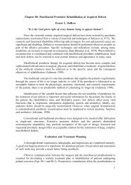
Classification of <strong>Flap</strong>s<br />
Improvement in skin-flap survival has resulted from improved flap designs that take<br />
advantage of the vascular anatomy. Adequate blood flow is so critical to survival that<br />
cutaneous flaps have been classified according to their blood supply (Daniel, 1975; Kerrigan<br />
et al, 1986) (Fig. 8-3).<br />
Random cutaneous flaps<br />
The blood supply to a random cutaneous flap is derived from musculocutaneous<br />
arteries near the base of the flap. Blood is delivered to the tip of the flap via the<br />
interconnecting subdermal plexus in the pedicle. The random cutaneous flap is commonly<br />
used in local reconstructions and can be rotated, transposed, advanced, or tubed.<br />
3
Length-to-width ratios of random cutaneous flaps have been recommended for various<br />
areas of the body. These differences reflect a regional variation of the neurovascular supply<br />
to the skin. Such a description can serve as a guide in designing random cutaneous flaps<br />
(Cook, 1986), but should not imply that a wider flap would extend survival length (Daniel,<br />
1975).<br />
Arterial cutaneous flaps<br />
Arterial cutaneous flaps (also called axial pattern flaps) typically have an improved<br />
survival relative to random cutaneous flaps. This advantage results from the incorporation of<br />
a direct cutaneous artery (recently classified as a septocutaneous artery by Daniel and<br />
Kerrigan, 1990) within its longitudinal axis. An island flap is an arterial flap with a pedicle<br />
consisting of nutrient vessels without the overlying skin. Island flaps can be useful to increase<br />
flexibility and reduce pedicle bulk in certain reconstructive procedures.<br />
Use of arterial cutaneous flaps is limited by the availability of direct cutaneous<br />
arteries. Examples of arterial cutaneous flaps used in head and neck reconstruction are the<br />
deltopectoral flap based on the anterior perforators of the internal mammary and the midline<br />
forehead flap based on the supratrochlear vessels.<br />
The surviving length of arterial flaps is related to length of the included direct<br />
cutaneous artery. Survival beyond the arterial portion of the flap is based on the subdermal<br />
plexus and is essentially a random cutaneous extension of the flap. <strong>Flap</strong> necrosis secondary<br />
to ischemia can be said to occur only in the random portion of the flap (destruction of the<br />
arterial pedicle making the entire flap random).<br />
Myocutaneous and fasciocutaneous flaps<br />
Myocutaneous flaps represent an additional modification to improve flap survival.<br />
Myocutaneous flaps are based on distal segmental vessels leaving the local vasculature<br />
(perforators and cutaneous vessels) intact. This requires incorporating muscle with the flap.<br />
Myocutaneous flaps are typically named for the donor muscle. Examples include the<br />
pectoralis myocutaneous flap based on the pectoral branch of the thoracoacromial artery and<br />
the latissimus dorsi myocutaneous flap based on the thoracodorsal artery.<br />
The increased blood flow and higher tissue oxygen tensions available with<br />
myocutaneous flaps (Gottrup et al, 1983, 1984) makes this design superior in the treatment<br />
of contaminated or infected defects. Improved phagocytotic and bactericidal activity of<br />
leukocytes is seen in myocutaneous flaps relative to random pattern flaps in the canine model<br />
(Eshima et al, 1990). These physiologic benefits contribute to the ability of myocutaneous<br />
flaps to resist bacterial inoculation more effectively than random pattern flaps.<br />
As is the case with arterial flaps, extending the surface area of the flap is often<br />
desirable in clinical situations. A random portion on the flap can be incorporated based on<br />
the subdermal plexus. This random extension is usually the portion of the flap most at risk<br />
of ischemic necrosis.<br />
4
Fasciocutaneous flaps use direct arterial (septocutaneous) vessels with the cutaneous<br />
branches at the level of the deep fascia, forming a plexus that supplies the subdermal plexus<br />
(Cormack and Lamberty, 1984). The appropriate size of fasciocutaneous flaps is less well<br />
defined than that of axial pattern flaps with their obvious arterial supply. Fasciocutaneous<br />
flaps appear to rely more on potential skin vascular territories. Four types of fasciocutaneous<br />
flaps have been described based on the pattern of blood supply incorporated into the fascial<br />
component of the flap. Examples include the parascapular flap and the radial forearm flap.<br />
Venous flaps<br />
<strong>Flap</strong>s with only an intact venous supply demonstrate the minimal nutritional<br />
requirements needed for flap survival. In flaps based on the dog saphenous or cephalic vein,<br />
survival occurred when the vein was intact on entering and exiting the flap, providing a flow<br />
through the venous system (Sasa et al, 1988). <strong>Flap</strong>s with proximal or distal ligation of the<br />
vein or with an arterial pedicle alone necrosed (Amarante et al, 1988; Baek et al, 1985). The<br />
surviving flaps showed no evidence of arterial blood flow as measured by injected<br />
microspheres until the third postoperative day (Sasa et al, 1988). Venous injections<br />
demonstrated little uptake in the flap, but capillary blood flow may not be ruled out in this<br />
model (Weinberg, 1988). Venous flaps performed in humans have been most successful in<br />
the distal extremities where multiple venous anastomoses and no valves are present (Chavoin<br />
et al, 1987).<br />
<strong>Physiology</strong> of Acutely Raised <strong>Flap</strong>s<br />
A number of changes detrimental to skin survival occur when a cutaneous flap is<br />
created. That flap survival occurs at all is a testimony to the minimal nutritional requirements<br />
of skin relative to the blood flow available in intact skin. The primary insult affect flap<br />
survival is impaired vascular supply and resultant ischemia. In the presence of adequate blood<br />
flow complete flap survival occurs. Nerve section and inflammation can also influence flap<br />
survival by affecting blood flow.<br />
Vascular<br />
Partial interruption of the vascular supply to the skin is the most obvious and critical<br />
change that occurs with elevation of a cutaneous flap. This interruption results in a local<br />
decrease in perfusion pressure to the skin. The decrease in perfusion pressure becomes more<br />
pronounced with increasing distance from the base of the flap (Cutting, 1982; Landis, 1927).<br />
When perfusion is reduced in one area, the adjacent vascular territories supplied by a separate<br />
perforating vessel can provide a low-pressure blood supply via the subdermal plexus. Because<br />
the nutritional requirements of skin are relatively low compared to the baseline skin blood<br />
flow, a number of vascular territories can be compromised before necrosis will result.<br />
In the arterial or myocutaneous portion of flaps the blood supply is usually adequate<br />
(Gottrup et al, 1984) and survival of the cutaneous covering is ensured. The survival length<br />
of the random portion of the flap depends on the physical properties of the supplying vessels<br />
(intravascular resistance) relative to the perfusion pressure (Daniel, 1975). Nutritional blood<br />
flow ceases and flap necrosis occurs when the perfusion pressure drops below the critical<br />
closing pressure of the arterioles in the subdermal plexus. In the past random cutaneous flaps<br />
5
were often designed relative to a desired length/width ratio - a wider base being needed to<br />
transfer a longer flap successfully. However, incorporation of additional vessels with the same<br />
perfusion pressure by widening the flap does not alter survival length (Daniel, 1975; Milton,<br />
1971).<br />
Myers (1986) has emphasized that "fresh flaps are always both viable and ischemic".<br />
Depending on the degree of ischemia and the amount of time before recovery of nutrient<br />
blood flow, the flap will either proceed toward necrosis or recovery. In the pig model, arterial<br />
and random flaps can tolerate an average of 13 hours of total avascularity and remain viable<br />
(Kerrigan and Daniel, 1982b). In the presence of less than total avascularity this period is<br />
probably much longer.<br />
In surviving flaps the reduced blood flow gradually increases. If the flap is placed in<br />
a favorable recipient site a fibrin layer forms within the first 2 days. Neovascularization of<br />
the flap begins 3 to 7 days after flap transposition. Early neovascularization has been detected<br />
at 4 days in the pig and rabbit models (Tsur et al, 1980; Verlander, 1964) and at 3 days in<br />
the rat model (Gatti et al, 1984). Revascularization adequate for division of the flap pedicle<br />
has been demonstrated by 7 days in animal models and man (Cummings and Trachy, 1985;<br />
Klingenstrom and Nylen, 1966; Tsur et al, 1980). Distal flap blood flow continues to increase<br />
during the second week despite what appears to be an adequate nutritional supply (Cummings<br />
and Trachy, 1985; Gottrup et al, 1984).<br />
Ischemia is one of many conditions that can induce angiogenesis (Abrams, 1983;<br />
Semashko et al, 1985). Angiogenic agents have been isolated from tumors and multiple<br />
tissues (Hom et al, 1988). In the presence of an angiogenic stimulus new capillaries arise<br />
from small venules in the recipient site and migrate toward the stimulus. Some capillaries join<br />
preexisting flap vessels (inosculation), but the majority of revascularization appears to involve<br />
direct ingrowth of recipient vessels into the flap (Smahel, 1977).<br />
The venous outflow from the skin is also impaired with flap elevation. Venous flow<br />
can occur through the subdermal plexus or via the single or paired venous channels that<br />
accompany the feeding artery in the pedicle. Complete venous occlusion in the early<br />
postelevation period may be more damaging to flap survival than inadequate arterial supply.<br />
Venous occlusion for 8 hours in the rat island skin flap model was incompatible with flap<br />
survival whereas 70% of flaps with a comparable arterial occlusion survived (Su et al, 1982).<br />
Fortunately, the subdermal plexus alone is often able to provide adequate venous outflow.<br />
Care must be taken, however, to preserve venous outflow in flaps pedicled solely on the<br />
feeding vessels.<br />
Impairment of lymphatic drainage with flap elevation also occurs. Reduction of the<br />
cutaneous lymphatic drainage results in an increase in interstitial fluid pressure that is<br />
compounded by increased leakage of intravascular protein associated with inflammation. The<br />
resulting edema formation can decrease capillary perfusion by increasing the intravascular<br />
resistance.<br />
6
Nerve section<br />
Both cutaneous and sympathetic nerves are severed in the process of flap elevation.<br />
Although loss of sensation may limit the usefulness of the flap after transfer, adrenergic<br />
denervation has implications for flap survival. When a sympathetic nerve is divided,<br />
catecholamines are released from the nerve terminal and the mechanism for catecholamine<br />
re-uptake is eliminated (Jurell et al, 1968; Palmer, 1970; Pearl, 1981). A local hyperadrenergic<br />
state exists, which produces vasoconstriction mediated by alpha-adrenergic receptors in the<br />
cutaneous vasculature.<br />
The vasoconstricting effect of sympathectomy further reduces the total flap blood flow<br />
(Kerrigan and Daniel, 1984; Pang et al, 1986c), which is already diminished by division of<br />
supplying vessels. This negatively affects the ratio of perfusion pressure to the critical closing<br />
pressure of the arterioles in the subdermal plexus, and a greater proportion of the distal flap<br />
is excluded from the blood supply. The stored transmitter is depleted within 24 to 48 hours<br />
(Jurell, 1986; Palmer, 1970) and blood flow increases as the concentration of norepinephrine<br />
declines (Pang et al, 1986c). In critical areas of the flap, however, the time to recovery of<br />
nutrient blood flow may be delayed sufficiently to produce additional necrosis.<br />
Inflammation/prostaglandins<br />
The surgical trauma associated with an acutely raised flap results in an inflammatory<br />
response. Inflammation consists of a vascular and cellular response to injury that prepares the<br />
tissue for the repair process. With injury, histamine, serotonin, and kinins are released into<br />
the extracellular compartment, markedly increasing the permeability of the microcirculation.<br />
The result is an increase in the concentration of proteins and cells within the extracellular<br />
space. This response can be beneficial as long as it is limited to nonbacterial inflammation,<br />
which begins prior to flap elevation (Liston, 1984; Macht and Frazier, 1980). The<br />
inflammation created by flap elevation may be deleterious because of the resultant edema<br />
formation.<br />
The action of the primary mediators of the inflammatory response (histamine,<br />
serotonin, and kinins) is short-lived. Following kinin formation and in the presence of<br />
complement, prostaglandins are synthesized by injured cells. Prostaglandins play an important<br />
role in the later stages of the inflammatory reaction while simultaneously initiating the early<br />
phases of injury repair.<br />
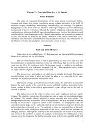
Prostaglandins are derived from 20-carbon essential fatty acids, which are incorporated<br />
in membrane phospholipids (Fig. 8-4). Activation of phospholipases results in the release of<br />
arachidonate from cell membrane phospholipids. Once released, arachidonate is metabolized<br />
by several distinct microsomal enzyme systems, one of which is cyclooxygenase. The action<br />
of cyclooxygenase results in the production of prostaglandin H 2 (PGH 2 ). PGH 2 is chemically<br />
unstable but can be transformed into a variety of products.<br />
Prostaglandin E 1 (PGE 1 ) and prostaglandin E 2 (PGE 2 ) can be synthesized from<br />
prostaglandin H 2 by isomerases in the vascular endothelium. Both PGE 1 and PGE 2 produce<br />
vasodilatation. Prostaglandin D 2 (PGD 2 ) is also formed by an isomerase reaction and is the<br />
principal cyclooxygenase product of the mast cell. Its effects on the cutaneous<br />
7
microvasculature are similar to PGE 1 . Prostacyclin (PGI 2 ) is a vasodilating agent and inhibitor<br />
of platelet aggregation that is derived from PGH 2 through the action of prostacyclin synthase.<br />
In the skin PGI 2 is primarily produced in the endothelial cells of blood vessels (Hauben and<br />
Aijlstra, 1984; Kaley et al, 1985). Prostacyclin is metabolized to 6-keto-PGF 1a .<br />
Thromboxane synthetase converts PGH 2 into thromboxane A 2 (TxA 2 ) and is primarily<br />
located in the platelets. Its effects include vessel constriction and promotion of platelet<br />
aggregation (Kay and Green, 1986). TxA 2 is unstable and rapidly converted into thromboxane<br />
B 2 (TxB 2 ). Prostaglandin F 2a (PGF 2a ) is derived from PGH 2 by a reductase reaction. PGF 2a<br />
does not appear to influence blood flow in segmental or perforating arteries but does result<br />
in venoconstriction at these levels. A marked increase in resistance is seen in cutaneous<br />
arteries, arterioles, and venules in the presence of PGF 2a (Nakano, 1973).<br />
The synthesis of prostaglandins and thromboxane can be altered by pharmacologic<br />
manipulation. The action of phospholipase A 2 can be inhibited by drugs that reduce the<br />
availability of Ca ++ . Glucocorticoids also affect phospholipase A 2 activity by inducing the<br />
synthesis of a protein that inhibits the enzyme (Campbell, 1990). Aspirin and other<br />
nonsteroidal antiinflammatory medications interfere with the cyclooxygenase enzyme, thus<br />
inhibiting the synthesis of PGH 2 .<br />
Recent studies have provided further insight into the activity of prostaglandins in<br />
ischemic flaps. Prostacyclin levels were found to increase 4 days after elevation of a porcine<br />
flank flap, to peak on day 7, and then to decrease up to postoperative day 21 (Hauben and<br />
Aijlstra, 1984). Elevation of a bipedicled rat dorsal flap resulted in elevated levels of PGE 2 ,<br />
PGF 2a , and TxB 2 , with a return to near-normal levels by day 7. Conversion to a single-pedicle<br />
flap ("delay") resulted in a blunted production of thromboxane and an elevated PGE 2 that<br />
lasted for at least 7 days. Elevation of an acute flap showed an elevation of PGE 2 , PGF 2a , and<br />
TxB 2 that was greater and more prolonged than seen with surgical delay (Murphy et al, 1985).<br />
Blood samples drawn from a rat hind limb rendered ischemic for 5 hours showed<br />
marked elevation of TxB 2 , 6-ketoprostaglandin F 2a (a metabolite of PGI 2 ), and PGE 2 (Feng<br />
et al, 1988). A difference between tissue tolerating reflow and tissue demonstrating no reflow<br />
was noted. Injection of 2% formic acid into the rat dorsal flap resulted in an increase of TxA 2<br />
and a small increase of PGE 2 . After flap elevation the flaps treated with formic acid<br />
demonstrated a decrease in TxA 2 and an increase in PGE 2 (Lawrence et al, 1984). It is clear<br />
from these studies that prostaglandins play a role in the inflammatory response after flap<br />
surgery. Whether these changes in prostaglandin levels represent a cause or a side effect of<br />
the observed phenomenon remains to be demonstrated.<br />
Reperfusion (free radicals)<br />
Return of blood flow to an ischemic flap under the influence of excess<br />
vasoconstriction due to excessive release of norepinephrine occurs in approximately 12 hours.<br />
With norepinephrine depletion and continued inflammatory response, blood flow can reach<br />
a maximum at 24 hours in the rat and pig models (Pang et al, 1986c; Sasaki and Pang, 1980).<br />
When oxygen becomes available with reperfusion, an additional menace to flap survival is<br />
produced, the free radical. This byproduct of reperfusion can cause damage at both the<br />
cellular and subcellular levels, contributing to postischemic tissue necrosis.<br />
8
Free radicals are extremely reactive compounds by virtue of an unpaired electron in<br />
their outer orbitals. Oxygen-free radicals are formed by the sequential univalent reduction of<br />
molecular oxygen. The superoxide anion radical (O 2- ) is formed by the addition of a single<br />
electron to molecular oxygen. Superoxide is a byproduct of adenosine triphosphate (ATP)<br />
production in the mitochondria and other oxidation reduction reactions (Southorn and Powis,<br />
1988). Polymorphonuclear cells are a second source of superoxide radicals, which are released<br />
in response to bacterial inflammation (Babior et al, 1973).<br />
A major source of free radicals in ischemic tissue is the enzyme xanthine oxidase<br />
(McCord, 1985) (Fig. 8-5). With ischemia, high-energy phosphate compounds are converted<br />
to hypoxanthine, which accumulates in the tissues. When oxygen becomes available with<br />
reperfusion, xanthine oxidase catalyzes the conversion of hypoxanthines into uric acid,<br />
producing superoxide in the process. This reaction is thought to be an important mechanism<br />
in postischemic tissue injury in skin flaps (McCord, 1986).<br />
Xanthine oxidase activity has been found in normal rat skin and increases its activity<br />
after venous occlusion and reperfusion (Im et al, 1984). Xanthine oxidase activity also<br />
increases after elevation of a dorsal rat flap with the highest levels being present distally<br />
(Angel et al, 1988). Tissue damage resulting from free radical production can occur from lipid<br />
peroxidation of the cellular membrane and denaturation of the intracellular matrix (Mulliken<br />
and Im, 1986; Southorn and Powis, 1988).<br />
Research Methods<br />
A large amount of literature is available on skin-flap physiology. The results of several<br />
studies give conflicting results. Experimental results are often difficult to interpret because<br />
of variations in choice of animal model, timing of treatment, route of drug administration,<br />
method of data collection, and repeatability of the study (Kerrigan and Daniel, 1982a). Some<br />
standardization of flap research methods would help resolve some of these difficulties.<br />
Guidelines for pharmacologic investigation of skin flaps were suggested by Kerrigan and<br />
Daniel (1982a). These recommendations include (1) postoperative treatment only, (2) control<br />
flaps on the same animal, (3) baseline fluorescein measurements, (4) double-blind<br />
experimental design, and (5) measurement of drug-induced changes in blood flow. No<br />
consensus has been reached regarding these or other guidelines.<br />
Two basic experimental designs have been used to investigate the consequences of a<br />
vascular insult on a surgical flap. In one design the blood supply to a flap is interrupted for<br />
varying amounts of time by occluding or otherwise interrupting flow through the vascular<br />
pedicle. The maximum amount of ischemic time the flap can survive in the experimental and<br />
control group is determined. This design is useful investigating the no-reflow phenomenon<br />
and ischemia tolerance. The second design involves flaps having a random extension in which<br />
the effect of experimental manipulation of blood flow or flap survival is compared to a<br />
control. From this basic framework a number of animal models and methods to assess blood<br />
flow and survival have been developed.<br />
9
Animal models<br />
The most commonly used model for flap research is the rat, a relatively inexpensive<br />
animal. A large amount of data is available for referencing. Unlike humans, rats are looseskinned<br />
and have a preponderance of skin supplied by direct cutaneous arteries. The<br />
abdominal flap is based on the epigastric vessels with an axial pattern on one side and a<br />
random extension as it crosses the midline or extends cranially. Petry and Wertham (1984)<br />
suggested that some of the survival variance in rat epigastric flaps is caused by an<br />
inconsistent incorporation of the lateral branch of the superficial epigastric artery. Dorsal flaps<br />
can be based caudad or cephalad. McFarlane et al (1965a) designed a dorsal rat flap so that<br />
it became necrotic when raised acutely but survived after a 2-week delay. The amount of<br />
necrosis in rat dorsal flaps can vary from 22% to 50% in the cranially based flaps (McFarlane<br />
et al, 1965a) and from 30% to 60% in caudally based flaps (Adamson et al, 1967). This<br />
variance seen in control animals often means that large numbers of animals are necessary in<br />
order to obtain meaningful results.<br />
The pig is another common animal model in flap research. Pigs are fixed-skinned<br />
animals with numerous musculocutaneous arteries and their cutaneous blood supply is more<br />
similar to that of man. Multiple flaps can be raised, allowing experimental and control flaps<br />
to be raised on the same animal. Kerrigan et al (1986) reviewed the flaps available on the pig.<br />
Random flaps included the dorsal flank flap, which had a predictable survival, the opportunity<br />
for up to 10 flaps per animal, and a position that enabled easy monitoring and care of the<br />
flap. Because a variable amount of panniculus carnosus can be included with the flap, it may<br />
not be completely random. This situation can be avoided by raising the flap superficial to the<br />
muscle. The random buttock flap has the advantage of not having a muscle component, but<br />
the experiment is limited to two flaps per animal.<br />
Arterial flaps on the pig flank correspond to the random flaps except that the pedicle<br />
is placed ventrally, preserving an arterial pedicle. The ventral flank flap, based 4 cm lateral<br />
to the nipple line, has the same advantages and disadvantages as its random counterpart. The<br />
arterial buttock flap has a large neurovascular pedicle, no muscle component, a large skin<br />
area, and a reliable survival (approximately 13 cm). Again, only two flaps per pig are<br />
possible.<br />
The pig is a satisfactory model for studying myocutaneous flaps because of the<br />
multiple perforating arteries supplying the skin. A variation of the myocutaneous flap model<br />
includes placing a catheter around the pedicle to allow pedicle occlusion at varying schedules<br />
(Cummings et al, 1985; Millican and Poole, 1985a). Kerrigan et al (1986) conclude that the<br />
latissimus dorsi flap based on the thoracodorsal artery is the best myocutaneous flap model<br />
in the pig. The gracilis myocutaneous flap had no reliable necrosis and a poor location. The<br />
rectus abdominis myocutaneous flap was faulted for having two dominant pedicles and a<br />
dependent position.<br />
A modification for the pig myocutaneous model was suggested by Haughey and Panje<br />
(1989). With their design up to 10 myocutaneous flaps are raised on a single animal.<br />
Problems with excess skin survival are reduced by limiting the size of the muscle block to<br />
4 x 4 cm. A random extension of skin 12 cm long is created beyond the muscle. A variation<br />
among the flaps can occur in that the skin can be loosely attached (gracilis, pectoralis major),<br />
10
separated by panniculus carnosus and subpannicular fat (latissimus dorsi), or separated by a<br />
dense deep fascia (biceps).<br />
The use of flaps created from pig skin to research changes in cutaneous surface area<br />
and thickness with tissue expansion has been criticized (Bartell and Mustoe, 1989). The<br />
biomechanical properties of pig skin were found to be at variance with human skin. Changes<br />
in cutaneous blood flow with tissue expansion are well studied in the pig model.<br />
Rabbits, like rats, are loose-skinned animals. Rabbits have been used as models in skin<br />
flap research when a larger skin area is desired in a random flap (Chu and Deshmukh, 1989).<br />
Forrest and Pang (1988) found that the skin over the latissimus dorsi muscle was supplied<br />
mainly by a direct cutaneous artery. The perforators present were few and could not support<br />
the flap. A similar result was found studying the pectoralis major myocutaneous flap (Nieto<br />
et al, 1985). The authors of both papers conclude that myocutaneous flap studies in the rabbit<br />
may have less relevance to human myocutaneous flaps than equivalent studies in the pig<br />
model.<br />
The canine was the first animal model used in skin flap research (Donovan, 1975). A<br />
number of studies using the canine model for investigation of flap physiology have been<br />
published and are discussed in this chapter. The dog is a loose-skinned animal and care must<br />
be used when investigating vascular changes in myocutaneous flaps. The biomechanical<br />
properties of canine skin have been found to be more similar to human skin than the<br />
properties of pig skin (Bartell and Mustoe, 1989). The expense and size of the model has<br />
limited its use when other models (rabbit, rat) can be substituted adequately.<br />
Perfusion measurement<br />
Direct observation of a flap is the most common method of assessing flap viability in<br />
clinical situations. Findings such as flap color, temperature, capillary refill, and bleeding at<br />
the distal edge give gross approximation of flap perfusion. Greater reliability is needed in<br />
clinical flaps having questionable viability and in the laboratory.<br />
Perfusion measurements are used in research to (1) quantify the effect of a particular<br />
agent on the blood flow to a flap, (2) obtain a baseline measurement to ensure that<br />
experimental and control flaps have an equivalent blood flow, and (3) predict the survival of<br />
a flap. Clinical uses of perfusion measurement have included monitoring the blood flow to<br />
a flap postoperatively and predicting flap viability at the time of surgery. Some of the<br />
techniques of perfusion measurement in skin flap research that are new or in frequent use are<br />
reviewed.<br />
Microspheres<br />
Microspheres are thought to be the most accurate means of estimating blood flow<br />
(Myers, 1986). The technique depends on three principles. First, the microspheres must be<br />
distributed to the tissues in direct proportion to their blood flow. For this to happen they must<br />
be well mixed and rheologically similar to red blood cells. Second, the microspheres must be<br />
trapped in the capillary bed in the first circulation. Finally, the systemic hemodynamics must<br />
not be affected by the embolization of the capillary bed (Pang et al, 1984).<br />
11
Microspheres are polystyrene beads of uniform size with isotopes placed inside. For<br />
measurement of capillary perfusion the beads are typically 15 microm in size to allow<br />
trapping in the capillary beds but passage through A-V shunts. Larger microspheres (50<br />
microm) can be used if trapping in the A-V shunts is desired. Microspheres are injected into<br />
the left ventricle, where they are mixed before being expelled with the blood and trapped in<br />
the tissue capillaries. The ratio of the blood flow in a specific tissue to the cardiac output<br />
equals the ratio of the number of microspheres trapped in that tissue to the total number of<br />
spheres injected. A blood sample is drawn at the time of microsphere injection to serve as a<br />
reference for calculating cardiac output and blood flow to the tissue being investigated (Pang<br />
et al, 1984).<br />
The microsphere technique was found to be linearly correlated with blood flow to the<br />
skin (Pang et al, 1984). When skin blood flow was low (approximately 0.03 mL/min), the<br />
repeatability of the technique was hindered. A blood flow this low is rarely seen in acute<br />
random flaps and would occur only when arterial spasm is at a maximum in the early<br />
postoperative period. By using different sets of microspheres, capillary blood flow can be<br />
measured simultaneously and consecutively to skin, muscle, and bone. A major disadvantage<br />
preventing its clinical use is the need to sample the tissue at the end of the experiment.<br />
Fluorescein<br />
Many vital dyes are available, including bromphenol, disulphine, patant blue, vicodan,<br />
and xylenol orange, but fluorescein is used most often. Sodium fluorescein dye (C 20 H 10 Na 2 O 5 ;<br />
molecular weight 376.3) is nontoxic at pharmacological doses of 10 to 15 mg/kg (Pang et al,<br />
1986a). LD-50 is 1000 mg/kg in laboratory animals. When exposed to ultraviolet light (< 510<br />
nanom), the dye will emit a yellow-green fluorescence. After intravenous injection the<br />
fluorescein moves quickly from the intravascular compartment to the extracellular space<br />
without penetrating the cell membranes. Staining occurs in tissues with a nutrient blood flow.<br />
Fluorescence can be detected visually with a Wood's light, photographically with the<br />
appropriate filters, or with a dermofluorometer.<br />
The visual fluorescein test is performed with a Wood's light, and the length of<br />
fluorescein staining is observed. With fluorescein photography a blue filter is placed over the<br />
flash and a yellow filter is placed over the lens. Both techniques require a relatively large<br />
dose of fluorescein (15 to 30 mg/kg). This dose can take 12 to 18 hours to clear, which limits<br />
how often the test can be performed. Both techniques are more difficult to perform in highly<br />
pigmented skin.<br />
Lower doses of fluorescein (1.5 mg/kg) can be used with a fiberoptic<br />
dermofluorometer. The dermofluorometer uses a fiberoptic cable to carry the ultraviolet light<br />
to the skin and transmit the induced fluorescence to a photodetector. A numerical output is<br />
generated, which can be read off the machine. For each estimation of blood flow, the skin<br />
fluorescence before and after fluorescein injection is measured. The rise in fluorescence of<br />
the skin under investigation and in a reference area are compared, and a dye fluorescence<br />
index (DFI) is calculated. By quantifying the fluorescence and lowering the fluorescein dose,<br />
blood flow can be examined at more frequent intervals.<br />
12
Areas in a skin flap with a rise in fluorescence that is approximately 30% of an area<br />
of normal skin (DFI = 30) would be expected to survive (Cummings et al, 1984; Sloan and<br />
Sasaki, 1985). This technique has been used to determine the optimal time for pedicle division<br />
of a regional flap (Gatti et al, 1984). An underestimation of actual skin flap survival with<br />
fluorescein has been noted when fluorescein is given early in the postoperative period. At 1<br />
hour after creating a skin flap the visual fluorescein test was found to underestimate skin flap<br />
survival by approximately 20% (Pang et al, 1986a). At 18 to 24 hours after surgery the flap<br />
survival was highly correlated with actual survival (Pang et al, 1986a; Sloan and Sasaki,<br />
1985). The underestimation of skin flap survival was thought to be due to postoperative<br />
arteriospasm in the distal portion of the flap (Pang et al, 1986a). Taking the changes in flap<br />
blood flow into account, Thomson and Kerrigan (1989) found a DFI of 7 was associated with<br />
flap survival when measured in the first 2 hours after surgery. A DFI of 27 was needed to<br />
ensure survival when measured 5 hours after flap elevation.<br />
Fluorescein uptake used as an indication of blood flow limits the measurement to<br />
intermittent readings. Fiberoptic fluorometry can also be used to monitor fluorescein washout<br />
or clearance (Denneny et al, 1986). Studies were done with arterial occlusion, venous<br />
occlusion, or pedicle occlusion for 30 minutes. With arterial occlusion a dramatic decrease<br />
in fluorescein elimination occurred and control levels returned with release of the clamp. With<br />
venous occlusion no fluorescein elimination occurred and partial occlusion led to prolonged<br />
elimination. The study concluded that by measuring fluorescein elimination, tissue perfusion<br />
could be monitored continuously.<br />
Laser doppler<br />
With the laser doppler a 2-mW helium-neon laser is used to produce a uniform light<br />
with a wavelength in air of 632.8 nanom. A fiberoptic cable is used to carry the light to the<br />
skin surface and to transmit the backscattered light to a photo-detector. Three different types<br />
of measurement can be obtained with a laser doppler. A laser doppler flow (LDF) signal is<br />
generated by measuring the movement of red blood cells. The doppler effect results in<br />
backscattered light from the surface of a stationary tissue plane having a different wavelength<br />
than light backscattered from a moving object (in this case a red blood cell). The number and<br />
average velocity of red blood cells determine the LDF value. A second measurement is laser<br />
photometry (LP). This signal is generated by the total intensity of backscattered light. At this<br />
wavelength light is mainly absorbed in the skin by the hemoglobin in red blood cells, so the<br />
LP value is inversely proportional to the blood volume of the tissue studied. In newer versions<br />
of the laser doppler the velocity of the red blood cells can be calculated (Phillips et al, 1989).<br />
Svensson et al (1985) used the LDF and LP outputs of the laser doppler to monitor<br />
free flaps and found that arterial and venous occlusion could be differentiated. With arterial<br />
or venous occlusion a dramatic decrease in the LDF occurred because of a lack of flow<br />
through the tissue. LP was noted to be unchanged or slightly increased with arterial occlusion,<br />
but dramatically decreased with venous occlusion, suggesting tissue engorgement. Phillips et<br />
al (1989) analyzed the LDF, blood volume, and velocity outputs of the laser doppler and<br />
found differences between arterial and venous occlusion in buttock island flaps raised in pigs.<br />
In this experiment the laser doppler detected decreased blood flow as early as 10 minutes<br />
after clamping the pedicle and could differentiate whether there was arterial or venous<br />
occlusion. At 60 minutes after arterial occlusion a 70% decrease in LDF, an 85% decrease<br />
13
in volume, but only a 10% decrease in velocity were observed. Sixty minutes after a venous<br />
occlusion a moderate decrease in LDF (70%) and velocity (50%), but only a small decrease<br />
in volume (15%) were detected.<br />
The quality of blood flow estimation with the laser doppler has had a mixed review.<br />
Marks et al (1984) felt that the laser doppler, like fluorescein, becomes more accurate at 24<br />
hours after flap elevation. Liu et al (1986) felt that the laser doppler was more likely to reflect<br />
nonnutritive blood flow in the immediate period after flap elevation. Sloan and Sasaki (1985)<br />
found the laser doppler to have an increased variability and had difficulty reproducing results.<br />
Reports of blood flow in nonperfused tissue have also been published (Fischer et al, 1985;<br />
Marks et al, 1984; Sloan and Sasaki, 1985). For this reason percentage change rather than<br />
absolute values are often followed when using the laser doppler clinically.<br />
Heden et al (1986) found the laser doppler to be as accurate as fluorescein in the rad<br />
dorsal flap if certain techniques were followed. This included immobilization of the skin to<br />
probe interface and monitoring a single site. The laser doppler has been used to monitor<br />
myocutaneous flaps (Cummings et al, 1984) and free flaps in humans. The laser doppler has<br />
the advantage of being relatively simple to use and is noninvasive and thus is an attractive<br />
option for continuous monitoring of revascularized tissue (Silverman et al, 1985).<br />
Metabolic monitoring<br />
Glinz and Clodius (1972) evaluated pH measurements in the subcutaneous tissue of<br />
pig pedicle flaps and found pH to be a reliable indicator of tissue necrosis. They found that<br />
tissues with a pH more than 0.35 units lower than adjacent normal tissue did not survive.<br />
Dickson and Sharpe (1985) studied pH changes in rat epigastric island flaps by placing a pH<br />
probe into the middle of the flap. They found a measurable fall in pH within a minute of<br />
clamping the pedicle. Arterial occlusion led to a faster pH fall to lower values than venous<br />
occlusion. In pig rectus abdominis flaps, pH measurement had similar results (Warner et al,<br />
1989). The pH changes in their study were nearly identical for the subcutaneous and muscular<br />
layers.<br />
Mahone and Lista (1988) used a pO 2 probe to monitor rabbit epigastric flaps. The<br />
measurement is based on the reduction of oxygen across an electrode pair. The pO 2 of the<br />
surrounding tissue is proportional to the current produced between the anode-cathode gap. In<br />
this study, arterial and venous occlusion could not be differentiated. Occlusion of the entire<br />
pedicle could be detected with an oxygen challenge test in which 100% oxygen was<br />
administered. If the pedicle was intact, measured pO 2 increased three to four fold. This<br />
increase was not present when the pedicle was occluded.<br />
Temperature monitoring of flaps has been criticized for having a slow response time<br />
and a small response (Warner et al, 1989). Monitoring of surface temperature is easy to obtain<br />
and requires simple equipment. Temperature probes can provide a continuous estimate of flap<br />
perfusion. <strong>Skin</strong> temperature is related to blood flow but not always in a predictable or reliable<br />
fashion. The temperature-blood flow relationship is influenced by core temperature, air<br />
temperature, humidity, light, and vasomotor responses. In the laboratory, Sloan and Sasaki<br />
(1985) found good correlation between temperature and survival, but noted that pedicle<br />
occlusion will be detected earlier by administration of cutaneous oxygen and fluorescein.<br />
14
Clearance<br />
The rate of removal of a particular substance can be used to estimate blood flow.<br />
Xenon, iodide, and sodium isotopes have been used in this regard. Hydrogen gas clearance<br />
and technetium are two techniques recently discussed in the literature. The use of fluorescein<br />
clearance has already been described.<br />
Measurement of hydrogen gas clearances uses electrodes placed into the dermis<br />
through a needle. A current is applied to one wire and the nearby hydrogen is ionized. The<br />
resulting microelectric current is measured by a second wire, and a clearance curve indicating<br />
the decline of hydrogen concentration is generated. Blood flow approximates the slope of the<br />
hydrogen clearance curve plotted against time. Koshu et al (1982) used a higher initial<br />
concentration of hydrogen to reduce the variability of the readings with hydrogen gas<br />
clearance. Suzuki et al (1985) found no influence on flap survival by the needle injections and<br />
the clearance of hydrogen correlated well with survival length and fluorescein staining. An<br />
advantage of the technique is the lack of a radioactive substance. The authors did not that<br />
there are several technical points that are important in obtaining an accurate and reliable<br />
measurement. These included the method of needle insertion and the temperature of the room<br />
and the skin.<br />
Technetium 99 pertechnetate clearance was used by Waterhouse et al (1986) to<br />
estimate blood flow. The isotope was injected intradermally and 240 readings were taken over<br />
a period of 40 minutes. Decreased clearance was associated with a decreased blood flow. The<br />
measured clearance rate was divided into a fast component occurring at 2 to 8 minutes<br />
postinjection and a slow component during the 28- to 40-minute readings. Young and Howell<br />
(1980) believe that isotope clearance is related to the depth of injection in pig skin. The fast<br />
component was related to clearance from the superficial papillary dermis, which comprises<br />
90% of skin blood flow and is involved with thermoregulation. The other 10% was found<br />
mostly in the deeper reticular dermis and was thought to comprise the slow component. In<br />
the study by Waterhouse et al (1986) a single clearance curve, implying loss of<br />
thermoregulatory blood flow, was seen in ischemic skin. Split-thickness skin grafts were also<br />
found to have a single clearance curve, suggesting that such grafts are revascularized as a<br />
single functional unit. This technique, however, was not felt to be useful in monitoring<br />
clinical flaps in the postoperative period.<br />
Measurement of interstitial changes<br />
Dynamic interstitial tissue compliance (change in volume or pressure) has been<br />
measured by injecting a small amount of fluid into a tissue and measuring the change in<br />
pressure (Odland and Cohen, 1988). The measurement reflects interstitial tissue pressure,<br />
which increases with ischemia and inflammation. Tissue pressure was found to be increased<br />
at all sites after flap elevation compared to normal skin ni the rat dorsal flap model. Distal<br />
location and increased time after flap elevation up to 18 hours was associated with further<br />
increases in interstitial pressure. Increased interstitial pressure is a potential factor in the noreflow<br />
phenomenon (Rosen et al, 1985) and in critical flaps limiting capillary nutrient blood<br />
flow and diffusion of nutrients into the interstitial space.<br />
15
Magnetic resonance imaging may be another way to investigate changes in the<br />
intercellular space after flap elevation. Interruption of the blood supply to the canine gracilis<br />
muscle flap resulted in a tendency for increased spin-spin relaxation time (T 2 ) but the sample<br />
was too small to be statistically significant (Greenberg et al, 1987).<br />
Attempts to Alter <strong>Skin</strong>-<strong>Flap</strong> Viability<br />
Kerrigan (1983) outlined extrinsic and intrinsic causes of skin-flap failure. Extrinsic<br />
reasons for flap necrosis are those not resulting from the design of the raised flap. Examples<br />
include systemic hypotension, infection, and pedicle compression. Often, these factors can be<br />
overcome in the clinical situation. The primary intrinsic factor affecting flap survival is<br />
inadequate blood flow. Numerous experimental attempts have been made to influence flap<br />
microcirculation and/or decrease the deleterious effects of inadequate flap blood flow. The<br />
most successful has been flap delay. Attempts to improve flap blood flow and cutaneous<br />
tolerance of ischemia have been less successful but continue to be active areas of research.<br />
Delay<br />
Four facts are accepted about the delay phenomenon. First, it requires surgical trauma;<br />
second, a large percentage of the neurovascular supply to the flap must be eliminated; third,<br />
delay results in increased flap survival at the time of tissue transfer; and fourth, the beneficial<br />
effects can last up to 6 weeks in the human (Pearl, 1984). To explain this phenomenon, three<br />
theories regarding the mechanism of delay have been developed: (1) delay improves the blood<br />
flow, (2) delay conditions the tissue to ischemia (McFarlane et al, 1965b), and (3) delay<br />
closes arteriovenous shunts (Reinsch, 1974). The most recent research supports a mechanism<br />
resulting in increased circulation to the flap, but how this occurs remains to be proved.<br />
Using microspheres Pang et al (1986b) found the percentage of arteriovenous shunt<br />
flow to be similar in delayed and acute flaps. The increased blood flow in delayed flaps was<br />
caused by an increase in total blood flow. The addition of systemic norepinephrine decreases<br />
the blood flow in delayed flaps to the level seen in acute flaps. The increased survival in the<br />
delayed flaps was thought to be caused by a decrease in vasoconstriction in the distal portion<br />
of the flap.<br />
<strong>Flap</strong>s delayed as little as 24 hours survive to a greater length (Sasaki and Pang, 1981)<br />
and can tolerate longer periods of ischemia (Weinberg et al, 1985, 1985). Using the<br />
microsphere technique, distal perfusion in the pig flank flap was found to increase with a<br />
delay up to 4 days. No further increase in perfusion was seen with continued delay up to 14<br />
days (Pang et al, 1986c). The early increase in blood flow was thought to occur too early to<br />
be caused by angiogenesis and a bipedicle flap design limited the effect of hypoxia as a<br />
stimulus. Pang et al (1986c) theorized that a modulation of the vasoactivity of the small<br />
arteries allowed delivery of more blood to the distal portion of the flap. This modulation<br />
could occur by release of vasoconstrictive substances (norepinephrine, thromboxane, and<br />
serotonin) during elevation of the bipedicled flap. Necrosis is not seen because the bipedicled<br />
flap has an adequate blood supply. Degeneration release of norepinephrine occurs soon after<br />
flap elevation and norepinephrine stores are largely depleted in the first 24 to 48 hours (Jurell,<br />
1986; Palmer, 1970). In bipedicle flaps, Cutting et al (1982) found that the catecholamine<br />
level started to rise 4 days after flap construction, whereas others (Jurell, 1986; Palmer, 1970)<br />
16
found the catecholamine levels to be depressed over a greater period. After depletion of the<br />
catecholamines a relative state of sympathectomy develops. Complete sympathetic denervation<br />
is unlikely in pedicled flaps because of the presence of sympathetic fibers in the periarteriolar<br />
tissue and between the media and adventitia of the vascular wall (Marshall, 1976; Somlyo and<br />
Somlyo, 1970). Because of the catecholamine depletion, conversion of the delayed flap to a<br />
single pedicle at this time is not accompanied by the same degree of vasoconstriction (Pang<br />
et al, 1986b).<br />
Early after elevation the vasculature to the flap has an increased sensitivity to the<br />
effects of adrenergic drugs (Pearl, 1981). Intravenous norepinephrine was shown to result in<br />
decreased blood flow to a myocutaneous flap in the porcine model despite increased blood<br />
flow to control skin (Moore et al, 1986). This represented a hypersensitivity to exogenous<br />
norepinephrine in this model. The investigators were also able to demonstrate a blunting of<br />
the norepinephrine-induced pressor effects by treatment with phenoxybenzamine, an alphaadrenergic<br />
blocking agent. The hypersensitivity to norepinephrine was seen at 2 and 5 days<br />
after flap elevation, which is similar to the period of 1 to 7 days after flap elevation when<br />
decreased tissue norepinephrine was found by Cutting et al (1982). This recovery from the<br />
hyperadrenergic state appears to play a role in the delay phenomenon.<br />
Development of vascular collaterals and reorientation of the major vascular channels<br />
is another mechanism for increasing blood flow to the distal portion of the single pedicle flap<br />
(Cutting et al, 1981; Guba, 1979). Using the rat dorsal flap model, Suzuki et al (1988) found<br />
the delay effect to be greater in narrow flaps as opposed to wide flaps. The longitudinal<br />
channeling was thought to be greater in the narrow flaps because more of the transverse<br />
vessels were cut. Longitudinal flow is also enhanced by vasodilating substances released by<br />
inflammation and mild ischemia (Suzuki et al, 1988). Pang et al (1986b) believed that the<br />
depletion of vasoconstricting substances played a role in the early stage of delay whereas<br />
locally released vasodilating substances were involved in the later stages.<br />
Increase blood supply<br />
Vasodilators<br />
Indirect. The intense vasoconstriction associated with release of norepinephrine in the<br />
early period after flap elevation would seem to hinder flap survival. As discussed above, one<br />
of the benefits of flap delay seems to be depletion of norepinephrine before the flap to be<br />
transferred is created. If this vasoconstriction could be blocked or reversed, the duration and<br />
severity of distal flap ischemia should be decreased. The result would be increased flap<br />
survival without the need for delay.<br />
Alpha-adrenergic blocking agents are directed against the catecholamine-induced<br />
vasoconstriction seen after flap elevation. Using the rat model, phenoxybenzamine resulted<br />
in improving flap survival in some studies (Finseth and Zimmerman, 1979; Myers and Cherry,<br />
1968; Wexler et al, 1975). Phenoxybenzamine and phentolamine ointments applied topically<br />
were also found to be effective in increasing flap survival in the rat model (Goshen et al,<br />
1985). Other investigators have been unable to reproduce beneficial effects in the rabbit or<br />
pig (Kerrigan and Daniel, 1982a; Myers, 1975). Depletion of norepinephrine stores before flap<br />
elevation with reserpine (Cutting et al, 1978; Jurrell and Jonsson, 1976; Kennedy et al, 1979;<br />
17
Kerrigan and Daniel, 1982a) and guanethidine (Aarts, 1980; Finseth and Adelberg, 1978;<br />
Hannigton-Kiff, 1974) has also met with mixed results and systemic toxicity.<br />
Direct. Direct vasodilators such as histamine, hydralazine, and topical<br />
dimethylsulfoxide have showed both beneficial effects and no effects on skin-flap survival<br />
(Kerrigan and Daniel, 1982a). Isoxsuprine is a phenylethylamine derivative of epinephrine<br />
having alpha-adrenergic receptor antagonist and beta-adrenergic receptor agonistic properties<br />
resulting in relaxation of vascular smooth muscle. In high doses it can decrease viscosity and<br />
inhibit platelet aggregation. This combination of actions and early experimental studies<br />
(Finseth and Adelberg, 1978; Finseth and Zimmerman, 1979) created optimism and a flurry<br />
of research on the effects of using isoxsuprine. Subsequent studies have shown minimal or<br />
no beneficial effects of isoxsuprine on skin-flap survival (Kerrigan and Daniel, 1984; Neligan<br />
et al, 1985; Pang et al, 1985; Wray and Young, 1984).<br />
Using the microsphere technique, isoxsuprine was found to increase blood flow in the<br />
area of the dominant artery in porcine myocutaneous and arterial flaps. No increase in blood<br />
flow was seen in the distal random portion of the flaps or in flap survival (Neligan et al,<br />
1985; Pang et al, 1985). Similar results were obtained with diazoxide, a non-diuretic thiazide<br />
and a direct dilator of arterial smooth muscle (Pang et al, 1985). The smaller vessels in the<br />
distal random portion of a flap were theorized to have a different sensitivity to vasodilator<br />
drugs than muscular or axial arteries. Manipulation of these distal vascular channels appears<br />
to be critical in increasing flap survival.<br />
Calcitonin gene-related peptide (CGRP) is a bioactive neuropeptide found in primary<br />
sensory neurons. A potent vasodilator, it is thought to stimulate smooth muscle relaxation by<br />
an endothelial-dependent mechanism. CGRP has been shown to improve blood flow in the<br />
rat epigastric flap (Knight et al, 1988), to improve survival in ischemic skin flaps in rats<br />
(Kjartansson et al, 1987a), and to delay onset of the no-reflow phenomenon (Westin and<br />
Heden, 1988). Pretreatment with capsaicin, which depletes neuropeptides from primary<br />
sensory neurons, results in a decreased survival of dorsal flaps in the rat (Kjartansson et al,<br />
1987b). These findings suggest a potential role for primary sensory neurons in cutaneous<br />
vascular control and flap survival.<br />
Acupuncture has also resulted in increased flap survival in the rat dorsal flap model<br />
(Jansen et al, 1989a). In the same model, electro-acupuncture increased blood flow in the<br />
same was as injection of CGRP (Jansen et al, 1989b). This supports the theory of a<br />
mechanism involving release of vasodilatory substances from sensory neurons causing the<br />
improved survival after acupuncture, but further study is needed.<br />
Topical application of vasodilating drugs directly to the skin-flap surface has had<br />
mixed results. Topical nitroglycerin (NTG) was found to increase survival in the rat<br />
abdominal flap and the porcine axial flank flap (Rohrich et al, 1984). NTG acts as a<br />
vasodilator with more potent venodilator than arteriodilator effects. Its action on veins was<br />
felt to contribute to the increased flap survival. When used on the cranially based dorsal rat<br />
flap, however, NTG did not result in improved survival (Nichter et al, 1985). On the other<br />
hand, intraperitoneal dimethyl sulfoxide significantly increased flap survival in the rat<br />
abdominal flap (Haller et al, 1987) despite its inconsistent record as a topical agent (Arturson<br />
and Khanna, 1970; Myers and Donovan, 1973).<br />
18
Calcium channel blockers are potent vasodilators that can potentially reduce necrosis<br />
by keeping calcium from entering ischemic cells. Because they are already used clinically for<br />
other disorders, they were thought to be promising for salvaging a critically ischemic flap<br />
(Myers, 1986). Nifedipine increased survival in rat dorsal flaps (Hira et al, 1990) but not in<br />
porcine dorsal random flank flap (Miller et al, 1985). The lack of beneficial effect in the pig<br />
model may have been caused by a decrease in systemic blood pressure. In the rat dorsal flap<br />
model, verapamil given by intraperitoneal injection also failed to increase flap survival over<br />
a control (Nichter and Sobieski, 1988). The failure of calcium channel blockers or other direct<br />
vasodilators to increase flap survival reproducibly indicates that mechanisms other than direct<br />
arterial dilatation are important in survival of the ischemic flap.<br />
Alter rheology<br />
In a homogeneous fluid that exhibits equal shear stress at different rates of shear, flow<br />
(Q) in a vessel can be approximated by the Poiseuille equation:<br />
Q = (deltaP * r 4 *pi)/l*8*n)<br />
where deltaP equals pressure gradient, r 4 equals the fourth power of the vessel radius,<br />
l equals vessel length, and n equals viscosity (Guyton, 1976). Although blood is a non-<br />
Newtonian fluid, the qualitative relationships in the equation remain applicable. In larger<br />
vessels of the circulation vessel radius is a dominant factor, but in the capillary<br />
microcirculation viscosity is more important. By decreasing the viscosity of blood it may be<br />
possible to increase flow to the distal random portion of the acutely raised flap and improve<br />
flap survival. Viscosity is influenced by the hematocrit, serum proteins, temperature, red blood<br />
cell deformability and aggregation, as well as other factors (Roth et al, 1988). Each of these<br />
factors can be potentially manipulated with a resultant change in viscosity.<br />
Hemodilution has been shown to decrease viscosity and have a beneficial effect on<br />
flap survival (Earle et al, 1974; Neilsen and Parkin, 1976; Ramasastry et al, 1985). Reducing<br />
blood viscosity by protein depletion also results in increased flap survival in rats (Ruberg and<br />
Falcone, 1978). Dextran solutions also result in decreased blood viscosity but a reproducible<br />
improvement in survival has not been attained (Goulain, 1967; Grabb and O'Neal, 1966).<br />
Pentoxifylline is a hemorrheologic agent used in the treatment of intermittent<br />
claudication. Chemically, it is a tri-substituted xanthine related to caffeine and theophylline.<br />
Pentoxifylline increases intracellular adenine triphosphate levels in red blood cells, which<br />
results in increased red blood cell deformability (Ehrly, 1976). Other effects include<br />
decreasing serum fibrinogen and platelet aggregability (Roth et al, 1988).<br />
A number of experiments have examined the effects of pentoxifylline on skin-flap<br />
survival. When given 7 to 10 days before flap elevation, pentoxifylline has resulted in<br />
increased flap survival in porcine dorsal flank flaps (Yessenow and Maves, 1989) and the rat<br />
dorsal flap (Roth et al, 19880. The increased survival was associated with a decrease in<br />
viscosity (Roth et al, 1988). Pentoxifylline needs to be administered for 2 to 4 weeks in order<br />
to achieve the desired effect Yessenow and Maves, 1989).<br />
19
Some studies have found increased survival with 24 hours or less of preoperative<br />
pentoxifylline (Hauben and Aijlstra, 1984; Monteiro et al, 1986; Nemiroff, 1988). Despite a<br />
50% increase in surviving length in treated flaps, no increase in distal blood flow could be<br />
detected using tagged red blood cells (Monteiro et al, 1986). Viscosity measurements also<br />
failed to show a difference from control (Roth et al, 1988). Direct measurements of red blood<br />
cell deformability after 24 hours of pentoxifylline treatment may provide an explanation for<br />
the increase in survival. Beneficial effects with limited preoperative dosing of pentoxifylline<br />
have not been uniform. No improvement in flap survival was seen in the rabbit caudally based<br />
dorsal flap (Chu and Deshmukh, 1989) and the porcine dorsal flank flap (Hodgson et al,<br />
1987).<br />
Fluosol-DA is a whole-blood substitute with low viscosity (particle size = 0.1 microm)<br />
and a high oxygen-carrying capacity (Chowdary et al, 1987b). When used alone Fluosol-DA<br />
(20%) failed to increase flap survival (Ramasastry et al, 1985). When combined with a highoxygen<br />
environment, a beneficial effect on flap survival has not been consistent in the rat<br />
model (Chowdary et al, 1987b; Ramasastry et al, 1985). Another fluorocarbon, oxypherol-ET,<br />
was found to increase survival in porcine flank flaps (Yessenow and Maves, 1988) but this<br />
has yet to be confirmed in other laboratories.<br />
Inflammation<br />
The surgical trauma associated with an acutely raised or delayed flap results in an<br />
inflammatory response. This response results in a local increase in blood flow, which could<br />
improve flap survival. Recent investigations have attempted to improve flap survival with<br />
different methods of creating an inflammatory response as well as to determine the<br />
mechanism by which inflammation produces a beneficial effect.<br />
Preoperative application of croton oil, which produces a superficial burn similar to a<br />
chemical peel, resulted in increased survival length in the dorsal rat flap (Liston, 1984).<br />
Increased survival was also seen after injecting 0.2% formic acid below the pannus in the rat<br />
dorsum (Lawrence et al, 1984). In contrast, application of a chemical peel to porcine dorsal<br />
flank flaps 48 hours before elevation did not result in improved survival length over control<br />
flaps (Gaughan et al, 1986). The untreated flaps in this study had a chemical peel applied to<br />
flaps 2 cm away on either side and may have benefited from the nearby inflammation. A<br />
similar mechanism was thought to be responsible for increased blood flow in skin flaps raised<br />
adjacent to previously delayed flaps (Jonsson et al, 1988).<br />
Low-power laser burns to the skin applied daily for 5 days either preoperatively or<br />
postoperatively resulted in increased flap survival in the dorsal rat flap (Kami et al, 1985).<br />
Examination of laser burn sites in unoperated skin showed increased blood flow measured by<br />
hydrogen clearance. Histologic exam of the burn sites showed hypovascular areas at the burn<br />
sites 1 hour after irradiation but increased blood vessel proliferation 2 days after irradiation.<br />
These studies demonstrate that the inflammatory response can be a stimulus for delay without<br />
sympathectomy or vascular division.<br />
Cyclooxygenase inhibitors, such as indomethacin, and ibuprofen have been shown to<br />
increase skin-flap viability (Robson et al, 1979; Sasaki and Pang, 1981). Glucocorticoids that<br />
inhibit phospholipase A 2 activity have increased flap survival in some studies (Kristensen et<br />
20
al, 1978; Mendelson and Woods, 1978; Mes, 1980). In the porcine model however,<br />
methylprednisolone was found to have no effect on survival or blood flow when given<br />
preoperatively or early in the postoperative period (Nakatsuka et al, 1985). Ibuprofen was also<br />
demonstrated to result in prolonged tolerance of ischemia (Douglas et al, 1987).<br />
Studies have also been performed using prostaglandins as experimental agents.<br />
Administration of prostacyclin has been shown to have a beneficial effect on flap survival in<br />
rate (Emerson and Sykes, 1981; Sasaki and Pang, 1981). Dilatation of the arterial pedicle and<br />
an increase in the total blood flow was seen in a nonischemic rabbit epigastric flap after local<br />
injection of prostacyclin (Knight et al, 1985). Low doses of alpha-cyclodexin clathrate (a<br />
stable PGE 1 ) had a beneficial effect on blood flow measured by hydrogen gas clearance and<br />
survival in a rabbit dorsal flap. At higher doses a resultant hypotension seemed to prevent an<br />
increase in blood flow to the flap (Suzuki et al, 1987). Low-dose PGI 2 has also been shown<br />
to enhance flap survival in the pig (Reus et al, 1984). In the rat model a topically effective<br />
analogue of PGE 2 was found to improve flap tolerance of 10 hours of pedicle occlusion<br />
(Silverman et al, 1989).<br />
Blocking TxA 2 synthesis has had mixed results. TxA 2 inhibition with dazmegrel, which<br />
should increase the effect of prostacyclin, resulted in no effect in the rat dorsal flap (Kay and<br />
Green, 1986). More recent studies have found inhibition of thromboxane synthetase to be<br />
successful in increasing ischemic flap viability (Ono et al, 1990) and tolerance of skin flaps<br />
to secondary ischemia (Mellow et al, 1990).<br />
Other<br />
A number of drugs with multiple effects that should alter the blood flow to the flap<br />
have been used with varying results. Ancrod, a defibrinogenating enzyme from the pit viper,<br />
has a selective affinity for fibrinogen and stimulates release of plasminogen activator with a<br />
resultant decrease in viscosity. The drug also causes increased endothelial generation of<br />
prostacyclin. When given postoperatively for 7 days, ancrod was unable to increase the viable<br />
surface area of porcine myocutaneous flaps (Moore and Cummings, 1988).<br />
Chlorpromazine's effects include alpha-adrenergic blockade, stabilization of cell<br />
membranes, serotonin antagonism, metabolic depression, and cooling. All these effects should<br />
be beneficial to the ischemic flap but the results from experimental studies have been mixed.<br />
No improvement in survival of the rat dorsal flap was seen when chlorpromazine was given<br />
30 minutes preoperatively and up to 7 days postoperatively. There was a significant<br />
improvement, however, when the postoperative treatment was continued for 14 days. The<br />
mechanism for this delayed beneficial effect is unknown (Bibi et al, 1986). Angel et al (1989)<br />
found chlorpromazine given preoperatively and 7 days postoperatively resulted in increased<br />
survival of dorsal rat skin flaps. Of chlorpromazine's effects, the increase in blood flow was<br />
believed to be more important than membrane stabilization. In contrast, chlorpromazine given<br />
preoperatively and 10 days postoperatively had no beneficial effect on flap survival in rats<br />
(Hoft et al, 1990).<br />
Application of a semipermeable membrane to the flap surface resulted in increased<br />
survival in the rat dorsal flap (Nicther et al, 1985). The improvement seen may be due to<br />
preservation of a moist environment and reduced depth of tissue loss (McGrath, 1981). In a<br />
21
study by Kaufman et al (1985) topical ointments were a hindrance to flap survival. Pressure<br />
dressings, however, resulted in increased survival due to improved contact between the flap<br />
and the recipient bed.<br />
The length of time a flap is dependent on its pedicle is determined by the rate of<br />
capillary ingrowth into the transferred tissue. Separation of porcine myocutaneous flaps from<br />
the recipient bed with silastic sheeting resulted in a prolongation of the time needed for<br />
revascularization (Millican and Poole, 1985b). In the same model, revascularization of muscle<br />
flaps required twice as much time as myocutaneous flaps demonstrating the importance of the<br />
dermal and subdermal vascular plexus. A potentially useful manipulation of neovascularization<br />
was demonstrated by Hom et al (1988). Increased flap survival and vascularity was seen when<br />
an endothelial growth supplement was applied in a sustained-release fashion.<br />
Tissue expansion has been demonstrated to increase the size of the transferred flap in<br />
experimental animals and man. Examination of expanded skin in the guinea pig has shown<br />
an increase in the thickness (Austad et al, 1982) and mitotic activity (Francies and Marks,<br />
1977) of the epidermal layer, indicating epidermal proliferation. Blood flow in expanded<br />
tissue is greater than in skin overlying a noninflated expander 1 hour after creation of a<br />
pedicled flap in the porcine model (Marks et al, 1986). The increased blood flow to expanded<br />
skin when compared to delay seems to be short-lived (<strong>Goding</strong> et al, 1988; Ricciardeli et al,<br />
1989). Apart from the acute changes seen with expander manipulation, flap viability and<br />
blood flow in expanded skin appears to be similar to that seen in delayed flaps (Sasaki and<br />
Pang, 1984).<br />
An interesting manipulation to increase survival of thin axial pattern skin flaps was<br />
proposed by Morrison et al (1990). In their study, the femoral artery and vein was implanted<br />
into the subdermal layer of skin in the rabbit model. After 8 to 12 weeks sufficient<br />
neovascularization had occurred to allow creation of a large skin flap based on the transferred<br />
pedicle. If confirmed in other laboratories, this technique may allow greater flexibility in the<br />
design of axial pattern flaps.<br />
Prolonged viability<br />
Protection against harmful agents<br />
The production of free radicals with reperfusion and the return of molecular oxygen<br />
to ischaemic tissue have been a recent focus of experiments attempting to improve flap<br />
survival. This research has focused on decreasing the production of free radicals and using<br />
agents that remove free radicals (free radical scavengers) from the immediate environment.<br />
Preoperative administration of allopurinol (a xanthine oxidase inhibitor) prevents the<br />
increased xanthine oxidase activity seen with acute flap elevation (Im et al, 1984). Improved<br />
survival of dorsal rat flaps has been accomplished with allopurinol when given at high doses<br />
(Angel et al, 1987; Pokorny et al, 1989), with lower doses having no effect (Angel et al,<br />
1987). The high doses required have led to concern about the use of allopurinol to increase<br />
flap survival in humans.<br />
22
A number of free radical scavengers are available to protect the tissues from<br />
destruction by free radicals. Superoxide dismutase (SOD), an intracellular free radical<br />
scavenger, catalyzes the conversion of superoxide to hydrogen peroxide (H 2 O 2 ) and molecular<br />
oxygen. When given systemically, SOD is an effective scavenger of the superoxide radical<br />
regardless of its source (McCord, 1986). SOD treatment has resulted in improved flap survival<br />
(Freeman et al, 1990; Pokorny et al, 1989; Zimmerman et al, 1987) and increased tolerance<br />
to ischemia in rat abdominal flaps (Marzella et al, 1988; Sagi et al, 1986). The beneficial<br />
effects of SOD appear to be partially suppressed when given in physiologic solutions, which<br />
may raise the pH of the ischemic tissue (Sagi et al, 1990). Administration of a copper chelator<br />
decreased flap survival in the rad abdominal flap model by deactivating endogenous SOD.<br />
This effect could be overcome by administration of relatively large doses of exogenous SOD<br />
(Freeman et al, 1990).<br />
Prolongation of the circulating half-life of SOD (normally 6 minutes) up to 30 hours<br />
(Boccu et al, 1982) can be accomplished by the attachment of polyethylene glycol to the<br />
molecule (PEG-SOD). Use of PEG-SOD improved flap survival of rad abdominal flaps<br />
compared to SOD and controls (Huang et al, 1987). Improved flap survival has also been<br />
demonstrated with a number of other naturally occurring compounds with free radical<br />
scavenging properties. These include deferoxamine (Angel et al, 1986a), vitamin E, vitamin<br />
A, vitamin C, glutathione (Hayden et al, 1987), various amino acids (Paniello et al, 1988) and<br />
amino-acid derivatives (Kim et al, 1990).<br />
The hydrogen peroxide formed by the dismutation of superoxide is not particularly<br />
harmful. In the presence of chelated metal complexes, however, hydroxyl radicals (OH-) are<br />
formed through a Fenton or Haber-Weiss reaction (Del Mastro, 1980). The hydroxyl radical<br />
is much more reactive and may be responsible for much of the damage inflicted by oxygenfree<br />
radicals (Southorn and Powis, 1988). Hydroxyl radical activity in skin-flap necrosis is<br />
implicated by the ability of dimethyl thiourea, a specific hydroxyl radical scavenger, in<br />
improving survival in rad abdominal flaps (Hayden et al, 1988).<br />
That iron acts as a catalyst producing free radicals in hematoma-induced flap necrosis<br />
is supported by Angel et al (1986b). In their experiment, blood instilled under ischemic rat<br />
dorsal flaps resulted in increased necrosis as compared to plasma or controls. Addition of<br />
deferoxamine, a specific iron chelator, to the instilled blood lessened the necrosis, whereas<br />
adding ferrous sulfate to instilled plasma increased the necrosis. Malonyldialdehyde, a product<br />
of free-radical-catalyzed lipid peroxidation, was increased in necrotic flaps overlying a<br />
hematoma, further implicating free radicals in this process.<br />
Increased tolerance of ischemia<br />
Increased oxygenation. Improvement in flap survival with hyperbaric oxygen<br />
treatment has been documented in the rat model (Kernahan et al, 1965; Perrins, 1975).<br />
Hyperbaric oxygen treatment, however, increases blood oxygen-carrying capacity by 20%, at<br />
the most (Kernahan et al, 1965). A greater effect of hyperbaric oxygen treatment may be to<br />
increase oxygen diffusion from surrounding perfused tissue to the ischaemic portion of the<br />
flap (Cutting, 1986). Increased flap survival occurs with treatments using 21% hyperbaric<br />
oxygen, implying that increased oxygen could be delivered with an increase in pressure alone<br />
(Tan et al, 1984).<br />
23
An 8-hour treatment of hyperbaric oxygen improved survival of rad dorsal flaps when<br />
given 48 hours before or 4 hours after flap elevation. No effect was seen with hyperbaric<br />
oxygen given 24 hours before or 48 hours after surgery (Nemiroff et al, 1985). This is<br />
consistent with earlier findings that a treatment delay of 24 hours or more after surgery results<br />
in little benefit from hyperbaric oxygen (Jurell and Kaijser, 1973). The effectiveness of<br />
hyperbaric oxygen given 48 hours preoperatively was thought to be the result of allowing<br />
enough time for increased fibroblastic activity and capillary ingrowth to occur. An experiment<br />
using a porcine model was less successful in improving flap survival. Hyperbaric oxygen<br />
given within 4 hours after surgery showed no difference in survival of flank, axial buttock,<br />
and random buttock flaps in the pig (Caffe and Gallagher, 1988).<br />
Nemiroff (1988) studied hyperbaric oxygen and pentoxifylline in the rat dorsal flap<br />
model. Both agents individually improved flap survival over a control group but had a<br />
synergistic effect when used in combination. This effect was felt to be a result of their<br />
separate but complementary mechanisms of action.<br />
Metabolic manipulation. Decreasing the metabolic requirements or increasing the<br />
metabolic reserves of skin are additional strategies for increasing flap survival. These<br />
approaches are based on the concept that flap necrosis occurs when tissue metabolic demand<br />
is greater than blood supply. Decreasing temperature is an effective way to reduce metabolic<br />
activity. Local hypothermia results in a dramatic decrease in blood flow to porcine<br />
myocutaneous flaps without hindering ultimate survival (<strong>Goding</strong> et al, 1991). A delay in<br />
necrotic changes has been noted earlier (Kiehn and Desprez, 1960). In both studies<br />
improvement in flap survival was not seen.<br />
Administration of adenosine triphosphate-magnesium chloride complex was used to<br />
increase survival in the rat abdominal flap (Zimmerman et al, 1987). The metabolic support<br />
provided by this compound was felt to delay the onset of irreversible cell damage.<br />
DIfluoromethylornithine (DFMO) was found to increase survival of abdominal flaps in rats<br />
(Perona et al, 1990). DFMO appears to reduce metabolic demand by inhibiting the synthesis<br />
of polyamines, which are required for protein synthesis and cellular proliferation.<br />
Impaired flaps<br />
Smoking tobacco is associated with an increased chance of flap necrosis in face-lift<br />
operations (Rees et al, 1984). Exposure to tobacco smoke resulted in increased flap necrosis<br />
of dorsal flaps in rats (Nolan et al, 1985) and hamsters (Craig and Rees, 1985). The<br />
deleterious effects of nicotine appear to be time dependent. Subcutaneous injection of nicotine<br />
resulted in decreased survival of rad dorsal flaps if given for 4 weeks preoperatively but not<br />
for 2 weeks (Forrest et al, 1987). Decreased distal and total capillary blood flow was seen as<br />
a result of exposure to nicotine for 5 weeks. The mechanism of tobacco or nicotine producing<br />
decreased flap survival is unknown, but may involve direct endothelial damage,<br />
vasoconstriction secondary to catecholamine release, or local concentrations of prostaglandins<br />
(Forrest et al, 1987).<br />
Radiation delivered in a single dose of 1000 rads did not effect survival of dorsal rat<br />
flaps when given within 2 days before or after flap elevation (Nemiroff et al, 1985). Radiation<br />
given 12 to 104 weeks before surgery resulted in decreased flap viability in a ventrally based<br />
24
porcine flank flap model (Young and Hopewell, 1983). A less important role for endarteritis<br />
obliterans in altered wound healing due to radiation has been proposed by two recent studies.<br />
In one, radiation of the recipient bed (5400 rads, completed 6 weeks preoperatively) was<br />
found to delay but not eliminate neovascularization of a rat epigastric flap (Clarke et al,<br />
1985). In the second study, an intact delay phenomenon was demonstrated in irradiated rat<br />
dorsal flaps (Fisher et al, 1984).<br />
The ischemic time occurring in the transfer of free flaps appears to hinder the flaps'<br />
ability to tolerate a second ischemic insult. Porcine ventral arterial island flaps initially<br />
exposed to 2 hours of ischemia followed by 12 hours of blood flow had a 50% survival rate<br />
after a subsequent 7.2 hours of "secondary ischemia" (Kerrigan et al, 1984). This is compared<br />
to 13 hours for the initial ischemic event needed to produce a 50% survival rate (Kerrigan and<br />
Daniel, 1982b).<br />
25