Vol 10 Part 14. An introduction to the immature stages of British Flies ...
Vol 10 Part 14. An introduction to the immature stages of British Flies ...
Vol 10 Part 14. An introduction to the immature stages of British Flies ...

Create successful ePaper yourself
Turn your PDF publications into a flip-book with our unique Google optimized e-Paper software.
Orthorrhapha-Brachycera which are mostly predaceous. In <strong>the</strong>se Brachycera <strong>the</strong><br />
pharynx is sclerotized and in some families (e.g. Dolichopodidae, Empididae, Stratiomyidae)<br />
may also be fused with <strong>the</strong> internal skele<strong>to</strong>n <strong>of</strong> <strong>the</strong> head. In <strong>the</strong> Cyclorrhapha<br />
<strong>the</strong> head consists <strong>of</strong> an outer membranous segment and <strong>the</strong> internal cephalopharyngeal<br />
skele<strong>to</strong>n, a characteristic feature <strong>of</strong> <strong>the</strong> familiar maggot type <strong>of</strong> larva. The cephalopharyngeal<br />
skele<strong>to</strong>n is normally divisible in<strong>to</strong> three main parts: <strong>the</strong> basal or pharyngeal<br />
sclerite (ps); <strong>the</strong> intermediate or hypopharyngeal sclerite (hs) (ten<strong>to</strong>ropharyngeal) and<br />
<strong>the</strong> mouth hooks or mandibles (m) (fig. 788). The pharyngeal sclerite consists <strong>of</strong> a pair<br />
<strong>of</strong> roughly U-shaped sclerites on ei<strong>the</strong>r side <strong>of</strong> <strong>the</strong> pharynx. The two arms <strong>of</strong> each <strong>of</strong><br />
<strong>the</strong>se sclerites are called <strong>the</strong> dorsal (de) and ventral cornua (vc) (singular cornu) or<br />
'wings' and <strong>the</strong> latter are fused <strong>to</strong> <strong>the</strong> pharynx on each side. The pharyngeal sclerites<br />
may be joined by a bridge anterodorsally and a pair <strong>of</strong> slender paras<strong>to</strong>mal bars may<br />
project from <strong>the</strong> anterior margin above <strong>the</strong> hypos<strong>to</strong>mal sclerite (fig. 788, p). Below <strong>the</strong><br />
basal part <strong>of</strong> <strong>the</strong> mandible <strong>the</strong>re may be a dental sclerite (ds) and beneath (Muscidae,<br />
fig. 911, ob, ar) or between (Calliphoridae, fig. 788) <strong>the</strong> apices <strong>of</strong> <strong>the</strong> mandibles o<strong>the</strong>r<br />
accessory oral sclerites (os) may be present. Papers by Roberts (1969- 197la) should be<br />
consulted for a detailed discussion <strong>of</strong> <strong>the</strong> mouthparts <strong>of</strong> larval Diptera in relation <strong>to</strong><br />
feeding habits. A knowledge <strong>of</strong> <strong>the</strong> functional morphology <strong>of</strong> mouth parts may provide<br />
important clues as <strong>to</strong> whe<strong>the</strong>r or not an unidentified larva is <strong>the</strong> cause <strong>of</strong> primary<br />
damage <strong>to</strong> its host.<br />
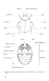
The antennae (fig. 2) are usually close <strong>to</strong> <strong>the</strong> anterior mandibular articulations near<br />
<strong>the</strong> anterodorsal corners <strong>of</strong> <strong>the</strong> genae (cheeks). They may be very small (e.g.<br />
<strong>An</strong>isopodidae, Bibionidae, Ptychopteridae, some Psychodidae, many Myce<strong>to</strong>philidae)<br />
and are normally subdivided in<strong>to</strong> three divisions (though in some Chironomidae seven<br />
may be evident). In Chaoboridae <strong>the</strong> antennae have evolved in<strong>to</strong> prehensile structures<br />
with apical spines used for capturing prey (fig. 45, an).<br />
The eyes are also located on <strong>the</strong> genae. In <strong>the</strong> Nema<strong>to</strong>cera and orthorrhaphous<br />
Brachycera <strong>the</strong> eyes are usually simple (double in many Chironomidae) but in some<br />
Culicidae and Chaoboridae a compound eye is present, in front <strong>of</strong> <strong>the</strong> simple eye<br />
(ocellus). Little work has been done on <strong>the</strong> eyes <strong>of</strong>larval Diptera but Roberts (1970b) is<br />
useful. <strong>An</strong> egg-burster or hatching spine may be present on <strong>the</strong> head <strong>of</strong> <strong>the</strong> first instar<br />
larva <strong>of</strong> some Diptera (fig. 984) (see Edwards, F. W., 1919; Madwar, 1934; Hin<strong>to</strong>n,<br />
1981 ). Egg-bursters in Diptera require fur<strong>the</strong>r investigation (see Smith, 1955a).<br />
Body<br />
This is very variable in shape. Most Nema<strong>to</strong>cera and some Brachycera are slender<br />
and subcylindrical; Therevidae, Scenopinidae and some Ephydridae and Canacidae<br />
are spindle-shaped (fusiform); Cyclorrhapha and Xylophagidae are markedly<br />
narrowed anteriorly; Fanniidae, Lonchopteridae, Platypezidae, Stratiomyidae and<br />
Xylomyidae are dorsoventrally flattened; Chaoboridae and Culicidae have a swollen<br />
thorax; Simuliidae are swollen posteriorly while some Syrphidae and parasitic groups<br />
are generally s<strong>to</strong>ut; Conopidae are pear shaped and Microdon (Syrphidae) species are<br />
hemispherical.<br />
Nema<strong>to</strong>cera usually have 12 body segments, o<strong>the</strong>r Diptera 11. Three <strong>of</strong> <strong>the</strong>se<br />
segments comprise <strong>the</strong> thorax and <strong>the</strong> remainder <strong>the</strong> abdomen. Variation in <strong>the</strong><br />
numbers <strong>of</strong> abdominal segments nearly always involves a decrease in Nema<strong>to</strong>cera and<br />
an increase in o<strong>the</strong>r Diptera.<br />
In <strong>the</strong> Chaoboridae, Culicidae and Simuliidae <strong>the</strong> three thoracic segments are fused.<br />
In <strong>the</strong> <strong>An</strong>isopodidae and Psychodidae subdivision <strong>of</strong> <strong>the</strong> segments, or pseudosegmentation,<br />
occurs (figs 40, 70, 71). The Therevidae and Scenopinidae have 20 segmental<br />
divisions.<br />
The cuticle <strong>of</strong>Diptera larvae is usually only weakly sclerotized and non-pigmented, a<br />
condition probably explained by <strong>the</strong>ir mainly concealed mode <strong>of</strong> life in a humid terrestrial<br />
atmosphere or by <strong>the</strong> adoption <strong>of</strong> an aquatic or semi-aquatic environment. Larvae<br />
26